Review
. 2016 Sep;283(Pt B):452-75.
doi: 10.1016/j.expneurol.2016.04.002. Epub 2016 Apr 9.

Inherited and acquired disorders of myelin: The underlying myelin pathology

Affiliations
Review

Inherited and acquired disorders of myelin: The underlying myelin pathology

Ian D Duncan et al. Exp Neurol. 2016 Sep.

Abstract

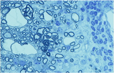
Remyelination is a major therapeutic goal in human myelin disorders, serving to restore function to demyelinated axons and providing neuroprotection. The target disorders that might be amenable to the promotion of this repair process are diverse and increasing in number. They range primarily from those of genetic, inflammatory to toxic origin. In order to apply remyelinating strategies to these disorders, it is essential to know whether the myelin damage results from a primary attack on myelin or the oligodendrocyte or both, and whether indeed these lead to myelin breakdown and demyelination. In some disorders, myelin sheath abnormalities are prominent but demyelination does not occur. This review explores the range of human and animal disorders where myelin pathology exists and focusses on defining the myelin changes in each and their cause, to help define whether they are targets for myelin repair therapy.

Keywords: Deficiency; Demyelination; Leukodystrophy; Multiple sclerosis; Myelin vacuolation; Toxin.


Figures

Fig. 1
Fig. 1
The normal oligodendrocyte and the development of myelin. a) A single oligodendrocyte, early in development has all the organelles required for lipid and protein production. Cisternae of rough endoplasmic reticulum are seen throughout the cell; the Golgi complex (GC) is present though not copious. Scattered mitochondria can be seen while a single lysosome (L) and peroxisome (P) are present. In the cytoplasmic tongue adjacent to the myelin sheath, microtubules (inset) are prominent. b) Most oligodendrocytes myelinate multiple axons as seen here in the developing cat spinal cord where the OL is in contact with and likely myelinates four axons. In a) and b) it is clear that myelination occurs first around larger diameter axons. c) In the mature white matter, myelinated axons are tightly packed. The correlation between axon diameter and myelin sheath thickness is obvious.
Fig. 2
Fig. 2
Myelin vacuolation does not always lead to myelin degeneration. a) In a case of canine mitochondrial DNA mutation, there is extensive vacuolation but no evidence of myelin breakdown or demyelination. b) In contrast in a feline model (FIDID), there is severe vacuolation, with evidence of degeneration of the inside loops of myelin adjacent to intact axons (arrow and enlarged in inset), and scattered demyelination and remyelination (arrowheads). Scale bar: 20 μm.
Fig. 3
Fig. 3
PLP mutations result in severe but variable deficiency in myelination. A. Sagittal and coronal sections of the brain of mature control and canine shp brains (over 18 months) show the marked deficiency in myelination of the mutant brain. B. In the rodent PLP mutants at 17–20 days of age compared to wildtype, there is a clear range of myelination from the mild hypomyelination seen in the mouse jprsh to the almost total absence of myelin in the PLP1 transgenic. In both the md rat and jp mouse the scattered myelin sheaths can be normal thickness and well compacted. C. In 1 μm sections of the spinal cord ventral column of the shp from 1 day to 2 years shows that the mutant lacks myelin at 1 day compared to control but myelin gradually increases with time. D.a. In the 80 day old long-lived md rat the majority of axons are non-myelinated but two larger ones have well compacted sheaths (D.a), while in other areas, sheaths are poorly compacted (D.b). On high power, while well compacted the mutant sheath in the md rat lacks an intraperiod line (D.d) compared to control (D.c). Wild type (WT), rumpshaker (jprsh), jimpy (jp), myelin deficient rat (md), PLP transgenic rat (PLP-tg). Scale bar: 20 μm (B), 10 μm (C).
Fig. 4
Fig. 4
Myelin loss in adrenoleukodystrophy. (A) Magnetic resonance imaging (MRI) scan showing the progression of demyelination in the parieto-occipital white matter over a period of three years in a child. At first, the patient had only neuropsychological deficits [(a), age seven; (b), age eight)], then showed a marked neurological deterioration at nine years of age (c). Note the progression of demyelinating lesions toward frontal lobes from (a) to (c). (B) ALD. Confluent demyelination of parietal and temporal white matter and posterior limb with some arcuate sparing in most superior parietal gyri. (Luxol fast blue-periodic acid Schiff, × 1).
Fig. 5
Fig. 5
The twitcher (twi) mouse develops progressive demyelination of the spinal cord and peripheral nerve. At 30 days of age the spinal cord appears normal (a) but a nerve root (arrow) seen at higher power (b) shows that demyelination has begun in the PNS but not CNS. By 50 days however the spinal cord is extensively demyelinated (c) with only the cortico-spinal tract (arrowhead) appearing to have retained myelin. In the area marked in (c) (arrow) there is clear demyelination of both the PNS and CNS (d). In the CNS, the white matter is invaded by unique, large macrophages known as globoid cells. These are seen in areas of severe demyelination (e and f). In some cases they are close to thinly demyelinated and myelinated axons that may be remyelinated or hypomyelinated (f). Toluidine blue. Scale bar: 0.2 mm (a, c), 20 μm (b, d–f).
Fig. 6
Fig. 6
Myelin vacuolation of the CNS in mitochondrial encephalomyelopathy. The spinal cord of a 4-week-old (a, b) and 15-week-old (c) affected dog shows variable but extensive myelin vacuolation, the severity of which is unrelated to age. In the 4-week-old dog, the myelin vacuolation is more severe in the thoracic cord (b) than the cervical cord (a); in panel b, there is little normal myelin (arrows). In the 15-week-old affected dog's thoracic cord, areas of myelin vacuolation occupy about 50% of the white matter (c). On higher power of adjacent normal (d) and vacuolated (e) areas of myelin, the splitting and vacuolation of myelin sheaths can be seen (toluidine blue). Higher power (f) of a vacuolated area clearly shows that the spongiform change is due to splitting of the myelin sheaths. Scale bar: a–c, 500 μm; d, e, 50 μm; f, 20 μm.
Fig. 7
Fig. 7
Mutation of the MBP gene in the Long Evans shaker (les) rat results in profound lack of myelin in the brain and loss of rudimentary myelin in the spinal cord. Staining of the brain of the les (b) rat fails to demonstrate myelin compared to controls (a). No detectable MBP immunostaining was evident throughout the CNS of the les rat (c, d), whereas levels of PLP appear to be only modestly reduced compared with that in controls (e, f) (44 day old les rats and controls). No MBP mRNA was detected throughout the les brain, whereas the expression and distribution of PLP mRNA appear similar to that in the control. g–i), Dark-field photomicrographs of 44-d-old sagittal brain sections from les (h, j) and control (g, i) rats hybridized with 35S-labeled probe for MBP (g–h) and PLP (i–j) mRNA. Robust expression of PLP mRNA in les verifies that normal numbers of mature oligodendrocytes are present in the mutant CNS. cb, Cerebellum; cc, corpus callosum; hf, hippocampal fimbria. k. An example of a myelinated axon in les. Although it is uncompact, the membrane sheaths in les form an organized multilamellar structure that is characteristic of a myelin sheath. Scale bar, 1 μm. l. Electron micrographs of the spinal cord from 2 week, 4 week, 3 month, and 9 month control and les animals. At 2 weeks, control rats begin to develop myelin sheaths that persist as the animal ages. In contrast, at 2 weeks, les animals develop thin, uncompact myelin sheaths. However, most of this attempt at myelination is lost by 4 weeks, and at later ages there is practically no myelin in the CNS. Scale bar, 1 μm.
Fig. 8
Fig. 8
Hypomyelination then demyelination in the taiep rat result from microtubule accumulation in oligodendrocytes. a) Sagittal sections of the brain of control and taiep rats at 1, 6 and 12 months show that the taiep rat has myelin at 1 month though less than control but myelin is gradually lost throughout the brain and brainstem. The spinal cord area of the taiep cord at 6 months (b) is less than control (a). This is most obvious in the dorsal column where by 12 months the fasciculus gracilis (asterisk) and corticospinal tract (arrowhead) have no obvious myelin (d). At 4 months, the ventral column of the taiep spinal cord (e) contains non-myelinated and hypomyelinated axons compared to controls (f). EM of the 4 month taiep spinal cord shows accumulation of microtubules in the cytoplasm of an OL (g) that are forming arrays, aligned with smooth ER (h). Processes of OLs contained densely packed microtubules (i). Accumulation of microtubules leads to failure of transport of certain proteins such as MAG that accumulate around the cell body of OLs in the taiep cerebellum (k) not seen in controls (j). Myelin is gradually lost in the optic nerve of taiep from 3 months (l), 6 months (m) and 12 months (n). Occasional examples of myelin breakdown are seen (o) and rare degenerating axons (p). Scale bar: 20 μm (e, f), 100 μm (j, k).
Fig. 9
Fig. 9
Myelin loss in the MS spinal cord can be severe. In the spinal cord from a patient with secondary progressive MS, almost two thirds of the cervical spinal cord is demyelinated. Perivascular cuffs of T cells (arrow in a) and in inset b) persist though inflammation is less likely than earlier in disease. c) EM of an area from a similar large plaque shows demyelinated axons embedded in gliotic scar. Scale bar: 1 mm (a).
Fig. 10
Fig. 10
EAE is an inflammatory, demyelinating disease with extensive cell infiltrate of T cells, B cells and macrophages from the periphery. All sections from relapsing–remitting EAE in the Biozzi mouse. a) Longitudinal section of the thoracic spinal cord showing both sub-pial inflammation (arrows) and perivascular cuffing of all vessels. b) There is clear cuffing of the ventral spinal artery of the spinal cord with cells migrating (*) toward the spinal cord. Inset of the area marked shows both plasma cells and T lymphocytes. A small area of demyelination (arrow) is enlarged in the right inset. c) Demyelination is extensive adjacent to a partially cuffed vessel and extends into the neuropil. d) Macrophages cuffing a vessel and migrating into the CNS on the left while those that have engulfed myelin are lipid filled and are surrounding a vessel on right, likely prior to returning to the circulation. e) In a Biozzi mouse in the third relapse, the loss of myelin is uneven with the left side of the cord and part of the dorsal column being severely affected. Three areas of the spinal cord (inset) are enlarged in f–h. The dorsal column (e-asterisk) contains both areas of demyelination (f) and adjacent areas with marked axon loss (g). In the ventral cord (↑) the hallmarks of EAE are seen (h) with demyelination, remyelination and axon degeneration. Scale bar: 10 μm (b-inset, f–h), 20 μm (b, c, d), 0.5 mm (e).
Fig. 11
Fig. 11
Topical hexachlorophene in the rat pup results in status spongiosus of the white matter. Four weeks after topical administration of hexachlorophene, the entire WM is vacuolated (a) but 2 weeks after stopping treatment (b), myelin appears to have recompacted and only scattered vacuoles remain.
Fig. 12
Fig. 12
The myelin changes in acute feline irradiated diet induced demyelination (FIDID). a) In early disease there is severe vacuolation of the ventral and lateral columns of the spinal cord, though not in the dorsal column and deep white matter adjacent to the grey matter (arrows). b) Higher power of the mid-line ventral column confirms the generalized vacuolation. c) Details of the cellular changes in b). Myelin breakdown with intact axons is widespread (arrows) as are demyelinated (arrowheads) and remyelinated axons (asterisks). d) One axon is present in a vacuolated myelin sheath (arrow) and in another (small arrow), the axon is bordered by myelin debris. e) Within the myelin debris, numerous macrophages are seen. f) The optic nerve of an affected cat is completely demyelinated. Toluidine blue 1 μm sections. Scale bar: 1 mm (a), 200 μm (b), 20 μm (c–f).
Supplementary Fig. 1
Myelin vacuolation is non-disease specific. In a mouse with EAE, myelin vacuoles are seen close to a vessel with perivascular cuffing and adjacent demyelination. Scale bar: 20 μm.

Comment in

References

    1. Adachi M., Schneck L., Cara J., Volk B.W. Spongy degeneration of the central nervous system (van Bogaert and Bertrand type; Canavan's disease). A review. Hum. Pathol. 1973;4:331–347. - PubMed
    1. Agamanolis D.P., Victor M., Harris J.W., Hines J.D., Chester E.M., Kark J.A. An ultrastructural study of subacute combined degeneration of the spinal cord in vitamin B12-deficient rhesus monkeys. J. Neuropathol. Exp. Neurol. 1978;37:273–299. - PubMed
    1. Almad A., Sahinkaya F.R., McTigue D.M. Oligodendrocyte fate after spinal cord injury. Neurotherapeutics. 2011;8:262–273. - PMC - PubMed
    1. Aubourg P. Axons need glial peroxisomes. Nat. Genet. 2007;39:936–938. - PubMed
    1. Back S.A., Rosenberg P.A. Pathophysiology of glia in perinatal white matter injury. Glia. 2014;62:1790–1815. - PMC - PubMed

Publication types