. 2016 Apr 12:13:6.
doi: 10.1186/s12987-016-0030-5.

Growth-factor reduced Matrigel source influences stem cell derived brain microvascular endothelial cell barrier properties

Affiliations

Growth-factor reduced Matrigel source influences stem cell derived brain microvascular endothelial cell barrier properties

Ronak Patel et al. Fluids Barriers CNS. .

Abstract

Background: Patient-derived induced pluripotent stem cells (iPSCs) are an innovative source as an in vitro model for neurological diseases. Recent studies have demonstrated the differentiation of brain microvascular endothelial cells (BMECs) from various stem cell sources, including iPSC lines. However, the impact of the culturing conditions used to maintain such stem cell pluripotency on their ability to differentiate into BMECs remains undocumented. In this study, we investigated the effect of different sources of Matrigel and stem cell maintenance medium on BMEC differentiation efficiency.

Methods: The IMR90-c4 iPSC line was maintained on mTeSR1 or in essential-8 (E-8) medium on growth factor-reduced (GFR) Matrigel from three different manufacturers. Cells were differentiated into BMECs following published protocols. The phenotype of BMEC monolayers was assessed by immunocytochemistry. Barrier function was assessed by transendothelial electrical resistance (TEER) and permeability to sodium fluorescein, whereas the presence of drug efflux pumps was assessed by uptake assay using fluorescent substrates.

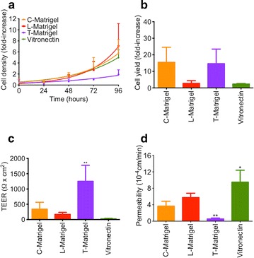
Results: Stem cell maintenance medium had little effect on the yield and barrier phenotype of IMR90-derived BMECs. The source of GFR-Matrigel used for the differentiation process significantly impacted the ability of IMR90-derived BMECs to form tight monolayers, as measured by TEER and fluorescein permeability. However, the Matrigel source had minimal effect on BMEC phenotype and drug efflux pump activity.

Conclusion: This study supports the ability to differentiate BMECs from iPSCs grown in mTeSR1 or E-8 medium and also suggests that the origin of GFR-Matrigel has a marked inpact on BMEC barrier properties.

Keywords: Barrier function; Blood–brain barrier; Drug transporters; Matrigel; Stem cells.


Figures

Fig. 1
Fig. 1
Effects of Matrigel origin on IMR90 iPS cell growth and BMEC differentiation using mTeSR medium. a Undifferentiated IMR90 cells maintained in mTeSR were dissociated as single cells seeded on Matrigel from BD Biosciences (B-Matrigel, blue), from Corning (C-Matrigel, orange), from ThermoFisher (L-Matrigel, red) or from Trevigen (T-Matrigel, purple) at 20 × 103 cells/cm2. After 24, 48 and 72 h following cell seeding, IMR90 iPSC colonies were dissociated and counted using a cell hemocytometer and Trypan blue exclusion assay. b BMEC yield after 8 days of differentiation obtained by calculating the ratio of cell density at 8 days of differentiation versus the initial cell density seeded at day 0, ** denotes P < 0.01 in comparison to B-Matrigel. c Representative immunostained micrographs of PECAM-1, VE-cadherin, Glut-1 Claudin 5 and Occludin expression in purified BMEC monolayers 48 h post-purification. Note the formation of a monolayer as marked by defined tight junction complexes in one focal plane and the absence of junctional cellular overlap. Scale bar = 20 µm
Fig. 2
Fig. 2
Matrigel origin impacts BMECs barrier properties. a, b TEER and sodium fluorescein permeabilities in IMR90-derived BMECs differentiated on B-, C- or T-Matrigel. Note the degraded barrier function of cells grown on C-Matrigel compared to the two other groups. * and ** denotes P < 0.05 and P < 0.01 in comparison to B-Matrigel. c Representative immunostained micrographs of P-gp, BCRP and MRP1 expression in purified BMECs monolayers. Scale bar = 40 µm
Fig. 3
Fig. 3
Matrigel origin has a minor impact on BMECs drug efflux activity. a Drug uptake of rhodamine 123, BODIPY FL-prazosin and CM-DCFDA in presence of 5 μM cyclosporine A (CsA, P-gp inhibitor), 1 μM Ko143 (BCRP inhibitor) or 10 μM MK571 (pan-MRP inhibitor), respectively. Fluorescence values in controls (no inhibitor) were arbitrarily set to 100 %, * and ** denote P < 0.05 and P < 0.01 in comparison to B-Matrigel. b Doxorubicin accumulation profile in presence of CsA, Ko143 or MK571. Doxorubicin net uptake was calculated as previously described
Fig. 4
Fig. 4
Essential-8 medium does not impact BMECs differentiation. a IMR90 iPSC growth curve on C-Matrigel (orange), L-Matrigel (red), T-Matrigel (purple) or vitronectin (green)-coated surfaces. IMR90 iPSCs were seeded as single cells at day 0 at a density of 20 × 103 cells/cm2 on the different types of matrices and maintained in E-8 medium. At each timepoint (day), IMR90 iPSC colonies were dissociated as single cells using Accutase and counted with a cell hemocytometer using 0.4 % Trypan blue as an exclusion dye. b Cell yield at day 8 of differentiation. Cell yield was determined by dividing average density at day 8 of differentiation by the initial cell seeding density at day 0. c, d TEER and sodium fluorescein permeability values on purified iPSC-derived BMECs monolayers 48 h after purification. Note the poor barrier properties in cells differentiated on Geltrex (L-Matrigel) or on vitronectin-N (vitronectin), * and ** denote P < 0.05 and P < 0.01 in comparison to C-Matrigel
Fig. 5
Fig. 5
Essential-8 medium. a Drug uptake profile of rhodamine 123, BODIPY-prazosin and CM-DCFDA in presence of CsA, Ko143 or MK571. Drug uptakes of the same efflux substrates in absence of inhibitors were used as controls. Fluorescence in controls was arbitrarily set to 100 %. b Doxorubicin drug uptake profile in presence of CsA, Ko143 or MK571, * and ** denote P < 0.05 and P < 0.01 in comparison to C-Matrigel

References

    1. Blanchette M, Daneman R. Formation and maintenance of the BBB. Mech Dev. 2015 - PubMed
    1. Palmiotti CA, Prasad S, Naik P, Abul KM, Sajja RK, Achyuta AH, et al. In vitro cerebrovascular modeling in the 21st century: current and prospective technologies. Pharm Res. 2014;31(12):3229–3250. doi: 10.1007/s11095-014-1464-6. - DOI - PMC - PubMed
    1. Obermeier B, Daneman R, Ransohoff RM. Development, maintenance and disruption of the blood–brain barrier. Nat Med. 2013;19(12):1584–1596. doi: 10.1038/nm.3407. - DOI - PMC - PubMed
    1. Abbott NJ. Blood–brain barrier structure and function and the challenges for CNS drug delivery. J Inherit Metab Dis. 2013;36(3):437–449. doi: 10.1007/s10545-013-9608-0. - DOI - PubMed
    1. Wilhelm I, Krizbai IA. In vitro models of the blood–brain barrier for the study of drug delivery to the brain. Mol Pharm. 2014;11(7):1949–1963. doi: 10.1021/mp500046f. - DOI - PubMed

Publication types

LinkOut - more resources