Clinical Trial
. 2016 Apr 12:7:11290.
doi: 10.1038/ncomms11290.

T-cell activation is an immune correlate of risk in BCG vaccinated infants

Affiliations
Clinical Trial

T-cell activation is an immune correlate of risk in BCG vaccinated infants

Helen A Fletcher et al. Nat Commun. .

Erratum in

  • Corrigendum: T-cell activation is an immune correlate of risk in BCG vaccinated infants.
    Fletcher HA, Snowden MA, Landry B, Rida W, Satti I, Harris SA, Matsumiya M, Tanner R, O'Shea MK, Dheenadhayalan V, Bogardus L, Stockdale L, Marsay L, Chomka A, Harrington-Kandt R, Manjaly-Thomas ZR, Naranbhai V, Stylianou E, Darboe F, Penn-Nicholson A, Nemes E, Hatherill M, Hussey G, Mahomed H, Tameris M, McClain JB, Evans TG, Hanekom WA, Scriba TJ, McShane H. Fletcher HA, et al. Nat Commun. 2016 May 6;7:11633. doi: 10.1038/ncomms11633. Nat Commun. 2016. PMID: 27151680 Free PMC article. No abstract available.

Abstract

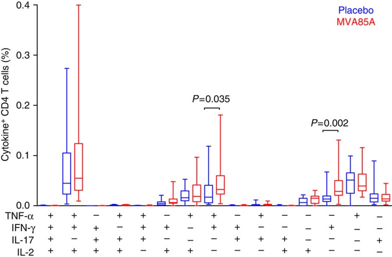
Vaccines to protect against tuberculosis (TB) are urgently needed. We performed a case-control analysis to identify immune correlates of TB disease risk in Bacille Calmette-Guerin (BCG) immunized infants from the MVA85A efficacy trial. Among 53 TB case infants and 205 matched controls, the frequency of activated HLA-DR(+) CD4(+) T cells associates with increased TB disease risk (OR=1.828, 95% CI=1.25-2.68, P=0.002, FDR=0.04, conditional logistic regression). In an independent study of Mycobacterium tuberculosis-infected adolescents, activated HLA-DR(+) CD4(+) T cells also associate with increased TB disease risk (OR=1.387, 95% CI=1.068-1.801, P=0.014, conditional logistic regression). In infants, BCG-specific T cells secreting IFN-γ associate with reduced risk of TB (OR=0.502, 95% CI=0.29-0.86, P=0.013, FDR=0.14). The causes and impact of T-cell activation on disease risk should be considered when designing and testing TB vaccine candidates for these populations.


Conflict of interest statement

H.M. was previously a shareholder in the Oxford-Emergent Tuberculosis Consortium (OETC), a joint venture established for the development of MVA85A (OETC no longer exists). The remaining authors declare no competing financial interests.

Figures

Figure 1
Figure 1. Cohort of infants from the MVA85A efficacy trial included in this immune correlates case–control study.
At 4–6 months of age, blood was collected from HIV-negative, HIV unexposed BCG-vaccinated infants with no active or chronic illnesses (including suspected TB), and with no household exposure to an adult who had active TB disease. Infants were then randomized to receive either MVA85A or placebo and followed for 2–3 years. TB cases (n=71) were defined by a positive culture for M. tuberculosis or the presence of M. tuberculosis DNA using a certified molecular diagnostic assay; or a stringent composite end point, which included symptoms, radiological signs and an exposure history. Control infants (n=213) had no evidence of TB exposure, no evidence of TB infection on interferon gamma release assay (IGRA) and were matched (3 controls to 1 case) on age, race, time on study and CDC weight percentile.
Figure 2
Figure 2. Distribution of significant immune variables in TB case and controls from the infant MVA85A vaccine trial.
Immune variables P<0.05 at D0 or D28 are shown. Infants are stratified according to TB case (red) or control (grey) status. The 50th percentile of the immune variable is indicated by a horizontal line. The shape of the point indicates whether an infant was in the placebo (round) or MVA85A (diamond) arm of the study. There were no significant differences in immune variables when infants were stratified by study arm. Combined low, medium and high immune responses at D0 (a,c,e) or D28 (b,d,f) were used to divide the all samples (both case and control groups) into thirds; medium third is indicated by the grey horizontal bar. The log frequency of live CD3+ cells expressing CD4, CD4 and HLA-DR or CD8 and HLA-DR, respectively. SFC/million PBMC is the number of IFN-γ spot-forming cells per million PBMC (only results from infants with a PHA response greater than 1,000 SFC/million are shown). Optical density was measured by ELISA at wavelength 405 nm.
Figure 3
Figure 3. BCG-specific CD4 T-cell response in whole blood.
Frequencies of cytokine-expressing BCG-specific CD4-positive T cells expressing IFN-γ, tumour-necrosis factor (TNF)-α, IL-2 or IL-17 measured by whole blood intracellular cytokine staining 28 days after administration of placebo (n=19) or MVA85A (n=17). Only P-values<0.05 shown. Mann–Whitney test, not adjusted for multiple comparisons.
Figure 4
Figure 4. Frequencies of HLA-DR-expressing CD4+ T cells in latently M. tuberculosis-infected adolescents.
Frequencies of HLA-DR-expressing CD4+ T cells in M. tuberculosis-infected adolescents who progressed to TB disease (red; progressors, n=61 samples from 30 progressors) compared with matched controls who did not progress to disease (grey; controls, n=132 samples from 59 controls).
Figure 5
Figure 5. Risk of TB disease over time by magnitude of immune response (TB cases only).
Hazard of disease over time for HLA-DR+ CD4+ T cell, BCG ELISpot, CD4+ T-cell and Ag85A IgG OD immune response magnitude is shown for TB case infants. All immune responses were measured at D0 before immunization with MVA85A. For each immune parameter, TB cases were stratified into subgroups divided into thirds according to immune response level (low blue, mid green and high yellow). The plots show the cumulative hazard of TB over time for the three immune level groups.
Figure 6
Figure 6. Ag85A IgG is boosted by MVA85A but is also increased in the placebo group from D0 to D28.
Ag85A-specific IgG stratified by vaccine group (MVA85A D0 n=79, MVA85A D28 n=107, placebo D0 n=88 and placebo D28 n=95). Lines represent median responses; round: placebo, diamond: MVA85A, red: case, grey: control. Although Ag85A IgG is boosted by MVA85A, Ag85A-specific IgG is significantly higher at D28 when compared with D0 in both the placebo and MVA85A groups.

Similar articles

Cited by

References

    1. Hawkridge A. et al.. Efficacy of percutaneous versus intradermal BCG in the prevention of tuberculosis in South African infants: randomised trial. BMJ 337, a2052 (2008). - PMC - PubMed
    1. Altare F. et al.. Impairment of mycobacterial immunity in human interleukin-12 receptor deficiency. Science 280, 1432–1435 (1998). - PubMed
    1. Jouanguy E. et al.. Interferon-gamma-receptor deficiency in an infant with fatal bacille Calmette-Guerin infection. N. Engl. J. Med. 335, 1956–1961 (1996). - PubMed
    1. Lichtenauer-Kaligis E. G. et al.. Severe Mycobacterium bovis BCG infections in a large series of novel IL-12 receptor beta1 deficient patients and evidence for the existence of partial IL-12 receptor beta1 deficiency. Eur. J. Immunol. 33, 59–69 (2003). - PubMed
    1. Newport M. J. et al.. A mutation in the interferon-gamma-receptor gene and susceptibility to mycobacterial infection. N. Engl. J. Med. 335, 1941–1949 (1996). - PubMed

Publication types

MeSH terms