. 2016 Jun;34(6):1692-701.
doi: 10.1002/stem.2376. Epub 2016 May 18.

Analysis of Bone-Cartilage-Stromal Progenitor Populations in Trauma Induced and Genetic Models of Heterotopic Ossification

Affiliations

Analysis of Bone-Cartilage-Stromal Progenitor Populations in Trauma Induced and Genetic Models of Heterotopic Ossification

Shailesh Agarwal et al. Stem Cells. 2016 Jun.

Erratum in

Abstract

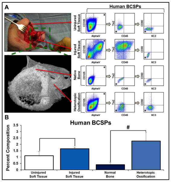
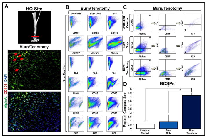
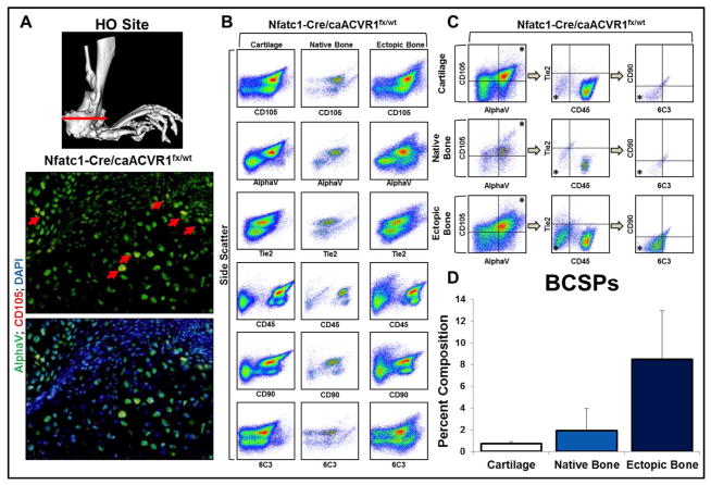
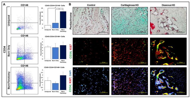
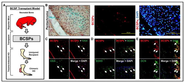
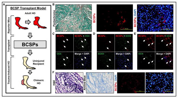
Heterotopic ossification (HO), the formation of extra-skeletal bone in soft tissues, is a pathologic process occurring after substantial burns or trauma, or in patients with type I bone morphogenetic protein (BMP) receptor hyperactivating mutations. Identifying the cells responsible for de novo bone formation during adulthood is of critical importance for therapeutic and regenerative purposes. Using a model of trauma-induced HO with hind limb Achilles' tenotomy and dorsal burn injury and a genetic nontrauma HO model (Nfatc1-Cre/caAcvr1(fl/wt) ), we demonstrate enrichment of previously defined bone-cartilage-stromal progenitor cells (BCSP: AlphaV+/CD105+/Tie2-/CD45-/Thy1-/6C3-) at the site of HO formation when compared with marrow isolated from the ipsilateral hind limb, or from tissue of the contralateral, uninjured hind limb. Upon transplantation into tenotomy sites soon after injury, BCSPs isolated from neonatal mice or developing HO incorporate into the developing lesion in cartilage and bone and express chondrogenic and osteogenic transcription factors. Additionally, BCSPs isolated from developing HO similarly incorporate into new HO lesions upon transplantation. Finally, adventitial cells, but not pericytes, appear to play a supportive role in HO formation. Our findings indicate that BCSPs contribute to de novo bone formation during adulthood and may hold substantial regenerative potential. Stem Cells 2016;34:1692-1701.

Keywords: Bone; Bone marrow stromal cells; Cell migration; Chondrogenesis; Experimental models; Pericytes; Progenitor cells.


Figures

Figure 1
Figure 1. Human HO and injured tissues are enriched for BCSPs when compared to uninjured soft tissue and native bone
A) FACS schematic demonstrating the identification of BCSPs (AlphaV+/CD105+/CD45-/Tie2-/CD90-/6C3-) from human tissues at the following sites: uninjured soft tissue, injured soft tissue, native bone, and heterotopic ossification; B) Quantification of BCSPs in human tissue at the site of injury as a percent of viable cells (Uninjured soft tissue (n=3): 1.12% (s.d. 0.33%); injured soft tissue (n=2): 1.66% (s.d. 0.11%); normal bone (n=2): 0.41% (s.d. 0.18%); heterotopic ossification (n=2): 2.26% (s.d. 0.68%)). Error bars demonstrate standard deviation. # represents p<0.05.
Figure 2
Figure 2. Bone progenitor cells are enriched at the site of future HO formation in a trauma model of HO
A) Micro-CT demonstrating tenotomy site and immunostaining demonstrating presence of BCSPs in tendon transection site three weeks after injury; B) FACS analysis characteristics of murine hindlimb soft tissue in: uninjured, burn only, and burn/tenotomy mice; C) FACS analysis demonstrating the identification of proposed BCSPs in murine hindlimb soft tissue in: uninjured, burn only, and burn/tenotomy mice; D) Quantification of BCSPs in hindlimb soft tissue in uninjured hindlimb, unburned mouse (0.57% +/− 0.31%), uninjured hindlimb, burned mouse: (0.88% +/− 0.88%), and injured hindlimb of burned mouse (4.17% +/− 0.34%). Error bars demonstrate standard deviation. * represents population analyzed in successive panels; # represents p<0.05; N=3 for all samples.
Figure 3
Figure 3. Bone progenitor cells are enriched at the site of future HO formation in a genetic model of HO
A) Micro-CT demonstrating site of section; immunostaining demonstrating presence of BCSPs in Nfatc1-Cre/caACVR1fl/wt mice; B) FACS analysis characteristics of murine cartilage, bone, and ectopic bone in Nfatc1-Cre/caACVR1fl/wt mice; C) FACS analysis demonstrating the identification of proposed BCSPs in murine cartilage, bone, and ectopic bone in Nfatc1-Cre/caACVR1fl/wt mice. Asterisk represents population analyzed in successive panels; D) Quantification of BCSPs in murine cartilage (0.73 +/− 0.18%), bone (1.93 +/− 2.02%), and ectopic bone (8.48 +/− 4.49%) in Nfatc1-Cre/caACVR1fl/wt mice as a percent of viable cells. N=2 mice per group.
Figure 4
Figure 4. Adventitial cells, but not pericytes, are enriched in HO
(A) Presence of adventitial CD45-/CD34+/CD146- cells (Uninjured: 3.66 +/− 1.22%, Burn/Uninjured: 6.53 +/− 2.07%, Burn/Tenotomy: 21.03 +/− 1.72%), pericyte CD45-/CD34−/CD146+ cells (Uninjured: 4.28% +/− 0.43%, Burn/Uninjured: 4.07 +/− 1.48%, Burn/Tenotomy: 4.84 +/− 1.11%), and CD45-/CD34+/CD146+ endothelial cells (Uninjured: 1.89 +/− 0.63%, Burn/Uninjured: 1.35 +/− 0.33%, Burn/Tenotomy: 2.94 +/− 0.70%) three weeks after injury (N=3/group); (B) Sites of uninjured tissue (control), developing cartilage, and osteoid with respective immunofluorescent staining for CD34 and Ki67.
Figure 5
Figure 5. Transplantation of BCSPs from neonatal mice into new tenotomy site
(A) Schematic showing transplantation of BCSPs from neonatal mice into tenotomy site; (B) BCSPs from neonatal mice were injected into tenotomy sites and observed in Alcian-blue staining cartilage; (C) BCSPs from neonatal mice injected into tenotomy sites express Osterix; (D) BCSPs from neonatal mice injected into tenotomy sites express Sox9; (E) BCSPs from neonatal mice injected into tenotomy sites express Osteocalcin.
Fig 6
Fig 6. Transplantation of BCSPs from the developing HO into new tenotomy site
(A) Schematic showing transplantation of BCSPs from developing HO into tenotomy site (B) BCSPs from the HO site injected into separate tenotomy sites were identified in the lesion; (C) BCSPs from the HO site injected into separate tenotomy sites express Osterix; (D) BCSPs from the HO site injected into separate tenotomy sites express Sox9; (E) BCSPs from the HO site injected into separate tenotomy sites express Osteocalcin; (F) BCSPs from the HO site injected into uninjured tendon form osteoid.

References

    1. Shore EM, et al. A recurrent mutation in the BMP type I receptor ACVR1 causes inherited and sporadic fibrodysplasia ossificans progressiva. Nature genetics. 2006;38:525–527. - PubMed
    1. Kan L, Kessler JA. Evaluation of the cellular origins of heterotopic ossification. Orthopedics. 2014;37:329–340. - PubMed
    1. Wosczyna MN, Biswas AA, Cogswell CA, Goldhamer DJ. Multipotent progenitors resident in the skeletal muscle interstitium exhibit robust BMP-dependent osteogenic activity and mediate heterotopic ossification. Journal of bone and mineral research: the official journal of the American Society for Bone and Mineral Research. 2012;27:1004–1017. - PMC - PubMed
    1. Medici D, et al. Conversion of vascular endothelial cells into multipotent stem-like cells. Nature medicine. 2010;16:1400–1406. - PMC - PubMed
    1. Davis TA, et al. Heterotopic ossification in complex orthopaedic combat wounds: quantification and characterization of osteogenic precursor cell activity in traumatized muscle. The Journal of bone and joint surgery. 2011;93:1122–1131. - PubMed

Publication types

MeSH terms