Review
. 2016 Apr 15;44(2):377-85.
doi: 10.1042/BST20150254.

Homing to solid cancers: a vascular checkpoint in adoptive cell therapy using CAR T-cells

Affiliations
Review

Homing to solid cancers: a vascular checkpoint in adoptive cell therapy using CAR T-cells

Ann Ager et al. Biochem Soc Trans. .

Abstract

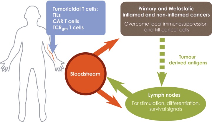
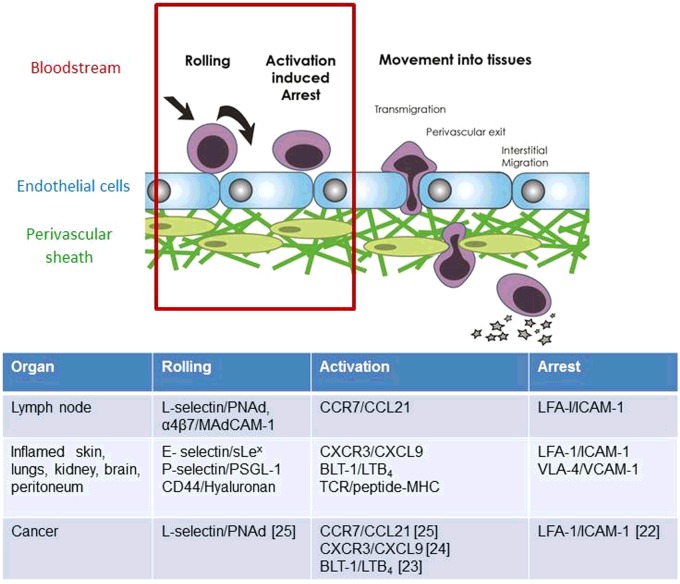
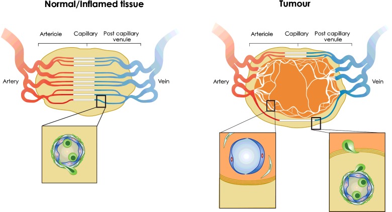
The success of adoptive T-cell therapies for the treatment of cancer patients depends on transferred T-lymphocytes finding and infiltrating cancerous tissues. For intravenously transferred T-cells, this means leaving the bloodstream (extravasation) from tumour blood vessels. In inflamed tissues, a key event in extravasation is the capture, rolling and arrest of T-cells inside blood vessels which precedes transmigration across the vessel wall and entry into tissues. This depends on co-ordinated signalling of selectins, integrins and chemokine receptors on T-cells by their respective ligands which are up-regulated on inflamed blood vessels. Clinical data and experimental studies in mice suggest that tumour blood vessels are anergic to inflammatory stimuli and the recruitment of cytotoxic CD8(+)T-lymphocytes is not very efficient. Interestingly, and somewhat counter-intuitively, anti-angiogenic therapy can promote CD8(+)T-cell infiltration of tumours and increase the efficacy of adoptive CD8(+)T-cell therapy. Rather than inhibit tumour angiogenesis, anti-angiogenic therapy 'normalizes' (matures) tumour blood vessels by promoting pericyte recruitment, increasing tumour blood vessel perfusion and sensitizing tumour blood vessels to inflammatory stimuli. A number of different approaches are currently being explored to increase recruitment by manipulating the expression of homing-associated molecules on T-cells and tumour blood vessels. Future studies should address whether these approaches improve the efficacy of adoptive T-cell therapies for solid, vascularized cancers in patients.

Keywords: chemokine receptors; endothelial cell anergy; extravasation; high endothelial venules; homing; integrins; selectins; tumour blood vessels; tumouricidal T-lymphocytes; vessel normalization.


Figures

Figure 1
Figure 1. A designer adoptive T-cell therapy for solid cancers
T-lymphocytes expressing conventional TCRs (TILs, TCRgm) or CARs at different stages of activation and differentiation are required to kill primary and metastatic cancers and to persist in cancer patients. Fully activated, tumouricidal T-cells expressing inflammation-associated homing molecules migrate from tumour blood vessels into primary cancers and sites of metastases (including sentinel lymph nodes) where they kill cancer cells. Tumouricidal T-cells migrating to non-cancerous tissues are unable to exert anti-cancer activity and are ineffective. Central memory T-cells expressing conventional TCRs, but not CARs, are re-activated by endogenously processed and presented tumour-derived antigens in tumour-draining lymph nodes before being redistributed to cancerous tissue. Central memory T-cells receive survival signals during normal recirculation through lymphoid organs. Recruitment of T-cells into non-inflamed cancers is promoted by patient conditioning which sensitizes the normally anergic tumour blood vessels to inflammatory mediators, increases the expression of homing-associated molecules and promotes recruitment of tumouricidal T-cells. Maturation of tumour blood vessels by activated T-cells in already inflamed tumours, promotes the development of HEV which recruit central memory T-cells into the tumour and shift the site of T-cell activation to cancerous tissues which avoids the loss of tumouricidal T-cells to non-cancerous tissues during their redistribution from the normal LN site of priming. γ-chain cytokines generate central memory and effector T-cells during T-cell expansion prior to adoptive transfer to cancer patients.
Figure 2
Figure 2. Homing addresses for T-cell entry into normal and cancerous tissues
T-lymphocyte recruitment from the bloodstream into tissues is regulated by a co-ordinated sequence of adhesive interactions which can be separated into distinct stages. The term ‘homing address’ (red box) describes the combination of rolling, activation and arrest receptors on T-cells and their respective ligands on the apical (inner) surface of blood vessel EC. Distinct homing addresses for T-cells according to the stage of T-cell activation, nature of the inflammatory stimulus and the organ involved have been identified and are listed. For example, the homing address on naïve and central memory T-cells for lymph nodes is L-selectin-CCR7-LFA-1 integrin. Specifically, L-selectin (CD62L) on T-cells binds to PNAd on the apical, inner surface of HEV blood vessels and allows T-cells to roll on the inner blood surface in the direction of blood flow. Rolling T-cells encounter immobilized CCL21 on the EC surface which engages CCR7 on T-cells and arrests rolling T-lymphocytes by activating LFA-1 integrin binding to ICAM-1. Activated Th1 CD4 T-cells entering inflamed tissues such as hypersensitivity lesions in the skin use a different homing address. Here the rolling receptor is E-selectin which is up-regulated on vascular EC by inflammatory cytokines. Inflammatory chemokines synthesized by inflamed tissues are reverse transcytosed across the blood vessel wall and immobilized on the apical surface of EC where they arrest rolling T-cells via LFA-1 and/or VLA4 integrins. Tumour blood vessels express homing-associated adhesion molecules and chemokines typical of inflamed tissues and lymph nodes and those which have been shown to regulate the recruitment of adoptively transferred tumouricidal T-cells from tumour blood vessels into tumours in syngeneic mice are cited.
Figure 3
Figure 3. T-cell recruitment from normal, inflamed and tumour blood vessels
(Left) In normal or inflamed tissues, the vascular network is highly ordered and T-cells are recruited from post-capillary venules where haemodynamic forces promote the margination of T-cells and other leucocytes to the outside stream of flowing blood. T-cell recruitment from HEV in lymph nodes or inflamed blood vessels in non-lymphoid tissues follows the multistep adhesion cascade described in Figure 2. Inset shows T-cell (green) binding to and in the process of transmigrating the EC lining and surrounding pericytic sheath which together comprise the blood vessel wall. (Right) Angiogenic blood vessels growing inside the tumour in response to hypoxia lack pericyte support and are highly disorganized, poorly perfused and leaky. They are non-responsive (anergic) to inflammatory stimuli but responsive to tumour-derived factors that prevent expression of homing molecules on tumour EC and induce FasL expression which directly kills tumouricidal T-cells. As a consequence intra-tumoural blood vessels are poor at recruiting effector CD8+ T-cells into the tumour [30]. In contrast, peritumoural blood vessels respond to inflammatory stimuli, up-regulate homing-associated molecules and recruit effector CD8+ T-cells to the peritumoural area. Cognate recognition of tumour antigen by conventional TCR regulates the movement of extravasated T-cells into the tumour to kill cancer cells. Insets show (left) immature, anergic tumour blood vessel lacking pericyte support inside the tumour and (right) mature peritumoural vessel supporting T-cell recruitment adjacent to a fibroblast-associated matrix which T-cells cross to enter the tumour. Chemokine production by cancer-associated fibroblasts can limit tumouricidal T-cell entry and killing of tumour tissue by arresting T-cells in the peritumoural area [30].

References

    1. Galon J., Costes A., Sanchez-Cabo F., Kirilovsky A., Mlecnik B., Lagorce-Pages C., Tosolini M., Camus M., Berger A., Wind P., et al. Type, density, and location of immune cells within human colorectal tumors predict clinical outcome. Science. 2006;313:1960–1964. doi: 10.1126/science.1129139. - DOI - PubMed
    1. Fridman W.H., Pages F., Sautes-Fridman C., Galon J. The immune contexture in human tumours: impact on clinical outcome. Nat. Rev. Cancer. 2012;12:298–306. doi: 10.1038/nrc3245. - DOI - PubMed
    1. Martinet L., Garrido I., Filleron T., Le Guellec S., Bellard E., Fournie J.J., Rochaix P., Girard J.P. Human solid tumors contain high endothelial venules: association with T- and B-lymphocyte infiltration and favorable prognosis in breast cancer. Cancer Res. 2011;71:5678–5687. doi: 10.1158/0008-5472.CAN-11-0431. - DOI - PubMed
    1. Martinet L., Le Guellec S., Filleron T., Lamant L., Meyer N., Rochaix P., Garrido I., Girard J.P. High endothelial venules (HEVs) in human melanoma lesions: major gateways for tumor-infiltrating lymphocytes. Oncoimmunology. 2012;1:829–839. doi: 10.4161/onci.20492. - DOI - PMC - PubMed
    1. Dudley M.E., Wunderlich J.R., Robbins P.F., Yang J.C., Hwu P., Schwartzentruber D.J., Topalian S.L., Sherry R., Restifo N.P., Hubicki A.M., et al. Cancer regression and autoimmunity in patients after clonal repopulation with antitumor lymphocytes. Science. 2002;298:850–854. doi: 10.1126/science.1076514. - DOI - PMC - PubMed

Publication types