

Benign sacrococcygeal teratoma incidentally found on routine scoliosis radiographs in a 12-year-old female: a case report
- PMID: 27069263
- PMCID: PMC4807676
Benign sacrococcygeal teratoma incidentally found on routine scoliosis radiographs in a 12-year-old female: a case report
Abstract
Objective: To describe the imaging characteristics of sacrococcygeal teratomas and to review appropriate diagnostic evaluation and management.
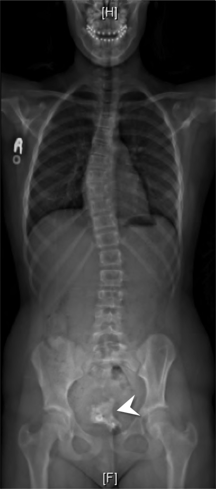
Clinical features: 12-year-old otherwise healthy, asymptomatic female with an incidental pelvic mass found on routine scoliosis radiographs.
Intervention and outcome: The pelvic mass was further evaluated by MRI and CT scan. Management consisted of successful surgical resection with no postoperative complications. Pathology confirmed a mature, benign, sacrococcygeal teratoma.
Conclusions: Chiropractors manage patients with scoliosis, which may include radiographic surveillance. Familiarity with the radiographic features of masses such as sacrococcygeal teratomas is important for prompt diagnosis and management.
Objectif: Décrire les caractéristiques d’imagerie des tératomes sacrococcygiens et examiner l’évaluation et la prise en charge du diagnostic approprié.
Caractéristiques cliniques: Jeune fille asymptomatique de 12 ans en bonne santé avec une masse pelvienne découverte fortuitement après des radiographies de routine pour la scoliose.
Intervention et résultats: La masse pelvienne a davantage été évaluée à partir d’images d’IRM et de TDM. La solution était la résection chirurgicale réussie sans complications postopératoires. La pathologie a confirmé un tératome sacrococcygien mature bénin.
Conclusions: Les chiropraticiens prennent en charge les patients atteints de scoliose, ce qui peut inclure la surveillance radiographique. La familiarité avec les caractéristiques radiographiques des masses, comme les tératomes sacrococcygiens, est importante pour leur diagnostic et la prise en charge rapide.
Keywords: pediatric; sacrococcygeal teratoma.
Figures


References
-
- Bale PM, Painter DM, Cohen D. Teratomas in childhood. Pathology. 1975;7:209–218. - PubMed
-
- Hong JH, Lee JK, Song SH, Hwang JH, So KA, Shin BK, Lee NW, Lee KW. Unilateral ovarian dermoid cyst accompanied by an ipsilateral paratubal cyst in a girl with Proteus Syndrome discovered by laparoscopic surgery. J PediatrAdolesc Gynecol. 2010;23:107–110. - PubMed
-
- Kocaoglu M, Frush DP. Pediatric presacral masses. Radiographics. 2006;26:833–857. - PubMed
-
- Comerci JT, Licciardi F, Bergh PA, Gregori C, Breen JL. Mature cystic teratoma: a clinicopathologic evaluation of 517 cases and review of the literature. Obstet Gynecol. 1994;84:22–28. - PubMed
Publication types
LinkOut - more resources
Full Text Sources