. 2016 Mar 23:11:1159-73.
doi: 10.2147/IJN.S102096. eCollection 2016.

Cerium oxide nanoparticles promote neurogenesis and abrogate hypoxia-induced memory impairment through AMPK-PKC-CBP signaling cascade

Affiliations

Cerium oxide nanoparticles promote neurogenesis and abrogate hypoxia-induced memory impairment through AMPK-PKC-CBP signaling cascade

Aditya Arya et al. Int J Nanomedicine. .

Retraction in

Abstract

Structural and functional integrity of the brain is adversely affected by reduced oxygen saturation, especially during chronic hypoxia exposure and often encountered by altitude travelers or dwellers. Hypoxia-induced generation of reactive nitrogen and oxygen species reportedly affects the cortex and hippocampus regions of the brain, promoting memory impairment and cognitive dysfunction. Cerium oxide nanoparticles (CNPs), also known as nanoceria, switch between +3 and +4 oxidation states and reportedly scavenge superoxide anions, hydrogen peroxide, and peroxynitrite in vivo. In the present study, we evaluated the neuroprotective as well as the cognition-enhancing activities of nanoceria during hypobaric hypoxia. Using polyethylene glycol-coated 3 nm nanoceria (PEG-CNPs), we have demonstrated efficient localization of PEG-CNPs in rodent brain. This resulted in significant reduction of oxidative stress and associated damage during hypoxia exposure. Morris water maze-based memory function tests revealed that PEG-CNPs ameliorated hypoxia-induced memory impairment. Using microscopic, flow cytometric, and histological studies, we also provide evidences that PEG-CNPs augmented hippocampus neuronal survival and promoted neurogenesis. Molecular studies revealed that PEG-CNPs promoted neurogenesis through the 5'-adenine monophosphate-activated protein kinase-protein kinase C-cyclic adenosine monophosphate response element-binding protein binding (AMPK-PKC-CBP) protein pathway. Our present study results suggest that nanoceria can be translated as promising therapeutic molecules for neurodegenerative diseases.

Keywords: cerium oxide nanoparticles; hypoxia; memory; neuroprotection; oxidative stress.


Figures

Figure 1
Figure 1
Schematic of experimental design. Notes: The animals were randomly divided into two groups (n=24 each); one group received PEG-CNPs injection (concentration 5 µg/kg BW, intraperitoneally) and the other group received normal saline (phosphate-buffered saline). Animals were habituated for 2 days by keeping them in the room containing MWM and placing them in water for 5 minutes each day. Next the animals were randomly divided into two groups. The second dose (out of the three) was administered on third day before beginning the Morris Water Maze experiments. Third dose of PEG-CNPs was injected on the sixth day, and after 7 days of training, in each quadrant, 12 animals from each group were subjected to probe trial, while the remaining 12 animals were exposed to hypobaric hypoxia (10 days). These groups were renamed as hypoxia (without PEG-CNPs injection) and hypoxia + PEG-CNPs groups. After 10 days of hypoxia, animals were again subjected to MWM learning for 2 days followed by probe trial. Abbreviations: CNPs, cerium oxide nanoparticles; MWM, Morris water maze; PEG, polyethylene glycol.
Figure 2
Figure 2
Characterization of PEG-CNPs. Notes: (A) Transmission electron micrographs of PEG-CNPs showing uniformly distributed spherical nanoparticles (scale bar: 20 nm). (B) Magnified view revealing diameter of PEG-CNPs in the size range of 3–5 nm. The selected area electron diffraction pattern confirms the ceria crystal structure in transmission electron microscopy. (C) X-ray diffraction analysis of the crystal lattice of PEG-CNPs showing prominent lattice planes {111}, {200}, and {220}. (D) Dynamic light scattering of CNPs in cerebrospinal fluid; mean hydrodynamic diameter was observed as 90 nm. (E) FT-IR spectra showing conjugation of PEG to CNPs. Abbreviations: CNPs, cerium oxide nanoparticles; PEG, polyethylene glycol; NPs, nanoparticles.
Figure 2
Figure 2
Characterization of PEG-CNPs. Notes: (A) Transmission electron micrographs of PEG-CNPs showing uniformly distributed spherical nanoparticles (scale bar: 20 nm). (B) Magnified view revealing diameter of PEG-CNPs in the size range of 3–5 nm. The selected area electron diffraction pattern confirms the ceria crystal structure in transmission electron microscopy. (C) X-ray diffraction analysis of the crystal lattice of PEG-CNPs showing prominent lattice planes {111}, {200}, and {220}. (D) Dynamic light scattering of CNPs in cerebrospinal fluid; mean hydrodynamic diameter was observed as 90 nm. (E) FT-IR spectra showing conjugation of PEG to CNPs. Abbreviations: CNPs, cerium oxide nanoparticles; PEG, polyethylene glycol; NPs, nanoparticles.
Figure 3
Figure 3
Assessment of polyethylene glycol-cerium oxide nanoparticles biodistribution using transmission electron microscopy. Notes: Electron micrograph of rat brain hippocampus region. (A) The electron-dense nanoparticles of polyethylene glycol-cerium oxide nanoparticles were observed after 24 hours of third dose. Arrows indicate the presence of CNPs in the hippocampus. Scale bar: 1 µm. (B) Magnified view of nanoparticles is indicated with their size (~4–5 nm) and selected area electron diffraction with characteristic diffraction fringes (inset). Scale bar: 100 nm. Abbreviation: CNPs, cerium oxide nanoparticles.
Figure 4
Figure 4
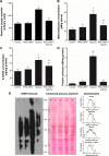
Evaluation of oxidative stress in the hippocampus region of rat brain. Notes: (A) ROS dichlorofluorescein diacetate fluorescence in arbitrary units as a measure of relative ROS content in the isolated rat hippocampi showed twofold higher ROS content in animals exposed to hypoxia. PEG-CNPs pretreatment significantly reduced the ROS content. Also, PEG-CNPs alone did not cause any increase in ROS. (B) MDA estimation using thiobarbituric acid reactive substances assay showed 2.4-fold higher MDA content in hypoxia, while PEG-CNPs pretreatment reduced the level of MDA to 1.4-fold in comparison to control. PEG-CNPs alone did not cause any elevation in MDA. (C) 8-OHdG concentration in animals exposed to hypoxia was 1.8-fold higher, while PEG-CNPs pretreatment in hypoxia reduced the concentration to 1.3-fold in comparison to control. PEG-CNPs alone did not elevate 8-OHdG. (D) Protein carbonylation estimation using enzyme-linked immunosorbent assay and (E) protein carbonylation estimation using oxyblot showed highest degree of protein carbonylation in hypoxia samples, while PEG-CNPs pretreatment prevented the carbonylation. Graphical view of immunoblot analyzed using ImageJ represents relative intensity at each level in the gel. Data represented as mean ± standard error of mean of three independent experiments (*P<0.01, **P<0.05). Abbreviations: 8-OHdG, 8-hydroxydeoxyguanosine; CNPs, cerium oxide nanoparticles; MDA, malondialdehyde; PEG, polyethylene glycol; ROS, reactive oxygen species; DNPH, dinitrophenylhydrazine.
Figure 5
Figure 5
Evaluation of spatial memory in rat after PEG-CNPs intervention. Notes: Spatial memory function test in rat using Morris water maze. (A) Graph showing latency to reach the hidden platform. Steeper slope in animals treated with PEG-CNPs, compared to controls indicated better retention of spatial memory. After hypoxia, the increase in latency was smaller in PEG-CNPs pretreated animals as compared to hypoxic animals. (B) Number of annulus crossings during the probe trial. Control animals showed five times mean annulus crossing, PEG-CNP treated animals showed six times, hypoxia-exposed animals showed only one annulus crossing, and PEG-CNPs + hypoxia animals showed four time annulus crossing. (C) Analysis of time spent in each quadrant during the probe trial showed that PEG-CNPs treated animals spent maximum time in target quadrant which was 40% higher than control, while minimum time was observed in animals subjected to hypoxia, which was 20% less than control; PEG-CNPs + hypoxia group spent 30% more time compared to control (*P<0.05, **P<0.01 Q1 – target quadrant containing hidden platform). (D) Representative path tracks of animals during spatial acquisition trials. Abbreviations: CNPs, cerium oxide nanoparticles; PEG, polyethylene glycol.
Figure 6
Figure 6
PEG-CNPs prevented apoptosis and induced neurogenesis. Notes: (A) Phase-contrast microphotographs showing reduced number of neurons in hypoxia-exposed animals and increased density of neurons in PEG-CNP treated cells exposed to hypoxia (scale bar: 20 µm). (B) Dot plots showing the neuronal population with BrdU incorporation. Lower left quadrant represents neuronal nonproliferating cells, lower right quadrant represents nonproliferating neuronal cells, upper left quadrant shows neuronal nonproliferating cells, and upper right quadrant shows neuronal proliferating cells. There were 51.95 BrdU-positive neuronal cells in the control group, 64.4% in PEG-CNPs group, 23.3% in hypoxia group, and 62.9% in PEG + hypoxia group. (C) Photomicrographs of dentate gyrus region of rat hippocampus stained with cresyl violet show neurodegeneration, while neuron density was higher in PEG-CNPs treated animals. At higher magnification, the number of pyramidal cells (deep violet stained) was higher in PEG-CNPs treated cells. Insets represent the histograms of blue channel with mean values representing relative number of cresyl violet positive cells (control – 107.5, PEG-CNPs – 109.19, hypoxia – 98.93, PEG-CNPs + hypoxia – 109.13). Arrows indicate neurodegeneration. Abbreviations: CNPs, cerium oxide nanoparticles; PEG, polyethylene glycol.
Figure 7
Figure 7
Evaluation of key molecular targets of PEG-CNPs using immunoblotting. Notes: Immunoblots show changes in expression or phosphorylation of neurogenic and apoptotic markers in the rat brain homogenates. AMPK expression was increased after PEG-CNPs treatment, while it was reduced in hypoxic exposure. Further, it was the highest in PEG-CNPs group. Phosphorylation of PKCζ and pCBP was increased in both PEG-CNPs and hypoxia + PEG-CNPs groups, while decreased phosphorylation was observed in hypoxia group compared with control. Caspase-3 and caspase-9 (cleaved forms) were upregulated in hypoxia alone, while PEGylated-CNP treatment reduced the concentration of both in comparison to control. Alpha tubulin was used as the loading control for all samples. Abbreviations: AMPK, 5′-adenine monophosphate-activated protein kinase; CBP, cyclic adenosine monophosphate response element-binding protein binding protein; CNPs, cerium oxide nanoparticles; PEG, polyethylene glycol; PKC, protein kinase C.
Figure 8
Figure 8
Mechanism of hippocampal neurogenesis and cell survival by PEG-CNPs. Notes: AMPK activates a molecular cascade via nuclear histone acyltransferase protein CBP and PKC, directing the adult neurogenesis. PEG-CNPs treatment also increased the amount of phosphorylated aPKCζ and further activated CBP by phosphorylation, thereby activating adult hippocampal neurogenesis. Another route that operated during hypoxia was activation of apoptotic events mediated via caspase-3 and caspase-9, which was also inhibited by PEG-CNPs pretreatment. Abbreviations: AMPK, 5′-adenine monophosphate-activated protein kinase; CBP, cyclic adenosine monophosphate response element-binding protein binding protein; CNPs, cerium oxide nanoparticles; PEG, polyethylene glycol; PKC, protein kinase C; ROS, reactive oxygen species.

References

    1. Sharp FR, Bernaudin M. HIF1 and oxygen sensing in the brain. Nat Rev Neurosci. 2004;5(6):437–448. - PubMed
    1. Wilson MH, Newman S, Imray CH. The cerebral effects of ascent to high altitudes. Lancet Neurol. 2009;8(2):175–191. - PubMed
    1. LaManna JC, Chavez JC, Pichiule P. Structural and functional adaptation to hypoxia in the rat brain. J Exp Biol. 2004;207(Pt 18):3163–3169. - PubMed
    1. Zhang J, Yan X, Shi J, Gong Q, Weng X, Liu Y. Structural modifications of the brain in acclimatization to high-altitude. PLoS One. 2010;5(7):e11449. - PMC - PubMed
    1. Zhang J, Zhang H, Li J, et al. Adaptive modulation of adult brain gray and white matter to high altitude: structural MRI studies. PLoS One. 2013;8(7):e68621. - PMC - PubMed

Publication types

MeSH terms