Review
. 2016:2016:4636859.
doi: 10.1155/2016/4636859. Epub 2016 Mar 16.

Vascular Remodelling and Mesenchymal Transition in Systemic Sclerosis

Affiliations
Review

Vascular Remodelling and Mesenchymal Transition in Systemic Sclerosis

Pier Andrea Nicolosi et al. Stem Cells Int. 2016.

Abstract

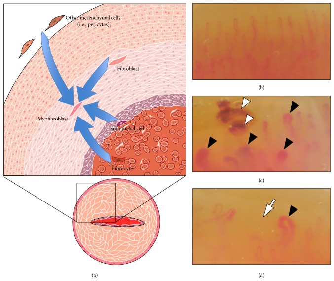
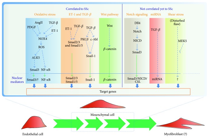
Fibrosis of the skin and of internal organs, autoimmunity, and vascular inflammation are hallmarks of Systemic Sclerosis (SSc). The injury and activation of endothelial cells, with hyperplasia of the intima and eventual obliteration of the vascular lumen, are early features of SSc. Reduced capillary blood flow coupled with deficient angiogenesis leads to chronic hypoxia and tissue ischemia, enforcing a positive feed-forward loop sustaining vascular remodelling, further exacerbated by extracellular matrix accumulation due to fibrosis. Despite numerous developments and a growing number of controlled clinical trials no treatment has been shown so far to alter SSc natural history, outlining the need of further investigation in the molecular pathways involved in the pathogenesis of the disease. We review some processes potentially involved in SSc vasculopathy, with attention to the possible effect of sustained vascular inflammation on the plasticity of vascular cells. Specifically we focus on mesenchymal transition, a key phenomenon in the cardiac and vascular development as well as in the remodelling of injured vessels. Recent work supports the role of transforming growth factor-beta, Wnt, and Notch signaling in these processes. Importantly, endothelial-mesenchymal transition may be reversible, possibly offering novel cues for treatment.


Figures

Figure 1
Figure 1
Vascular remodelling and capillaroscopic pattern in Systemic Sclerosis (SSc). (a) Stenoocclusive remodelling in SSc microvasculature (bottom right) is believed to result from an abnormal reparative attempt triggered by chronic endothelial damage, which drives intima-media hyperplasia and increased ECM production within the vessel wall. Mesenchymal cells, specifically myofibroblasts with a highly secretory phenotype, are the main final effectors responsible for these structural changes. Myofibroblasts in SSc vessels can originate from multiple cellular sources (upper left), either of mesenchymal origin, such as pericytes or fibroblast, or of nonmesenchymal origin, such as endothelial cells. (b)–(d) Capillaroscopic pattern in normal subjects (b) and scleroderma patients at magnification 200x ((c): “active” SSc pattern; (d): “late” SSc pattern). Note the heterogeneity in the architecture and morphology of SSc capillaries with frequent ectasias (black arrowheads). In the “active” scleroderma pattern there are plenty of giant capillaries (i.e., more than 50 μm of diameter) and microhaemorrhages (white arrowheads), with mild loss of capillaries. In the “late” scleroderma pattern giant capillaries and microhaemorrhages are less frequent, but a severe loss of capillaries is evident, with extensive avascular areas (white arrows).
Figure 2
Figure 2
Pathways involved in the EndoMT. The scheme summarized the putative pathways involved in the EndoMT highlighting which are already correlated or not with SSc. The activation of specific nuclear mediator leads to activation of target genes that are correlated with the increase of mesenchymal markers (such as Col I, α-SMA, and Twist 1) and/or decrease of endothelial markers (such as CD31, VE-Cad, and Fli-1). The activation of these pathways could lead endothelial cells to acquire initially mesenchymal characteristics and later on to acquire myofibroblastic features.

References

    1. Bhattacharyya S., Wei J., Varga J. Understanding fibrosis in systemic sclerosis: shifting paradigms, emerging opportunities. Nature Reviews Rheumatology. 2012;8(1):42–54. doi: 10.1038/nrrheum.2011.149. - DOI - PMC - PubMed
    1. Wigley F. M. Vascular disease in scleroderma. Clinical Reviews in Allergy and Immunology. 2009;36(2-3):150–175. doi: 10.1007/s12016-008-8106-x. - DOI - PubMed
    1. Saketkoo L. A., Distler O. Is there evidence for vasculitis in systemic sclerosis. Current Rheumatology Reports. 2012;14(6):516–525. doi: 10.1007/s11926-012-0296-9. - DOI - PubMed
    1. Farina G., York M., Collins C., Lafyatis R. dsRNA activation of endothelin-1 and markers of vascular activation in endothelial cells and fibroblasts. Annals of the Rheumatic Diseases. 2011;70(3):544–550. doi: 10.1136/ard.2010.132464. - DOI - PMC - PubMed
    1. Lenna S., Farina A. G., Martyanov V., et al. Increased expression of endoplasmic reticulum stress and unfolded protein response genes in peripheral blood mononuclear cells from patients with limited cutaneous systemic sclerosis and pulmonary arterial hypertension. Arthritis and Rheumatism. 2013;65(5):1357–1366. doi: 10.1002/art.37891. - DOI - PMC - PubMed

LinkOut - more resources