Review
. 2016:2016:8291260.
doi: 10.1155/2016/8291260. Epub 2016 Mar 16.

Neural Conversion and Patterning of Human Pluripotent Stem Cells: A Developmental Perspective

Affiliations
Review

Neural Conversion and Patterning of Human Pluripotent Stem Cells: A Developmental Perspective

Alexandra Zirra et al. Stem Cells Int. 2016.

Abstract

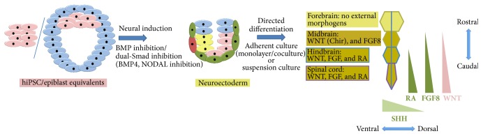
Since the reprogramming of adult human terminally differentiated somatic cells into induced pluripotent stem cells (hiPSCs) became a reality in 2007, only eight years have passed. Yet over this relatively short period, myriad experiments have revolutionized previous stem cell dogmata. The tremendous promise of hiPSC technology for regenerative medicine has fuelled rising expectations from both the public and scientific communities alike. In order to effectively harness hiPSCs to uncover fundamental mechanisms of disease, it is imperative to first understand the developmental neurobiology underpinning their lineage restriction choices in order to predictably manipulate cell fate to desired derivatives. Significant progress in developmental biology provides an invaluable resource for rationalising directed differentiation of hiPSCs to cellular derivatives of the nervous system. In this paper we begin by reviewing core developmental concepts underlying neural induction in order to provide context for how such insights have guided reductionist in vitro models of neural conversion from hiPSCs. We then discuss early factors relevant in neural patterning, again drawing upon crucial knowledge gained from developmental neurobiological studies. We conclude by discussing open questions relating to these concepts and how their resolution might serve to strengthen the promise of pluripotent stem cells in regenerative medicine.


Figures

Figure 1
Figure 1
Developmental stages of mouse embryo. First row (left to right), from the secondary oocyte the blastomere develops (2-cell, 4-cell, 8-cell, and 16-cell stages) to give rise to the early blastocyst formed of trophectoderm (cells that express Cdx2) and inner cell mass cells (that express Oct3/4). Later, the inner cell mass gives rise to the epiblast (cells that express Oct3/4 and Nanog) and endoderm (expressing Oct3/4 and GATA4). Second row (right to left), in the late mouse blastocyst Cdx2 positive cells give rise to the extraembryonic ectoderm and ectoplacental cone. At the same time the endoderm divides into an embryonic endoderm and an extraembryonic endoderm. The epiblast and the extraembryonic ectoderm form a cavity lined by embryonic endoderm. From the embryonic endoderm the distal visceral endoderm is formed (DVE). Third row (left to right), the DVE migrates proximally and will be known as the anterior visceral endoderm (AVE). The final image (third row, right) shows the development of the primitive streak (mesodermal cells) at the opposite (posterior) pole from the AVE. N.B. There are 2 different types of endoderm called extraembryonic and embryonic; these differ in their potency and give rise to distinct cellular derivatives. All timelines are given for mouse and human embryonic development.
Figure 2
Figure 2
Neural induction, neurulation, and neural patterning overview. (a) Neural induction: neuroectoderm (neural plate) differentiation happens under the influence of the AVE. The mesodermal cells start migrating in all directions and envelop the embryo between the endoderm and the ectoderm. At the distal pole of the embryo the node develops, to further act as the “trunk organiser.” (b) Neurulation: from the neural plate, cells start to proliferate and invaginate in order to form the neural tube and neural crest which derives from the dorsomedial borders of the neural folds. (c) Neural patterning: cells from the neural tube start to differentiate into precursors for forebrain, midbrain, hindbrain, and spinal cord according to a rostrocaudal axis. All timelines are given for mouse and human embryonic development.
Figure 3
Figure 3
Molecular pathways in neural induction. (a) The epiblast (depicted in pink) expresses Nodal. The epiblast through Nodal stimulates (pink arrow) the expression of BMP4 (depicted in blue) in the extraembryonic ectoderm (blue cells). The extraembryonic ectoderm, by the action of BMP4, stimulates (blue arrow) the WNT (depicted in pink) pathway in the epiblast that in turn further activates (pink arrow) Nodal expression. Thus, there is a positive feedback loop between Nodal, BMP, and WNT. Colour scheme: arrows corresponds to the related tissue/morphogen. (b) The DVE (depicted in red) expresses Cerberus1 and Lefty (also depicted in red) to inhibit Nodal expression, therefore downregulating Nodal in its proximity. It also expresses Dickkopf (depicted in red), a protein that inhibits WNT3 signals close to the DVE. Downregulating Nodal and WNT also inhibits BMP4 expression close to the DVE. Thus, there is a gradient of Nodal, WNT, and BMP with a high expression rostrally and low expression caudally. FGF8 (pink and blue), expressed both in the epiblast (pink) and extraembryonic ectoderm (blue), also inhibits BMP4 contributing to the gradient. Colour scheme: arrows corresponds to the secreted inhibitory molecules/tissue source (DVE). They show the consequence of the negative feedback that creates the morphogen gradients in the R-C axis. (c) The DVE migrates into the AVE and the gradients are thus remodelled with low Nodal, WNT, and BMP expression ventrally and high dorsally. (d) Due to these gradients the neuroectoderm is formed at the ventral pole of the epiblast. Colour scheme: arrows corresponds to the secreted inhibitory molecules/tissue source (AVE). They show the consequence of the negative feedback that creates the morphogen gradients in the D-V axis. All timelines are given for mouse and human embryonic development.
Figure 4
Figure 4
Neural patterning. (a) Rostrocaudal gradients of Nodal, BMP4, RA (retinoic acid), and FGF8 important in rostrocaudal patterning. (b) The interplay between different factors encoding forebrain (PAX6 and OTX2), midbrain (PAX6, OTX2, and EN1/PAX2), and hindbrain (EN1/PAX2, GBX2, and FGF8). The forebrain-midbrain barrier is defined by the mutually exclusive expression of PAX6 (forebrain) and EN1/PAX2 (midbrain), while the midbrain-hindbrain boundary by OTX2 (midbrain) and GBX2 (hindbrain). OTX2 and GBX2 are regulated by FGF8 expression. (c) Dorsoventral patterning with dorsal gradients for BMP4 and WNT and with a ventral gradient of SHH (Sonic hedgehog). (d) Transverse section through the neural tube depicting various neurons specified by the gradient of SHH from the floor plate and the BMP4 and WNT from the roof plate: V0–3: interneurons and MN: motor neurons.
Figure 5
Figure 5
Methods for directed differentiation: hPSCs can be directed to undergo neural conversion by applying developmental principles of inhibiting BMP4 and/or Nodal. From neuroectoderm, differentiation of different neuraxial regions can be achieved by recapitulating developmental morphogenetic instruction: forebrain (default), midbrain (WNT, FGF8 activation), hindbrain (WNT, FGF8, and others, RA), and spinal cord (WNT, FGF8, and others, RA). These gradients are shown on the right of the figure for the rostrocaudal axis. Another important morphogenetic cue used in directed differentiation is SHH for its ventralising effect within the D-V axis. All timelines are given for mouse and human embryonic development.

References

    1. Andoniadou C. L., Martinez-Barbera J. P. Developmental mechanisms directing early anterior forebrain specification in vertebrates. Cellular and Molecular Life Sciences. 2013;70(20):3739–3752. doi: 10.1007/s00018-013-1269-5. - DOI - PMC - PubMed
    1. Becker C. G., Diez Del Corral R. Neural development and regeneration: it's all in your spinal cord. Development. 2015;142(5):811–816. doi: 10.1242/dev.121053. - DOI - PubMed
    1. Kicheva A., Briscoe J. Developmental pattern formation in phases. Trends in Cell Biology. 2015;25(10):579–591. doi: 10.1016/j.tcb.2015.07.006. - DOI - PubMed
    1. Williams L. A., Davis-Dusenbery B. N., Eggan K. C. SnapShot: directed differentiation of pluripotent stem cells. Cell. 2012;149(5):1174–1174.e1. doi: 10.1016/j.cell.2012.05.015. - DOI - PubMed
    1. Han S. S. W., Williams L. A., Eggan K. C. Constructing and deconstructing stem cell models of neurological disease. Neuron. 2011;70(4):626–644. doi: 10.1016/j.neuron.2011.05.003. - DOI - PubMed

LinkOut - more resources