. 2016 Apr 11:6:15.
doi: 10.1186/s13395-016-0087-5. eCollection 2016.

Characterization of a multiprotein complex involved in excitation-transcription coupling of skeletal muscle

Affiliations

Characterization of a multiprotein complex involved in excitation-transcription coupling of skeletal muscle

Manuel Arias-Calderón et al. Skelet Muscle. .

Abstract

Background: Electrical activity regulates the expression of skeletal muscle genes by a process known as "excitation-transcription" (E-T) coupling. We have demonstrated that release of adenosine 5'-triphosphate (ATP) during depolarization activates membrane P2X/P2Y receptors, being the fundamental mediators between electrical stimulation, slow intracellular calcium transients, and gene expression. We propose that this signaling pathway would require the proper coordination between the voltage sensor (dihydropyridine receptor, DHPR), pannexin 1 channels (Panx1, ATP release conduit), nucleotide receptors, and other signaling molecules. The goal of this study was to assess protein-protein interactions within the E-T machinery and to look for novel constituents in order to characterize the signaling complex.

Methods: Newborn derived myotubes, adult fibers, or triad fractions from rat or mouse skeletal muscles were used. Co-immunoprecipitation, 2D blue native SDS/PAGE, confocal microscopy z-axis reconstruction, and proximity ligation assays were combined to assess the physical proximity of the putative complex interactors. An L6 cell line overexpressing Panx1 (L6-Panx1) was developed to study the influence of some of the complex interactors in modulation of gene expression.

Results: Panx1, DHPR, P2Y2 receptor (P2Y2R), and dystrophin co-immunoprecipitated in the different preparations assessed. 2D blue native SDS/PAGE showed that DHPR, Panx1, P2Y2R and caveolin-3 (Cav3) belong to the same multiprotein complex. We observed co-localization and protein-protein proximity between DHPR, Panx1, P2Y2R, and Cav3 in adult fibers and in the L6-Panx1 cell line. We found a very restricted location of Panx1 and Cav3 in a putative T-tubule zone near the sarcolemma, while DHPR was highly expressed all along the transverse (T)-tubule. By Panx1 overexpression, extracellular ATP levels were increased both at rest and after electrical stimulation. Basal mRNA levels of the early gene cfos and the oxidative metabolism markers citrate synthase and peroxisome proliferator-activated receptor gamma coactivator 1-alpha (PGC1α) were significantly increased by Panx1 overexpression. Interleukin 6 expression evoked by 20-Hz electrical stimulation (270 pulses, 0.3 ms each) was also significantly upregulated in L6-Panx1 cells.

Conclusions: We propose the existence of a relevant multiprotein complex that coordinates events involved in E-T coupling. Unveiling the molecular actors involved in the regulation of gene expression will contribute to the understanding and treatment of skeletal muscle disorders due to wrong-expressed proteins, as well as to improve skeletal muscle performance.

Keywords: Dihydropyridine receptor; Excitation-transcription coupling; Multiprotein complex; Nucleotide receptors; Pannexin 1; Skeletal muscle plasticity.


Figures

Fig. 1
Fig. 1
Co-immunoprecipitation evidences for a multiprotein complex involved in excitation-transcription coupling. a Characterization of triad-enriched fractions derived from rat or mouse muscles. Several proteins were detected by immunoblot: dihydropyridine receptor (DHPR), ryanodine receptor 1 (RyR1), sarco/endoplasmic reticulum Ca2+-ATPase (SERCA), heat shock protein 70 (HSP70), pannexin 1 (Panx1), P2Y2 receptor (P2Y2R) and caveolin-3 (Cav3). b, c Several protein components for the excitation-transcription machinery co-precipitate both in mouse adult muscle triad preparation (b) and in rat myotube extracts (c). The voltage sensor (DHPR), the ATP-release channel (Panx1), and a receptor for extracellular ATP (P2Y2R) co-precipitate suggesting a multiprotein complex. The scaffold protein dystrophin (Dys) co-precipitate with DHPR, Panx1, and P2Y2R. Protein immunodetection in the whole lysate and pre-clearing samples are shown as positive and negative controls, respectively (b, c)
Fig. 2
Fig. 2
Isolation of protein complexes containing DHPR, P2Y2R, and Panx1 from skeletal triads using 2D blue native SDS-PAGE. a Silver staining of a mouse triad sample resolved by BN/SDS-PAGE. Several multiprotein complexes are observed as dots in a same vertical lane. bd Immunoblot for complex putative interactors in triad-enriched samples derived from rat (b), BalbC mouse (c), or C57 mouse (d) previously resolved by BN/SDS-PAGE. Co-localization in the same vertical lane (as marked with arrowheads) suggests a multiprotein complex containing DHPR, Panx1, P2Y2R, and Cav3
Fig. 3
Fig. 3
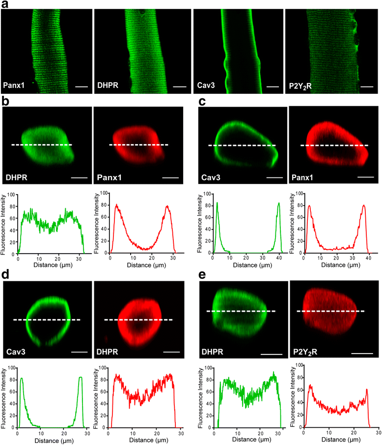
Subcellular distribution of DHPR, P2Y2R, Panx1, and Cav3 in isolated skeletal fibers. a Representative confocal immunofluorescence images for putative complex constituents in mouse FDB-isolated fibers. b–e Double-immunostaining and co-localization assays for different protein complex constituents. Protein distribution at the fiber z-axis was performed by line scan and z-projection reconstruction. Quantitation of fluorescence intensity in the area marked with a dashed line is shown under each image. Scale bars = 10 μm
Fig. 4
Fig. 4
Association between DHPR, P2Y2R, Panx1, and Cav3 demonstrated by proximity ligation assay technology. a Protein-protein proximity was demonstrated using the PLA probe technology in a whole fiber, as indicated (DHPR with Panx1, DHPR with P2Y2R, DHPR with Cav3, Panx1 with Cav3, and P2Y2R with Cav3). Z-stacks were collected by confocal microscopy (35–40 slices, 1 μm each), and then pixels in all the stacks were collapsed into one single illuminated image. b Positive PLA dot distribution in the median plane of the fibers for the protein-protein proximity assessed (DHPR with Panx1, DHPR with P2Y2R, DHPR with Cav3, Panx1 with Cav3, and P2Y2R with Cav3). Images correspond to the six medial slices collapsed in a single image. Scale bar = 10 um
Fig. 5
Fig. 5
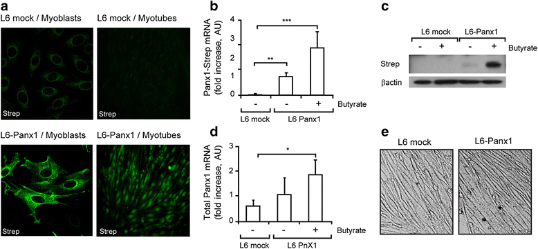
Characterization of an L6 skeletal cell line overexpressing Panx1-Strep-tag. a Representative immunofluorescence images of recombinant Panx1 overexpression, either in myoblast or myotube differentiation stages, using a Strep-tag antibody. Overexpression was induced with 5 mM sodium butyrate ON. b Panx1-Strep-tag mRNA levels were analyzed by qPCR using primers against the Strep epitope (n = 3). c Panx1 protein expression was assessed by immunoblot using Strep-tag antibodies. d The mRNA level of whole Panx1 (both endogenous and Panx1-strep) was analyzed by qPCR using primers against Panx1 (n = 3). e L6 cells overexpressing Panx1-Strep-tag properly differentiate to myotubes. Representative images of light microscopy are shown. In b and d, values are expressed as mean ± SEM. *p < 0.05; **p < 0.01; ***p < 0.001
Fig. 6
Fig. 6
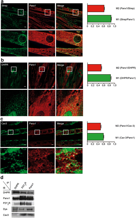
Multiprotein complex is properly assembled in L6 myotubes overexpressing Panx1. Double-staining immunofluorescence was performed using two different primary antibodies in the same sample, as detailed: anti-Strep/anti-Panx1 (a), anti-DHPR/anti-Panx1 (b), and anti-Cav3/anti-Panx1 (c). Bar graphs next to the image panels show Manders’ coefficients for the co-localization analysis. Scale bar = 10 μm. White boxes indicate area that is then magnified in the row below. Bars correspond to the average of the data obtained from three independent experiments, where three regions of interest (ROI) were evaluated per image for the Manders’ coefficient quantification. d Co-immunoprecipitation assay was performed in L6-Panx1 myotubes. Anti-Panx1, anti-DHPR, and anti-P2Y2R antibodies were used to immunoprecipitate whole lysates from L6-Panx1 cells. Panx1, DHPR, and P2Y2R all co-immunoprecipitated in these cells together with Dys and Cav3. Representative blot of the three experiments is shown
Fig. 7
Fig. 7
Nearness between DHPR, Panx1, P2Y2R, and Cav3 reinforced by proximity ligation assay technology. In situ proximity ligation assay (PLA) probes shown in red a well-defined closeness between DHPR and Panx1 (a), DHPR and Cav3 (b), DHPR and P2Y2R (c), Panx1 and Cav3 (d), and P2Y2R and Cav3 (e) in L6-Panx1 myotubes. No interaction was observed between DHPR and the nuclear envelope protein Lap2, as expected (f). To visualize cell limits and morphology, exogenous Panx1-Strep was stained in the same preparations using an anti-strep antibody (green). Scale bars = 10 μm
Fig. 8
Fig. 8
Panx1 overexpression increases resting levels of extracellular ATP and gene expression in muscle L6 cells. a Extracellular ATP resting levels are increased in L6 muscle cells overexpressing Panx1 (L6-Panx1), both in myoblast and in myotube differentiation stages (n = 3). b ATP release evoked by electrical stimulation (20 Hz, 270 pulses, 0.3 ms each) is increased in Panx1 overexpressing cells. ATP release induced by ES is graphed as fold increase compared to non-stimulated cells (n = 3). c mRNA resting levels of E-T coupling related genes (cfos, citrate synthase, PGC1α, and interleukin 6) in either mock or Panx1-overexpressing L6 myotubes,were analyzed by qPCR (n = 3-4). d Interleukin 6 mRNA levels evoked by ES in both mock and Panx1-overexpressing L6 myotubes were analyzed by qPCR (n = 3–4). GAPDH was used as a housekeeper gene for normalization. The dashed line (bd) marks the value of “1” to indicate the level of expression of IL6 in cells without ES, against which the results are normalized. Values are expressed as mean ± SEM. *p < 0.05; **p < 0.01; ***p < 0.001. All the assays were performed after overnight 5 mM sodium butyrate incubation, to improve Panx1 overexpression as detailed in Fig. 5
Fig. 9
Fig. 9
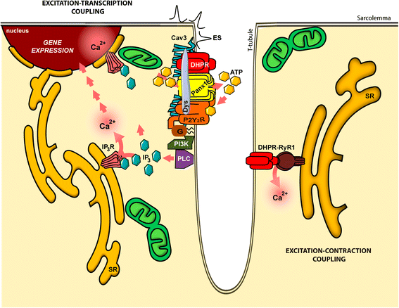
Hypothetical model of the multiprotein complex related to the excitation-transcription coupling in skeletal muscle. This model summarizes the findings discussed in this work and in our previous publications [9, 14, 17, 19, 20]. A multiprotein complex is assembled at the beginning of the T-tubule membrane, including dihydropyridine receptor (DHPR, Cav1.1) as the voltage sensor, pannexin 1 (Panx1) as the ATP release channel, the purinergic metabotropic P2Y2 receptor (P2Y2R), and caveolin-3 (Cav3), and dystrophin (Dys) as the scaffold proteins. The membrane depolarization sensed by DHPR induce the ATP release through Panx1. The heterotrimeric G protein attached to P2Y2R sequentially activate, upon ATP binding, phosphoinositide-3 kinase (PI3K) and phospholipase-C (PLC) to produce IP3 and activates calcium release through IP3R, for the slow calcium transient. This IP3-dependent slow calcium transient is involved in the gene expression control of several genes by the excitation-transcription coupling mechanism. The ryanodine receptor-1 (Ryr1), that is known to interact with DHPR, is involved in the excitation-contraction coupling, related with the fast calcium transients, but whether this protein is part of the same multiprotein complex involved in the excitation-transcription coupling has not yet been elucidated

References

    1. Bassel-Duby R, Olson EN. Signaling pathways in skeletal muscle remodeling. Annu Rev Biochem. 2006;75:19–37. doi: 10.1146/annurev.biochem.75.103004.142622. - DOI - PubMed
    1. Pette D. Training effects on the contractile apparatus. Acta Physiol Scand. 1998;162(3):367–76. doi: 10.1046/j.1365-201X.1998.0296e.x. - DOI - PubMed
    1. Pilegaard H, Ordway GA, Saltin B, Neufer PD. Transcriptional regulation of gene expression in human skeletal muscle during recovery from exercise. Am J Physiol Endocrinol Metab. 2000;279(4):E806–14. - PubMed
    1. Spina RJ, Chi MM, Hopkins MG, Nemeth PM, Lowry OH, Holloszy JO. Mitochondrial enzymes increase in muscle in response to 7–10 days of cycle exercise. J Appl Physiol. 1996;80(6):2250–4. - PubMed
    1. Bacou F, Rouanet P, Barjot C, Janmot C, Vigneron P, d’Albis A. Expression of myosin isoforms in denervated, cross-reinnervated, and electrically stimulated rabbit muscles. Eur J Biochem. 1996;236(2):539–47. doi: 10.1111/j.1432-1033.1996.00539.x. - DOI - PubMed

Publication types

MeSH terms

LinkOut - more resources