

Management of Oehler's Type III Dens Invaginatus Using Cone Beam Computed Tomography
- PMID: 27069697
- PMCID: PMC4812223
- DOI: 10.1155/2016/3573612
Management of Oehler's Type III Dens Invaginatus Using Cone Beam Computed Tomography
Abstract
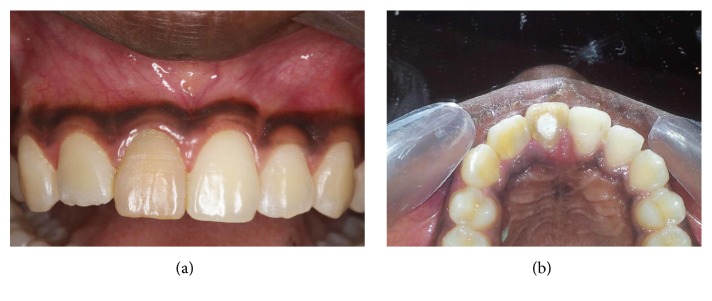
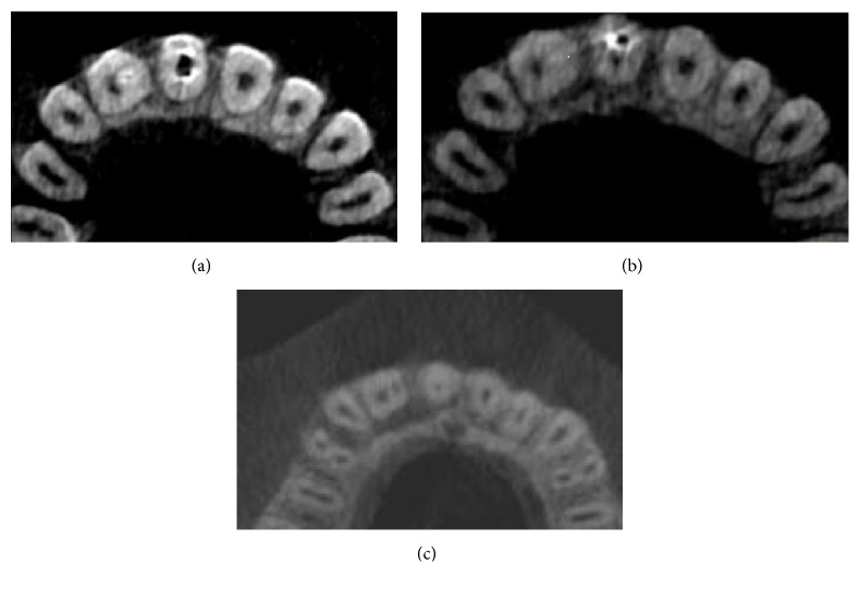
Dens Invaginatus is a dental malformation that poses diagnostic difficulties in the clinical context. This anomaly may increase the risk of pulp disease and can potentially complicate endodontic procedure due to the aberrant root canal anatomy. Compared to conventional radiographs, three-dimensional images obtained with Cone Beam Computed Tomography (CBCT) are invaluable in the diagnosis of the extent of this anomaly and in the appropriate treatment planning. Oehler's classification (1957) for Dens Invaginatus (DI) into three types depending on the depth of the invagination has been used for treatment planning. Of the three types Type III DI is characterized by infolding of the enamel into the tooth up to the root apex and is considered as the most severe variant of DI and hence the most challenging to treat endodontically, due to the morphological complexities. This report describes a case of Oehler's Type III DI in a necrotic permanent maxillary lateral incisor in which CBCT images played a key role in diagnosis and treatment planning. The case was managed successfully by a combination of nonsurgical and surgical endodontic therapy with orthograde and retrograde thermoplastic gutta percha obturation.
Figures


References
-
- Shafer W. G., Hine M. K., Levy B. M. Shafer's Textbook of Oral Pathology. 6th. section I. Elsevier; 2009.
-
- Jefferson Marion J. C., Maria Mesquita L., Duque T. M., Souza Filho F. J. Endodontic treatment of three types of dens invaginatus: report of four cases. Dental Press Endodontics. 2012;2(2):71–79.
LinkOut - more resources
Full Text Sources
Other Literature Sources
Miscellaneous
