. 2016 Apr;8(4):796-809.
doi: 10.18632/aging.100943.

Naturally occurring mitochondrial-derived peptides are age-dependent regulators of apoptosis, insulin sensitivity, and inflammatory markers

Affiliations

Naturally occurring mitochondrial-derived peptides are age-dependent regulators of apoptosis, insulin sensitivity, and inflammatory markers

Laura J Cobb et al. Aging (Albany NY). 2016 Apr.

Abstract

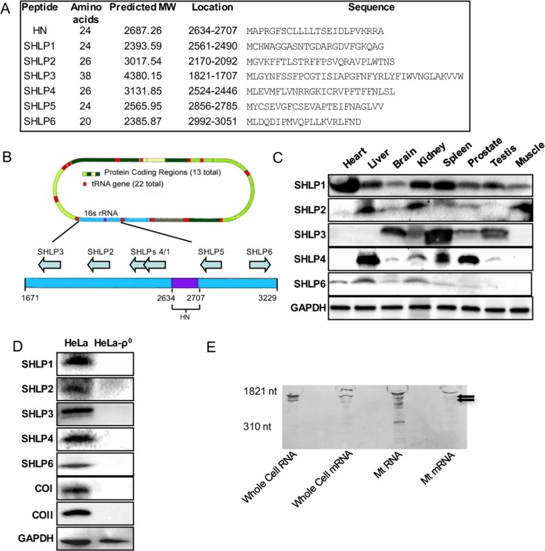
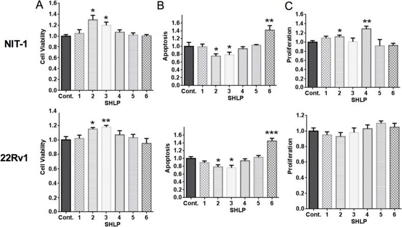
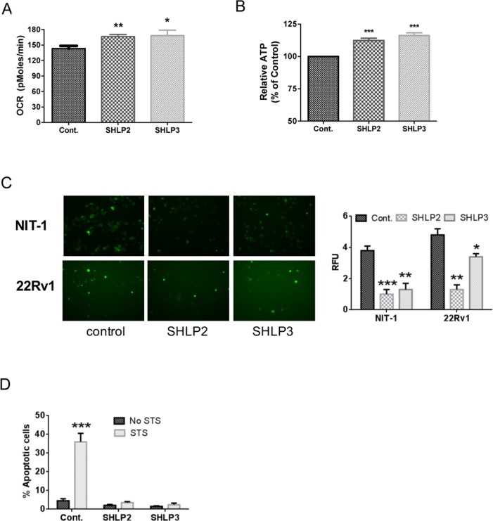
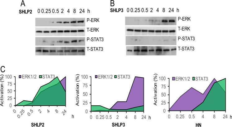
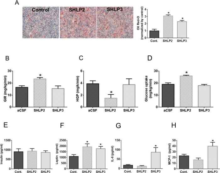
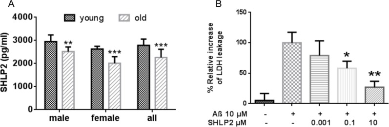
Mitochondria are key players in aging and in the pathogenesis of age-related diseases. Recent mitochondrial transcriptome analyses revealed the existence of multiple small mRNAs transcribed from mitochondrial DNA (mtDNA). Humanin (HN), a peptide encoded in the mtDNA 16S ribosomal RNA region, is a neuroprotective factor. An in silico search revealed six additional peptides in the same region of mtDNA as humanin; we named these peptides small humanin-like peptides (SHLPs). We identified the functional roles for these peptides and the potential mechanisms of action. The SHLPs differed in their ability to regulate cell viability in vitro. We focused on SHLP2 and SHLP3 because they shared similar protective effects with HN. Specifically, they significantly reduced apoptosis and the generation of reactive oxygen species, and improved mitochondrial metabolism in vitro. SHLP2 and SHLP3 also enhanced 3T3-L1 pre-adipocyte differentiation. Systemic hyperinsulinemic-euglycemic clamp studies showed that intracerebrally infused SHLP2 increased glucose uptake and suppressed hepatic glucose production, suggesting that it functions as an insulin sensitizer both peripherally and centrally. Similar to HN, the levels of circulating SHLP2 were found to decrease with age. These results suggest that mitochondria play critical roles in metabolism and survival through the synthesis of mitochondrial peptides, and provide new insights into mitochondrial biology with relevance to aging and human biology.

Keywords: SHLP; aging; humanin; mitochondria; small ORFs.


Conflict of interest statement

Conflict of interest statement

LC, CL, NB, and PC are consultants and stockholders of CohBar Inc.

Figures

Figure 1
Figure 1. Identification and validation of small open reading frames (sORFs) within the mitochondrial 16S ribosomal RNA (rRNA) gene
(A) Assigned names, size, location, and predicted sequences of SHLPs. (B) Location of SHLP ORFs within 16S rRNA. (C) Relative expression of SHLPs (1–4 and 6) in C57BL/6 mouse tissues relative to GAPDH (used as a loading control). (D) Expression of SHLPs (1–4 and 6), mitochondrial complexes (COI and COII) and nuclear GAPDH proteins in HeLa parental and HeLa-ρ0 cells. (E) Northern blot of whole cell RNA, whole cell mRNA, mitochondrial RNA, and mitochondrial mRNA using a SHLP6 probe identifies several bands smaller than the 16S rRNA indicated by the arrows.
Figure 2
Figure 2. Effect of SHLPs on cell growth and death
NIT-1 β and 22Rv1 cells were cultured in serum-free (SF) media with 100 nM SHLP or control peptides and assessed for (A) cell viability (using the MTS assay) after 72 h; (B) apoptosis after 24 h; and (C) cell proliferation by BrdU incorporation after 24 h. All data are presented as means ± SEM, and significance was determined by Student's t-tests. *P < 0.05; **P < 0.01; ***P < 0.001.
Figure 3
Figure 3. SHLP2 and SHLP3 modulate mitochondrial function
The effects of exogenous SHLP2 and SHLP3 on mitochondria were assessed in 22Rv1 cells incubated with 100 nM control peptide, SHLP2, or SHLP3 for 24 h by measuring (A) oxygen consumption rate (OCR) performed on a Seahorse XF24 Extracellular Flux Analyzer and (B) ATP production. (C) Reactive oxygen species (ROS) production as assessed by DHE (dihydroethidium) fluorescence in NIT-1 (top) and 22RV1 (bottom) cells after incubation with 100 nM control peptide, SHLP2, or SHLP3 overnight. All data are presented as means ± SEM. (D) NIT-1 β cells were pre-incubated with 100 nM SHLP2 or SHLP3 for 5 h, followed by incubation with 10 μM staurosporine (STS) for 24 h. Apoptosis (pre-G1 peak) was assessed by FACS (fluorescence-activated cell sorting) analysis. *P < 0.05; **P < 0.01; ***P < 0.001.
Figure 4
Figure 4. ERK and STAT-3 activation by SHLP2 and SHLP3
Levels of phospho- and total STAT3 and ERK were assessed in NIT-1 β cells treated with 100 nM (A) SHLP2 or (B) SHLP3. (C) Quantification of ERK and STAT3 levels after treatment with SHLP2, SHLP3, and HN. HN data were derived from Hoang et al., 2009.
Figure 5
Figure 5. In vitro and in vivo metabolic effects of SHLP2 and SHLP3 on insulin action and pro-inflammatory biomarker expression
(A) 3T3-L1 pre-adipocyte differentiation was assessed by Oil Red-O staining after incubation with insulin in the presence of control peptide, SHLP2, or SHLP3. (B–D) SHLP2 or SHLP3 were infused by ICV at a rate of 0.16 μg/kg/min into conscious SD rats (n = 6) and glucose flux was studied acutely under systemic pancreatic insulin clamp and physiologic hyperinsulinemic-euglycemic clamp. Glucose infusion rate (B), hepatic glucose production (C), and peripheral glucose uptake (D) were measured. (E–F) C57BL/6 mice (n = 5) were treated with control, SHLP2, or SHLP3 peptides (2 mg/kg/dose, BID, IP) for 5 days. Serum insulin (E), leptin (F), interleukin-6 (G), and monocyte chemotactic protein 1 (MCP-1) (H) levels were measured using LINCOplexTM analysis. All data are presented as means ± SEM. *P < 0.05.
Figure 6
Figure 6. Circulating SHLP2 levels decrease with age in mice and protect against amyloid-beta (Aβ) toxicity
(A) Plasma SHLP2 levels in young and old male and female C57BL/6 mice (n = 10 in each group) were assessed by ELISA. (B) Mouse primary cortical neurons were treated with vehicle control, 10 μM Aβ1–42 alone, or co-treated 10 μM Aβ1–42 with 1 nM, 100 nM, or 10 μM SHLP2. Aβ1–42-induced cytotoxicity was measured using a LDH (lactate dehydrogenase) cytotoxicity assay kit.

References

    1. Mercer TR, Neph S, Dinger ME, Crawford J, Smith MA, Shearwood AMJ, Haugen E, Bracken CP, Rackham O, Stamatoyannopoulos JA, Filipovska A, Mattick JS. The Human Mitochondrial Transcriptome. Cell. 2011;146:645–658. - PMC - PubMed
    1. Hashimoto Y, Niikura T, Tajima H, Yasukawa T, Sudo H, Ito Y, Kita Y, Kawasumi M, Kouyama K, Doyu M, Sobue G, Koide T, Tsuji S, et al. A rescue factor abolishing neuronal cell death by a wide spectrum of familial Alzheimer's disease genes and Abeta. Proc Natl Acad Sci U S A. 2001;98:6336–6341. - PMC - PubMed
    1. Ho L, Titus AS, Banerjee KK, George S, Lin W, Deota S, Saha AK, Nakamura K, Gut P, Verdin E, Kolthur-Seetharam U. SIRT4 regulates ATP homeostasis and mediates a retrograde signaling via AMPK. Aging (Albany NY) 2013;5:835–849. doi: 10.18632/aging.100616. - DOI - PMC - PubMed
    1. Guo B, Zhai D, Cabezas E, Welsh K, Nouraini S, Satterthwait AC, Reed JC. Humanin peptide suppresses apoptosis by interfering with Bax activation. Nature. 2003;423:456–461. - PubMed
    1. Ikonen M, Liu BR, Hashimoto Y, Ma LQ, Lee KW, Niikura T, Nishimoto I, Cohen P. Interaction between the Alzheimer's survival peptide humanin and insulin-like growth factor-binding protein-3 regulates cell survival and apoptosis. P Natl Acad Sci USA. 2003;100:13042–13047. - PMC - PubMed

Publication types