. 2016 Jun;22(6):1031-7.
doi: 10.3201/2206.160192. Epub 2016 Jun 15.

Infection, Replication, and Transmission of Middle East Respiratory Syndrome Coronavirus in Alpacas

Infection, Replication, and Transmission of Middle East Respiratory Syndrome Coronavirus in Alpacas

Danielle R Adney et al. Emerg Infect Dis. 2016 Jun.

Abstract

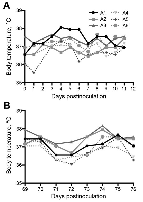
Middle East respiratory syndrome coronavirus is a recently emerged pathogen associated with severe human disease. Zoonotic spillover from camels appears to play a major role in transmission. Because of logistic difficulties in working with dromedaries in containment, a more manageable animal model would be desirable. We report shedding and transmission of this virus in experimentally infected alpacas (n = 3) or those infected by contact (n = 3). Infectious virus was detected in all infected animals and in 2 of 3 in-contact animals. All alpacas seroconverted and were rechallenged 70 days after the original infection. Experimentally infected animals were protected against reinfection, and those infected by contact were partially protected. Necropsy specimens from immunologically naive animals (n = 3) obtained on day 5 postinfection showed virus in the upper respiratory tract. These data demonstrate efficient virus replication and animal-to-animal transmission and indicate that alpacas might be useful surrogates for camels in laboratory studies.

Keywords: MERS-CoV; Middle East respiratory syndrome coronavirus; Vicugna pacos; alpacas; experimental infection; immunity; infection; replication; respiratory infections; transmission; viruses; zoonoses.


Figures

Figure 1
Figure 1
Body temperatures of 6 alpacas (A1–A6) experimentally infected with Middle East respiratory syndrome coronavirus after A) initial challenge and B) after rechallenge on day 70 postinfection.
Figure 2
Figure 2
Virus shedding (nasal swab specimens) in 6 alpacas experimentally infected with Middle East respiratory syndrome coronavirus (A1–A3) and co-housed with infected animals (A4–A6). A, B) initial challenge; C, D) rechallenge with addition of 3 immunologically naive alpacas (A7–A9). Individual animal results (A, C) and group means (B, D) are shown. Dotted vertical lines indicate detection limit of the assay.
Figure 3
Figure 3
Virus titers from tissues collected from 3 immunologically naive alpacas (A7–A9) challenged with Middle East respiratory syndrome coronavirus and sampled at necropsy on day 5 postinfection. Dotted line indicates detection limit of the assay.
Figure 4
Figure 4
Signs of mild upper respiratory inflammation, encephalitis, and virus antigen detection in respiratory epithelium of alpacas experimentally infected with Middle East respiratory syndrome coronavirus. A) Turbinate from alpaca A8 showing normal respiratory epithelium on the right with goblet cells (blue cells). Epithelium on the left has undergone squamous metaplasia (arrows) and is focally eroded with mild subepithelial inflammation (original magnification ×100). B) Virus antigen in apparently intact respiratory epithelium of alpaca A8 detected by immunohistochemical analysis and lack of subepithelial inflammation (original magnification ×200 [top] and ×400 [bottom]). C) Erosion in turbinate epithelium from alpaca A8 showing leukocytosis in underlying blood vessels (original magnification ×400). D) Perivascular infiltration of lymphocytes and monocytes in the brain of alpaca A9 (original magnification ×200). Hematoxylin and eosin stain in panels A, B, and D; immunohistochemical stain in panel C.

References

    1. Zaki AM, van Boheemen S, Bestebroer T, Osterhaus A, Fouchier R. Isolation of a novel coronavirus from a man with pneumonia in Saudi Arabia. N Engl J Med. 2012;367:1814–20. 10.1056/NEJMoa1211721 - DOI - PubMed
    1. World Health Organization. Middle East respiratory syndrome coronavirus (MERS-CoV), 2015. [cited 2016 Feb 22]. http://www.who.int/emergencies/mers-cov/en/
    1. Azhar EI, El-Kafrawy SA, Farraj SA, Hassan AM, Al-Saeed MS, Hashem AM, et al. Evidence for camel-to-human transmission of MERS coronavirus. N Engl J Med. 2014;370:2499–505. 10.1056/NEJMoa1401505 - DOI - PubMed
    1. Memish ZA, Cotten M, Meyer B, Watson SJ, Alsahafi AJ, Al Rabeeah AA, et al. Human infection with MERS coronavirus after exposure to infected camels, Saudi Arabia, 2013. Emerg Infect Dis. 2014;20:1012–5. 10.3201/eid2006.140402 - DOI - PMC - PubMed
    1. Haagmans BL, Al Dhahiry SH, Reusken CB, Raj VS, Galiano M, Myers R, et al. Middle East respiratory syndrome coronavirus in dromedary camels: an outbreak investigation. Lancet Infect Dis. 2014;14:140–5. 10.1016/S1473-3099(13)70690-X - DOI - PMC - PubMed

MeSH terms

LinkOut - more resources