

Made to measure - keeping Rho kinase at a distance
- PMID: 27070834
- PMCID: PMC4905274
- DOI: 10.1080/21541248.2016.1173770
Made to measure - keeping Rho kinase at a distance
Abstract
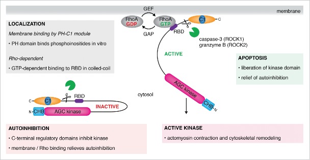
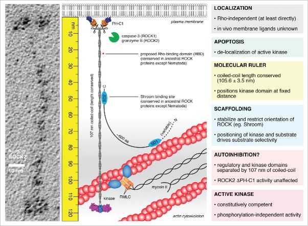
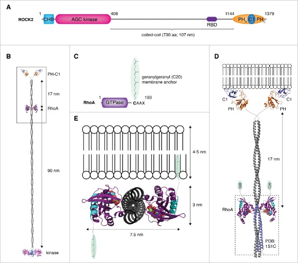
The Rho-associated coiled-coil containing kinases (ROCK) were first identified as effectors of the small GTPase RhoA, hence their nomenclature. Since their discovery, two decades ago, scientists have sought to unravel the structure, regulation, and function of these essential kinases. During that time, a consensus model has formed, in which ROCK activity is regulated via both Rho-dependent and independent mechanisms. However, recent findings have raised significant questions regarding this model. In their recent publication in Nature Communications, Truebestein and colleagues present the structure of a full-length Rho kinase for the first time. In contrast to previous reports, the authors could find no evidence for autoinhibition, RhoA binding, or regulation of kinase activity by phosphorylation. Instead, they propose that ROCK functions as a molecular ruler, in which the central coiled-coil bridges the membrane-binding regulatory domains to the kinase domains at a fixed distance from the plasma membrane. Here, we explore the consequences of the new findings, re-examine old data in the context of this model, and emphasize outstanding questions in the field.
Keywords: GTPase; ROCK; RhoA; coiled-coil; cytoskeleton; kinase; membrane anchor; molecular ruler; stress fibers.
Figures
Similar articles
-
A molecular ruler regulates cytoskeletal remodelling by the Rho kinases.Nat Commun. 2015 Dec 1;6:10029. doi: 10.1038/ncomms10029. Nat Commun. 2015. PMID: 26620183 Free PMC article.
-
Rho-associated coiled-coil containing kinases (ROCK): structure, regulation, and functions.Small GTPases. 2014;5:e29846. doi: 10.4161/sgtp.29846. Epub 2014 Jul 10. Small GTPases. 2014. PMID: 25010901 Free PMC article. Review.
-
Rho-associated coiled-coil kinase (ROCK) signaling and disease.Crit Rev Biochem Mol Biol. 2013 Jul-Aug;48(4):301-16. doi: 10.3109/10409238.2013.786671. Epub 2013 Apr 19. Crit Rev Biochem Mol Biol. 2013. PMID: 23601011 Review.
-
Structure of a highly conserved domain of Rock1 required for Shroom-mediated regulation of cell morphology.PLoS One. 2013 Dec 9;8(12):e81075. doi: 10.1371/journal.pone.0081075. eCollection 2013. PLoS One. 2013. PMID: 24349032 Free PMC article.
-
Ginseng (Panax quinquefolius) attenuates leptin-induced cardiac hypertrophy through inhibition of p115Rho guanine nucleotide exchange factor-RhoA/Rho-associated, coiled-coil containing protein kinase-dependent mitogen-activated protein kinase pathway activation.J Pharmacol Exp Ther. 2011 Dec;339(3):746-56. doi: 10.1124/jpet.111.182600. Epub 2011 Aug 29. J Pharmacol Exp Ther. 2011. PMID: 21875949
Cited by
-
ROCK inhibition with Y-27632 reduces joint inflammation and damage in serum-induced arthritis model and decreases in vitro osteoclastogenesis in patients with early arthritis.Front Immunol. 2022 Aug 11;13:858069. doi: 10.3389/fimmu.2022.858069. eCollection 2022. Front Immunol. 2022. PMID: 36032152 Free PMC article.
-
Rho-Kinase Planar Polarization at Tissue Boundaries Depends on Phospho-regulation of Membrane Residence Time.Dev Cell. 2020 Feb 10;52(3):364-378.e7. doi: 10.1016/j.devcel.2019.12.003. Epub 2020 Jan 2. Dev Cell. 2020. PMID: 31902655 Free PMC article.
-
Overexpression of Na+/H+ exchanger 1 specifically induces cell death in human iPS cells via sustained activation of the Rho kinase ROCK.J Biol Chem. 2019 Dec 20;294(51):19577-19588. doi: 10.1074/jbc.RA119.010329. Epub 2019 Nov 13. J Biol Chem. 2019. PMID: 31723030 Free PMC article.
-
Ligand-induced conformational rearrangements regulate the switch between membrane-proximal and distal functions of Rho kinase 2.Commun Biol. 2020 Nov 27;3(1):721. doi: 10.1038/s42003-020-01450-x. Commun Biol. 2020. PMID: 33247217 Free PMC article.
-
CDC42 binds PAK4 via an extended GTPase-effector interface.Proc Natl Acad Sci U S A. 2018 Jan 16;115(3):531-536. doi: 10.1073/pnas.1717437115. Epub 2018 Jan 2. Proc Natl Acad Sci U S A. 2018. PMID: 29295922 Free PMC article.
References
-
- Leung T, Manser E, Tan L, Lim L. A novel serine/threonine kinase binding the Ras-related RhoA GTPase which translocates the kinase to peripheral membranes. J Biol Chem 1995; 270:29051-4; PMID:7493923; http://dx.doi.org/10.1074/jbc.270.42.25107 - DOI - PubMed
-
- Ishizaki T, Maekawa M, Fujisawa K, Okawa K, Iwamatsu A, Fujita A, Watanabe N, Saito Y, Kakizuka A, Morii N, et al.. The small GTP-binding protein Rho binds to and activates a 160 kDa Ser/Thr protein kinase homologous to myotonic dystrophy kinase. EMBO J 1996; 15:1885-93; PMID:8617235. - PMC - PubMed
-
- Shimizu Y, Thumkeo D, Keel J, Ishizaki T, Oshima H, Oshima M, Noda Y, Matsumura F, Taketo MM, Narumiya S. ROCK-I regulates closure of the eyelids and ventral body wall by inducing assembly of actomyosin bundles. J Cell Biol 2005; 168:941-53; PMID:15753128; http://dx.doi.org/10.1083/jcb.200411179 - DOI - PMC - PubMed
-
- Thumkeo D, Keel J, Ishizaki T, Hirose M, Nonomura K, Oshima H, Oshima M, Taketo MM, Narumiya S. Targeted disruption of the mouse rho-associated kinase 2 gene results in intrauterine growth retardation and fetal death. Mol Cell Biol 2003; 23:5043-55; PMID:12832488; http://dx.doi.org/10.1128/MCB.23.14.5043-5055.2003 - DOI - PMC - PubMed
Publication types
MeSH terms
Substances
Grants and funding
LinkOut - more resources
Full Text Sources
Other Literature Sources