Comparative Study
. 2016 Apr 26;113(17):4824-9.
doi: 10.1073/pnas.1517558113. Epub 2016 Apr 11.

Contrasting variability patterns in the default mode and sensorimotor networks balance in bipolar depression and mania

Affiliations
Comparative Study

Contrasting variability patterns in the default mode and sensorimotor networks balance in bipolar depression and mania

Matteo Martino et al. Proc Natl Acad Sci U S A. .

Abstract

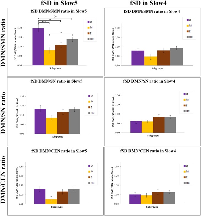
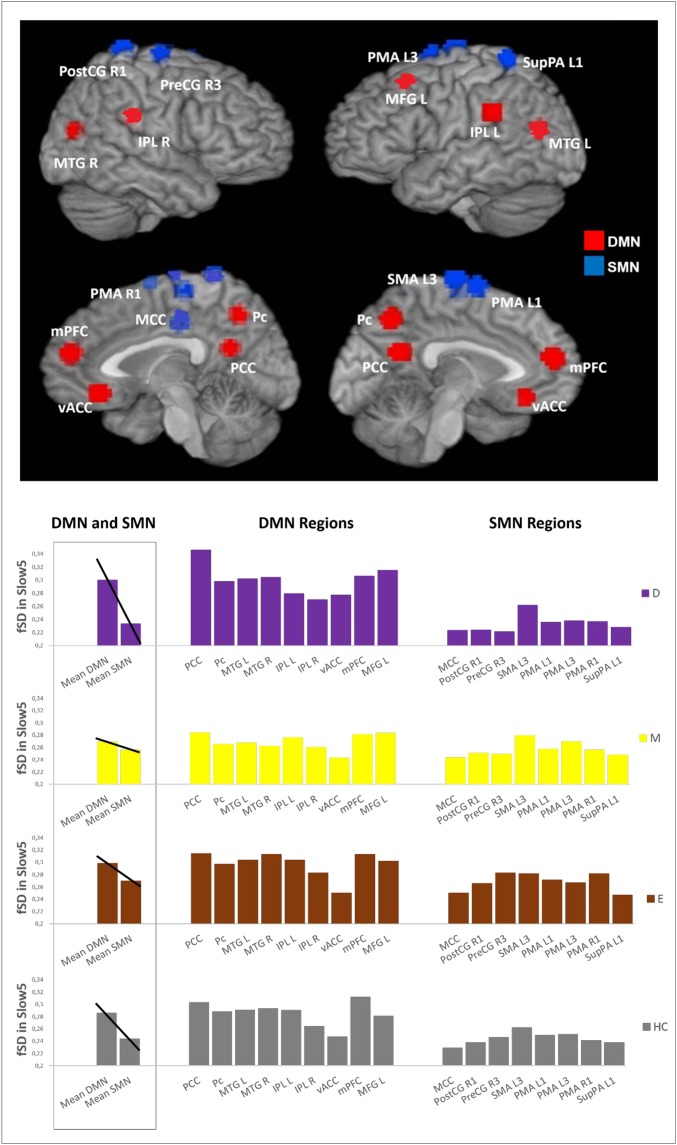
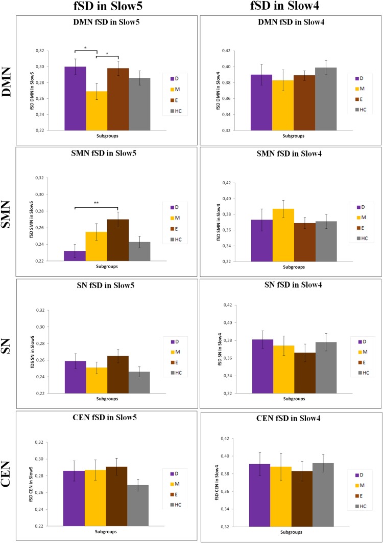
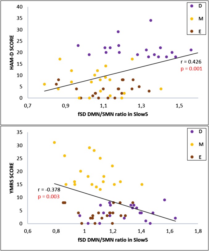
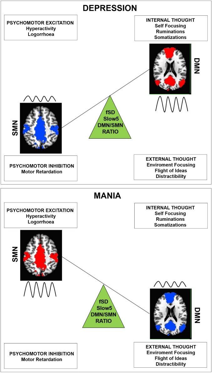
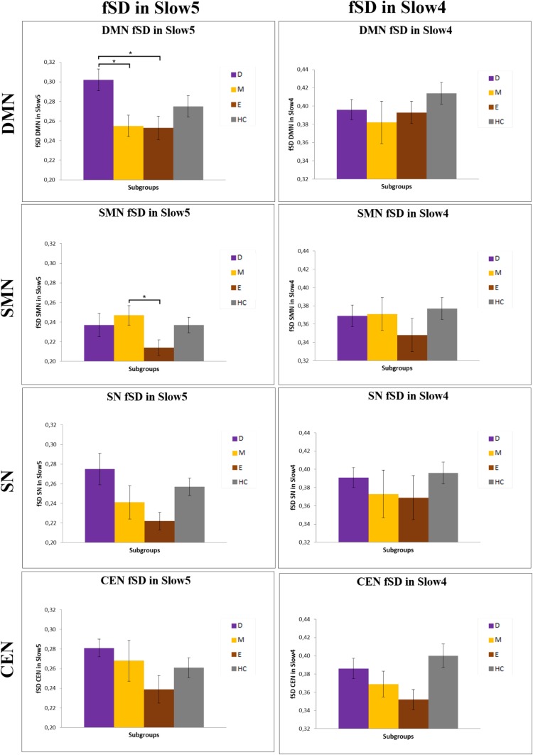
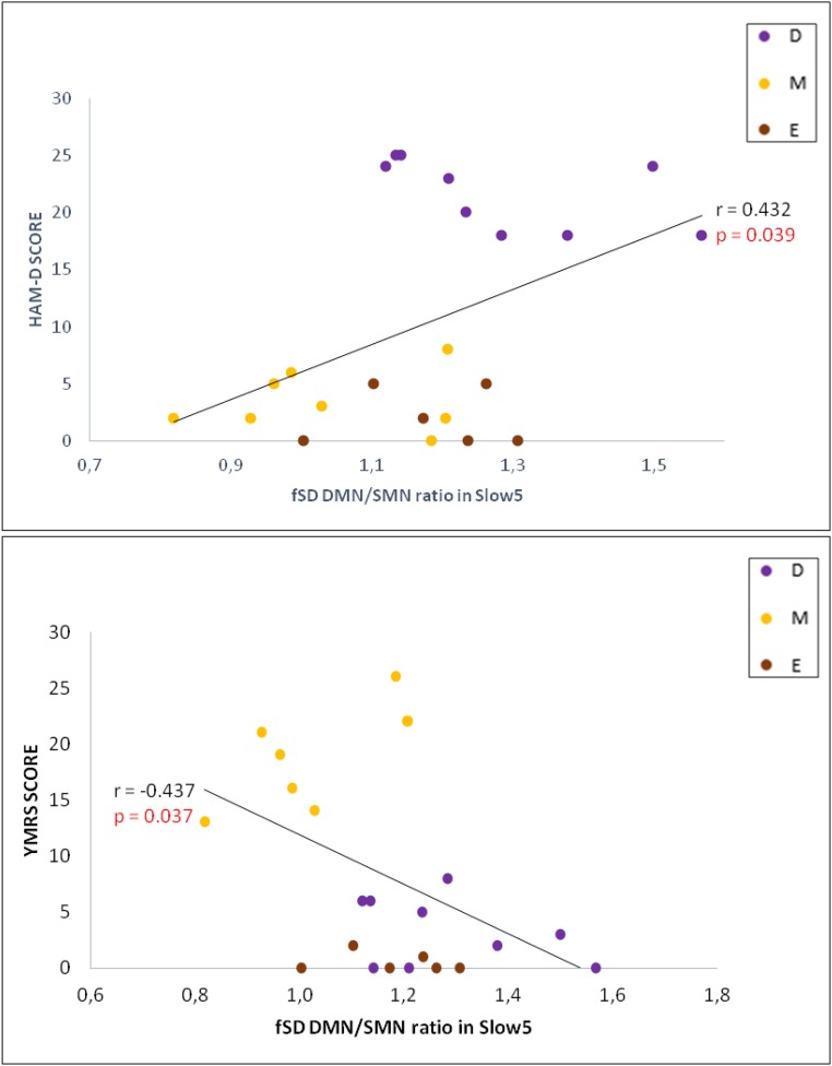
Depressive and manic phases in bipolar disorder show opposite constellations of affective, cognitive, and psychomotor symptoms. At a neural level, these may be related to topographical disbalance between large-scale networks, such as the default mode network (DMN) and sensorimotor network (SMN). We investigated topographical patterns of variability in the resting-state signal-measured by fractional SD (fSD) of the BOLD signal-of the DMN and SMN (and other networks) in two frequency bands (Slow5 and Slow4) with their ratio and clinical correlations in depressed (n = 20), manic (n = 20), euthymic (n = 20) patients, and healthy controls (n = 40). After controlling for global signal changes, the topographical balance between the DMN and SMN, specifically in the lowest frequency band, as calculated by the Slow5 fSD DMN/SMN ratio, was significantly increased in depression, whereas the same ratio was significantly decreased in mania. Additionally, Slow5 variability was increased in the DMN and decreased in the SMN in depressed patients, whereas the opposite topographical pattern was observed in mania. Finally, the Slow5 fSD DMN/SMN ratio correlated positively with clinical scores of depressive symptoms and negatively with those of mania. Results were replicated in a smaller independent bipolar disorder sample. We demonstrated topographical abnormalities in frequency-specific resting-state variability in the balance between DMN and SMN with opposing patterns in depression and mania. The Slow5 DMN/SMN ratio was tilted toward the DMN in depression but was shifted toward the SMN in mania. The Slow5 fSD DMN/SMN pattern could constitute a state-biomarker in diagnosis and therapy.

Keywords: bipolar disorder; default mode network; neuronal variability; sensorimotor network.


Conflict of interest statement

The authors declare no conflict of interest.

Figures

Fig. 1.
Fig. 1.
The DMN/SMN, DMN/SN, and DMN/CEN ratios in fSD Slow5 and Slow4 in the various subgroups. Results of the ANOVA and Games–Howell post hoc test of fSD of the DMN/SMN, DMN/SN, and DMN/CEN ratios in Slow5 and Slow4 between the various subgroups. Corrected *P < 0.05, **P < 0.01, ***P < 0.001. D, depressive patients; E, euthymic patients; HC, healthy controls; M, manic patients.
Fig. 2.
Fig. 2.
Differences in the DMN/SMN balance in the various subgroups. The Upper part of the figure is a global view of the DMN (red) and SMN (blue) regions. The Lower Left of the figure is the mean of the fSD values in Slow5 of the DMN and SMN, together with a visual trend of the balance between the DMN and SMN, for each subgroup. The Lower Right of the figure is the mean fSD values in Slow5 of the various regions belonging to the DMN and SMN, for each subgroup. D, depressive patients; E, euthymic patients; HC, healthy controls; IPL L, inferior parietal lobule left; IPL R, inferior parietal lobule right; M, manic patients; MCC, middle cingulate cortex; MFG L, middle frontal gyrus left; mPFC, medial preFrontal cortex; MTG L, middle temporal gyrus left; MTG R, middle temporal gyrus right; Pc, precuneus; PCC, posterior cingulate cortex; PMA L1, premotor area left; PMA L3, premotor area left; PMA R1, premotor area right; PostCG R1, postcentral gyrus right; PreCG R3, precentral gyrus right; SMA L3, supplementar motor area left; SupPA L1, superior parietal area left; vACC, ventral anterior cingulate cortex.
Fig. S1.
Fig. S1.
Differences in DMN, SMN, SN, and CEN fSD Slow5 and Slow4 in the various subgroups. Mean values of fSD in Slow5 and Slow4 of DMN, SMN, SN, and CEN in the various subgroups; *P < 0.05, **P < 0.01. D, depressive patients; E, euthymic patients; HC, healthy controls; M, manic patients.
Fig. 3.
Fig. 3.
Clinical correlations. Pearson correlation (after bootstrapping) between fSD of the DMN/SMN ratio in Slow5 and the HAM-D and YMRS in BD. D, depressive patients; E, euthymic patients; M, manic patients.
Fig. 4.
Fig. 4.
Schema of DMN/SMN disbalance in depression and mania. The changes in the fSD of the DMN/SMN ratio in Slow5 (green triangle). The model represents the hypothetical relationship between changes in DMN/SMN balance and the most typical clinical presentation of BD depression and mania. Changes of the relative weight of the lower frequency Slow5 band (wave) could affect the balance between different resting-state networks, in the various phases of BD. In depression, the increase of the ratio could tilt the network disbalance toward the DMN (red and higher amplitude of the wave) at the expense of the SMN (blue and lower amplitude of the wave), which may lead to internal thought (focused on internal contents at the expense of the external contents) and psychomotor inhibition. In mania, the decrease of the ratio could tilt the network disbalance toward the SMN (red and higher amplitude of the wave) at the expense of the DMN (blue and lower amplitude of the wave), which may lead to external thought (focused on external contents at the expense of the internal contents) and psychomotor excitation.
Fig. S2.
Fig. S2.
The DMN/SMN, DMN/SN, and DMN/CEN ratios in fSD Slow5 and Slow4 in the various subgroups of the independent sample. Results of the ANOVA and Games–Howell post hoc test of fSD of the DMN/SMN, DMN/SN, and DMN/CEN ratios in Slow5 and Slow4 between the various subgroups. Corrected *P < 0.05; D, depressive patients; E, euthymic patients; HC, healthy controls; M, manic patients.
Fig. S3.
Fig. S3.
Differences in DMN, SMN, SN, and CEN fSD Slow5 and Slow4 in the various subgroups of the independent sample. Mean values of fSD in Slow5 and Slow4 of DMN, SMN, SN, and CEN in the various subgroups; *P < 0.05. D, depressed patients; E, euthymic patients; HC, healthy controls; M, manic patients.
Fig. S4.
Fig. S4.
Clinical correlations in the independent sample. Pearson correlation (after bootstrapping) between fSD of the DMN/SMN ratio in Slow5 and the HAMD and YMRS in BD. D, depressive patients; E, euthymic patients; M, manic patients.

References

    1. American Psychiatrich Association (2013) Diagnostic and Statistical Manual for Mental Disorders. 5th ed. (DSM-5). (American Psychiatrich Association, Washington, DC)
    1. Kraepelin E. Clinical Psychiatry. Macmillan; New York: 1902.
    1. Northoff G. Spatiotemporal psychopathology I: No rest for the brain's resting state activity in depression? Spatiotemporal psychopathology of depressive symptoms. J Affect Disord. 2016;190:854–866. - PubMed
    1. Northoff G. Spatiotemporal Psychopathology II: How does a psychopathology of the brain's resting state look like? Spatiotemporal approach and the history of psychopathology. J Affect Disord. 2016;190:867–879. - PubMed
    1. Minassian A, et al. The quantitative assessment of motor activity in mania and schizophrenia. J Affect Disord. 2010;120(1-3):200–206. - PMC - PubMed

Publication types