. 2016 Apr 26;113(17):4723-8.
doi: 10.1073/pnas.1519128113. Epub 2016 Apr 11.

Heterozygous colon cancer-associated mutations of SAMHD1 have functional significance

Affiliations

Heterozygous colon cancer-associated mutations of SAMHD1 have functional significance

Matilda Rentoft et al. Proc Natl Acad Sci U S A. .

Erratum in

Abstract

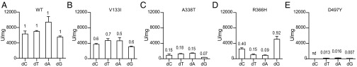
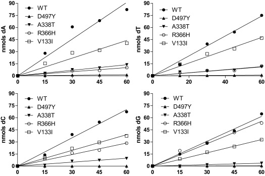
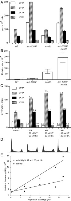
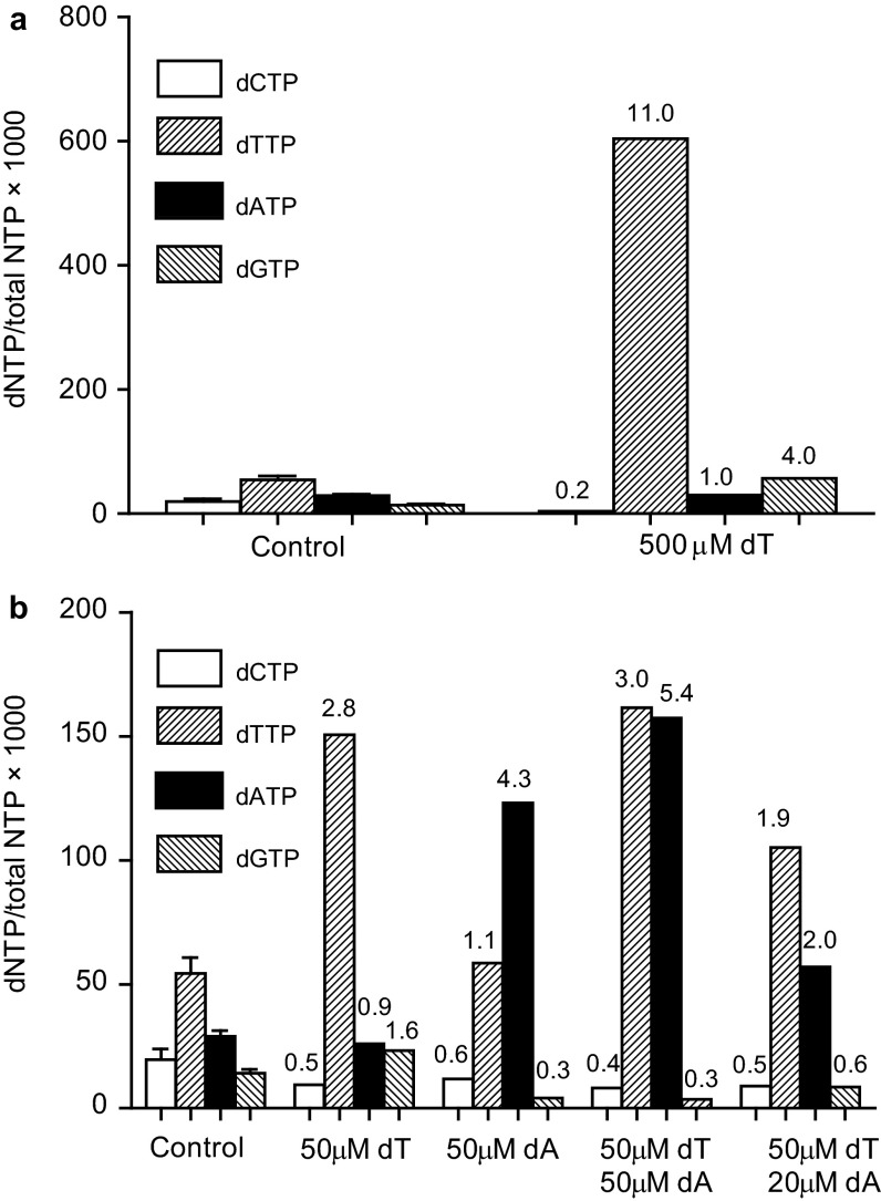
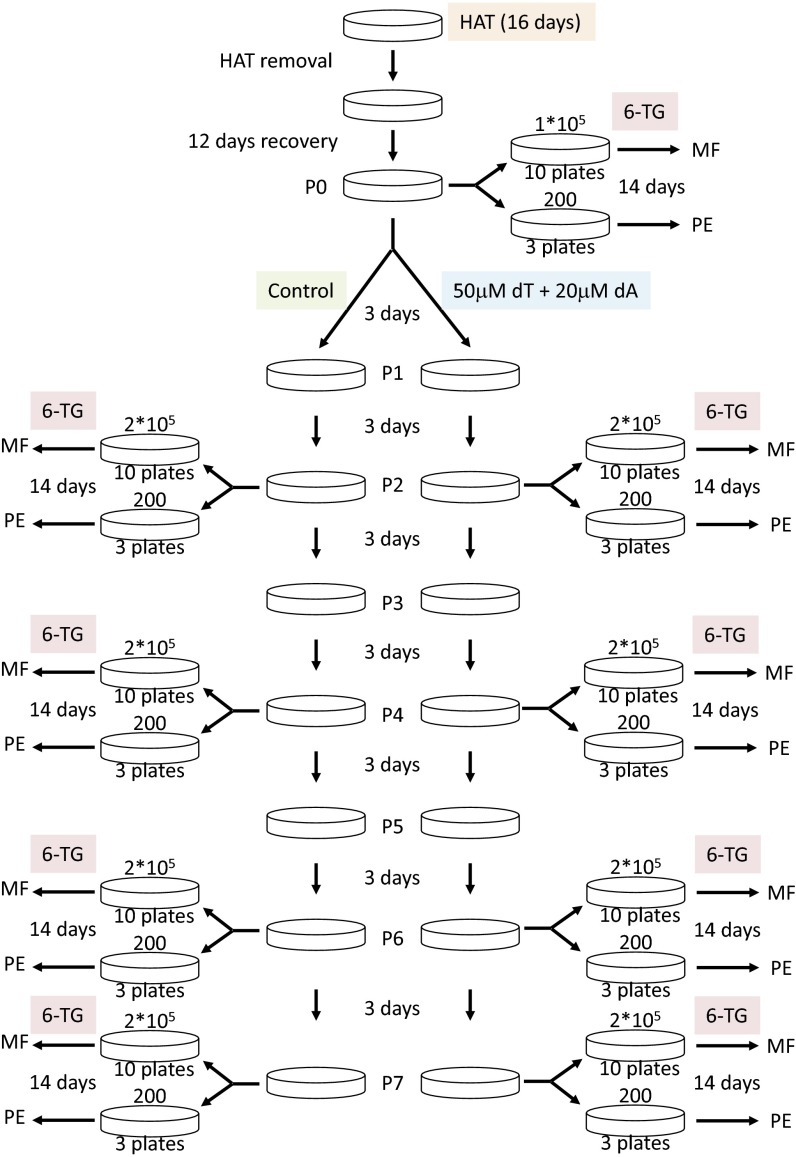
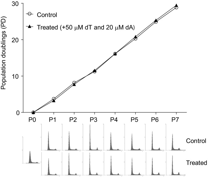
Even small variations in dNTP concentrations decrease DNA replication fidelity, and this observation prompted us to analyze genomic cancer data for mutations in enzymes involved in dNTP metabolism. We found that sterile alpha motif and histidine-aspartate domain-containing protein 1 (SAMHD1), a deoxyribonucleoside triphosphate triphosphohydrolase that decreases dNTP pools, is frequently mutated in colon cancers, that these mutations negatively affect SAMHD1 activity, and that several SAMHD1 mutations are found in tumors with defective mismatch repair. We show that minor changes in dNTP pools in combination with inactivated mismatch repair dramatically increase mutation rates. Determination of dNTP pools in mouse embryos revealed that inactivation of one SAMHD1 allele is sufficient to elevate dNTP pools. These observations suggest that heterozygous cancer-associated SAMHD1 mutations increase mutation rates in cancer cells.

Keywords: DNA replication fidelity; colon cancer; dNTP pools.


Conflict of interest statement

The authors declare no conflict of interest.

Figures

Fig. S1.
Fig. S1.
Analyses of purified SAMHD1 proteins. A total of 1 µg of purified SAMHD1 (a) and SAMHD1 mutant proteins V133I (b), A338T (c), R366H (d), and D497Y (e) were separated by 12.5% SDS/PAGE and stained with Coomassie Brilliant Blue.
Fig. 1.
Fig. 1.
In vitro dNTPase activity of SAMHD1 is altered by cancer-associated mutations. Purified SAMHD1 (A) and SAMHD1 mutant proteins V133I (B), A338T (C), R366H (D), and D497Y (E) were incubated with 1 mM dCTP, dTTP, dATP, or dGTP separately with 1 mM GTP as the activator, and the deoxynucleoside products (dC, dT, dA, and dG) were analyzed by HPLC. Numbers indicate detected deoxynucleoside products in mutants compared with WT SAMHD1. Error bars indicate SD. nd, not detectable.
Fig. S2.
Fig. S2.
In vitro dNTPase activity assays of SAMHD1 protein and the four mutants over a time course of 60 min. All reactions were performed by using the same sample of each protein. Reactions with a total volume of 300 μL [10 mM Tris⋅HCl (pH 7.5), 50 mM NaCl, 5 mM MgCl2, 1 mM GTP, 1 mM dNTP, and 0.5 μM SAMHD1] were incubated at 25 °C. Aliquots were collected at 0, 15, 30, 45, and 60 min, and the dN products were quantified. Lines were fitted by using the linear regression function in GraphPad Prism.
Fig. 2.
Fig. 2.
dNTP levels in mouse embryos are affected by SAMHD1 copy number. dNTP levels were measured in E13.5 mouse embryos that were WT (33 embryos), lacking one copy of SAMHD1 (13 embryos), or lacking both copies of SAMHD1 (18 embryos). Results are presented in a boxplot where the central box spans the first to the third quartile, the whiskers represent minimum and maximum values, and the segment inside the box is the median. Outliers are represented by circles. The significance value was calculated by using the Wilcoxon rank sum test.
Fig. 3.
Fig. 3.
Minor alterations of dNTP pools further elevate mutation rates in MMR-deficient cells. (A and B) Amount of each dNTP (A) and mutation rates (B) in the WT, msh2Δ, rnr1-Y285F, and msh2Δ rnr1-Y285F yeast strains. (C) Amount of each dNTP normalized to the total NTP pool in untreated HCT116 cells (control), HCT116 cells incubated in the presence of 50 μM thymidine and 20 μM deoxyadenosine for 20 h (20 h), and in HCT116 cells incubated in the presence of 50 μM thymidine and 20 μM deoxyadenosine for 20 h, after which an additional 50 μM thymidine and 20 μM were added. dNTP pools were measured after 1 h (+1 h) and after 4 h (+4 h). (D) Flow cytometry histograms of the HCT116 cells used for dNTP pool measurements in C. (E) Mutation frequencies and PDs of the HCT116 cells incubated in the presence or absence of 50 μM thymidine (dT) and 20 μM deoxyadenosine (dA).
Fig. S3.
Fig. S3.
MLH1 promoter methylation in the eight tumors with SAMHD1 mutations. Methylation levels of the MLH1 promoter in percentage are presented as a line plot. The x axis represents 50 CpG sites covering the region 1,468 bp upstream to 57,352 bp downstream of the transcriptional start site (marked by “1”). Red lines specify the variable region where an increased methylation level indicates silencing of the MLH1 gene. WT represents data from nine normal colon samples. The V133I variant is found in two tumors and denoted V133I_1 and V133I_2.
Fig. S4.
Fig. S4.
dNTP pools in HCT116 cells grown 1 h in the presence of thymidine (dT) and deoxyadenosine (dA) in the following concentrations: 500 μM thymidine (A) and 50 μM thymidine; 50 μM deoxyadenosine; 50 μM thymidine and 50 μM deoxyadenosine; and 50 μM thymidine and 20 μM deoxyadenosine (B).
Fig. S5.
Fig. S5.
Schematic representation of the workflow of the experiment for calculating the mutation rates of the HPRT locus of the HCT116 cells in the presence or absence of 50 μM thymidine (dT) and 20 μM deoxyadenosine (dA). 6-TG, 6-thioguanine; HAT, hypoxanthine-aminopterin-thymidine; MF, mutation frequency; PE, plating efficiency. For details, see SI Experimental Procedures.
Fig. S6.
Fig. S6.
Population doubling times and corresponding flow cytometry histograms of the HCT116 cells grown in the absence (control) or presence of 50 μM thymidine and 20 μM deoxyadenosine (treated). Each passage was 3 d.

References

    1. Schmitt MW, et al. Sequencing small genomic targets with high efficiency and extreme accuracy. Nat Methods. 2015;12(5):423–425. - PMC - PubMed
    1. Wang Y, et al. Clonal evolution in breast cancer revealed by single nucleus genome sequencing. Nature. 2014;512(7513):155–160. - PMC - PubMed
    1. Fox EJ, Loeb LA. Cancer: One cell at a time. Nature. 2014;512(7513):143–144. - PMC - PubMed
    1. Loeb LA, Springgate CF, Battula N. Errors in DNA replication as a basis of malignant changes. Cancer Res. 1974;34(9):2311–2321. - PubMed
    1. Schmitt MW, Prindle MJ, Loeb LA. Implications of genetic heterogeneity in cancer. Ann N Y Acad Sci. 2012;1267:110–116. - PMC - PubMed

Publication types

Substances