. 2016 Apr 26;113(17):E2355-62.
doi: 10.1073/pnas.1524398113. Epub 2016 Apr 11.

Soluble guanylate cyclase as an alternative target for bronchodilator therapy in asthma

Affiliations

Soluble guanylate cyclase as an alternative target for bronchodilator therapy in asthma

Arnab Ghosh et al. Proc Natl Acad Sci U S A. .

Abstract

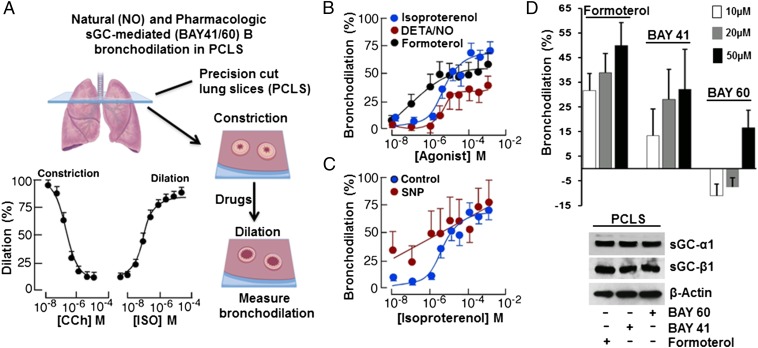
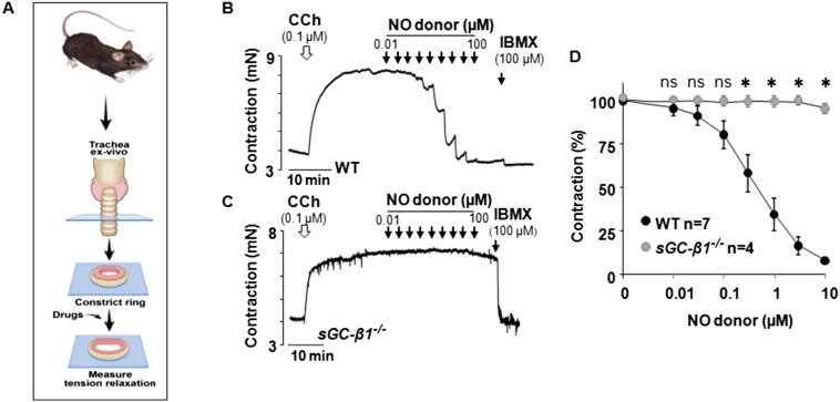
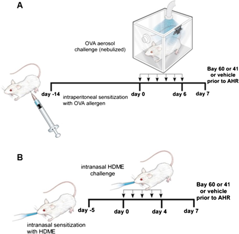
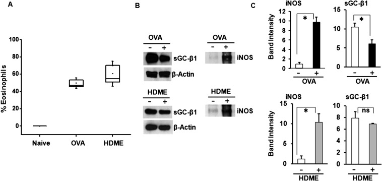
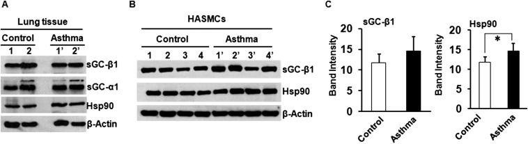
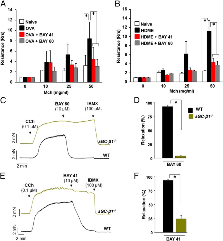
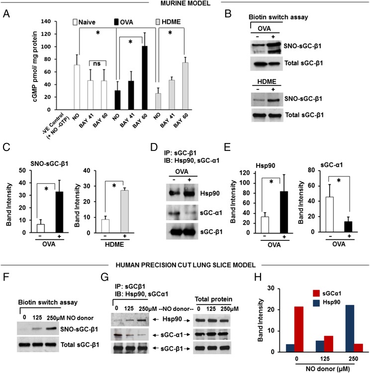
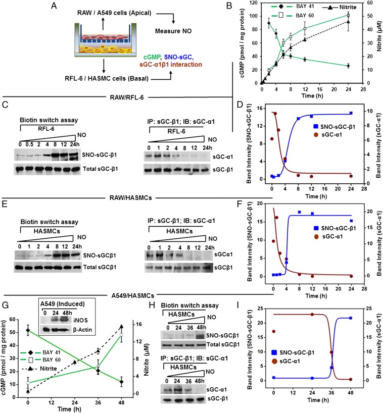
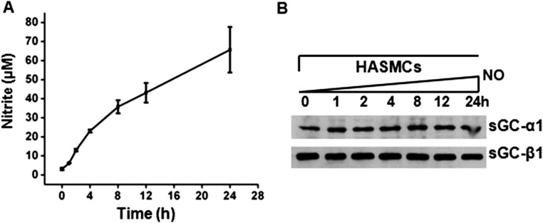
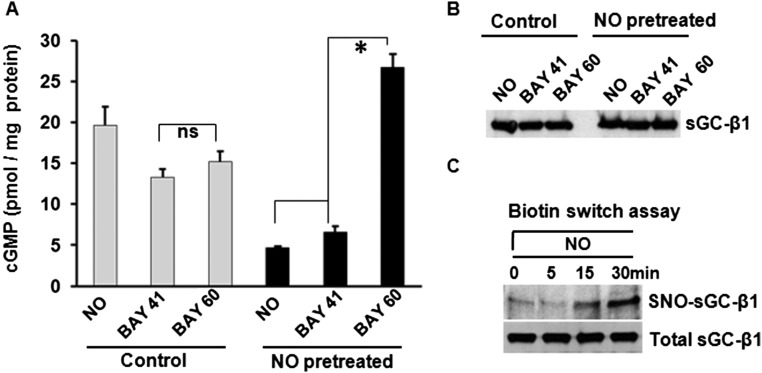
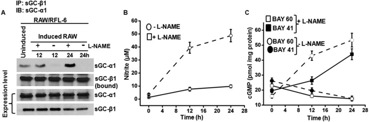
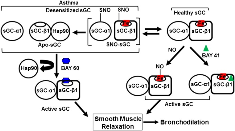
Asthma is defined by airway inflammation and hyperresponsiveness, and contributes to morbidity and mortality worldwide. Although bronchodilation is a cornerstone of treatment, current bronchodilators become ineffective with worsening asthma severity. We investigated an alternative pathway that involves activating the airway smooth muscle enzyme, soluble guanylate cyclase (sGC). Activating sGC by its natural stimulant nitric oxide (NO), or by pharmacologic sGC agonists BAY 41-2272 and BAY 60-2770, triggered bronchodilation in normal human lung slices and in mouse airways. Both BAY 41-2272 and BAY 60-2770 reversed airway hyperresponsiveness in mice with allergic asthma and restored normal lung function. The sGC from mouse asthmatic lungs displayed three hallmarks of oxidative damage that render it NO-insensitive, and identical changes to sGC occurred in human lung slices or in human airway smooth muscle cells when given chronic NO exposure to mimic the high NO in asthmatic lung. Our findings show how allergic inflammation in asthma may impede NO-based bronchodilation, and reveal that pharmacologic sGC agonists can achieve bronchodilation despite this loss.

Keywords: S-nitrosylation; bronchoconstriction; bronchodilation; heme protein; nitric oxide.


Conflict of interest statement

The authors declare no conflict of interest.

Figures

Fig. 1.
Fig. 1.
NO donors and sGC stimulators and activators bronchodilate human lung slices. (A) Small airways in human PCLS were contracted with CCh followed by addition of the indicated compounds and image collection and processing to determine bronchiole lumen area, expressed as percent compared with baseline. (B) NO donor (DETA/NO) caused a dose-dependent bronchodilation in a manner similar to the β-agonist Formoterol. (C) Coadministering a subthreshold dose of NO donor (SNP, 1 µM) enhanced Isoproterenol (ISO) dilation of PCLS. (D) Differing capacity of the BAY sGC activators to dilate PCLS relative to Formoterol, with the final sGC-α1 and β1 expression levels in the slices compared below. For A, n = 3; for B–D, n = 2; mean ± SD; four to seven slices per condition.
Fig. S1.
Fig. S1.
Mouse trachea response to NO requires sGC. (A) Tracheal rings prepared from normal (WT) or sGC-β1−/− mice were precontracted with CCh and then their relaxation in response to compounds was measured. (B and C) Representative traces indicating the relaxation achieved to the indicated concentrations of an NO donor (DEA-NO) and subsequently to the NO-independent relaxing agent IBMX (100 µM) to achieve maximal dilation. (D) Averaged data derived from replicate experiments with wild-type n = 7 rings and sGC-β1−/− n = 4 (*P < 0.05, by one-way ANOVA; ns, not statistically significant).
Fig. S2.
Fig. S2.
Mice models of allergic asthma. (A and B) Mice were treated to develop an inflammatory asthma toward either OVA or HDME and then received a single intratracheal administration of vehicle or BAY drug (50 µL; 30 µg/kg BAY 41–2272 or 90 µg/kg BAY 60–2770) at 30 min before testing airway resistance.
Fig. S3.
Fig. S3.
Changes in mouse lung associated with the allergic inflammatory asthma. (A) Percentage of eosinophils in bronchial lavage fluid from naïve mice and from mice with allergic asthma toward OVA or HDME (n = 3). (B) Comparative sGC-β1 and iNOS protein expression levels in lung supernatants and insoluble particulate fractions, respectively, from naïve and asthmatic mice. (C) Densitometric quantification of the iNOS and sGCβ1 protein expression. n = 3 each for naïve or allergic asthmatic mice (OVA/HDME) for iNOS and n = 4 for naive or OVA/HDME mice for sGC-β1 (*P < 0.05, by one-way ANOVA; ns, not statistically significant).
Fig. S4.
Fig. S4.
Expression of sGC and hsp90 in human lung tissue and in HASMCs derived from control or asthmatic lungs. (A) Expression of sGC-α1, β1, and hsp90 in control and asthmatic lung tissue. (B) Expression of sGC-β1 and hsp90 in control and asthmatic HASMCs. (C) Densitometric quantification of the sGC-β1 and hsp90 expression as depicted in B. Values are mean ± SD, and n = 4 each for control or asthmatic HASMCs (*P < 0.05, by one-way ANOVA).
Fig. 2.
Fig. 2.
sGC agonists abolish airway hyper-response in two models of allergic asthma. Mice were treated to develop an inflammatory asthma toward either OVA or HDME and then received an intratracheal administration of vehicle or BAY drug (50 µL; 30 µg/kg BAY 41–2272 or 90 µg/kg BAY 60–2770) at 30 min before testing airway resistance. (A and B) Airway resistance recorded for groups of naïve and asthmatic mice in response to methacholine bronchoconstrictor (Mch), showing the hypersensitive response of asthmatic mice was alleviated by either BAY compound. n = 6 for OVA-challenged mice at dosage 0 and 50 of Mch and n = 3 at other doses and for control mice. For HDME model, n = 4 for treated or control mice. Values are mean ±SD and are normalized with respect to their 0 Mch (*P < 0.05, by one-way ANOVA). (C and D) Test of sGC agonist specificity. Representative traces of mechanical tension versus time for tracheal rings from normal (WT) or sGC-β1−/− mice that were precontracted with CCh, then given BAY 60–2770 or BAY 41–2272, with IBMX. (E and F) Averaged data derived from replicate experiments with n = 5 rings for WT or sGC-β1−/− using BAY 60–2770 and n = 6 with BAY 41–2272 (*P < 0.05, by Mann–Whitney test two-tailed).
Fig. 3.
Fig. 3.
Asthmatic mouse lungs and NO-treated human PCLS contain altered sGC. (A) Activation profiles of lung supernatant sGC in response to NO donor (SNAP), BAY 41–2272, and BAY 60–2770. cGMP values are mean ± SD, n = 3 experiments. (B) Relative levels of total and SNO-sGC-β1 in lung supernatants from asthmatic and naïve mice. (C) Quantification of the SNO-sGC-β1 levels. (D) Change in sGC-β1 association with sGC-α1 and hsp90 as determined by immunoprecipitation. (E) Quantification of associated sGC-α1 and hsp90. Values are mean ± SD and n = 3 (for C) to 5 (for E) each for naive, OVA, or HDME mice. (*P < 0.05, by one-way ANOVA; ns, not statistically significant). (F) Groups of human PCLS (six to seven slices) were cultured overnight with three concentrations of NO donor (NOC-18) and their SNO-sGC and total sGC levels were determined. (G) Relative sGC-β1 associations with sGC-α1 versus hsp90 as determined by immunoprecipitation. (H) Densitometric quantification of the sGC-α1 or hsp90 associated with sGC-β1 from two independent experiments, as described in G.
Fig. 4.
Fig. 4.
Immuno-activated inflammatory cells alter sGC in responder cells. (A) Transwell coculture containing combinations of NO-producing cells (RAW/A549) and sGC-containing cells (RFL-6/HASMC) mimics the spatial relationship in the bronchial airways. (B) NO production by IFN-γ/LPS-activated RAW cells in the Transwell cultures, as indicated by nitrite accumulation, and the corresponding change in RFL-6 sGC activity over time in response to BAY 41–2272 or BAY 60–2770. Values are mean ± SD from three experiments. (C) Corresponding SNO-sGC-β1 buildup in the RFL-6 cells during the coculture (Left) and loss in sGC-α1β1 heterodimerization (Right) with time. (D) Quantification of SNO-sGC-β1 levels and sGC-α1 association versus time from two independent experiments. (E) Corresponding SNO-sGC-β1 build-up in HASMC (Left) in response to NO from activated RAW cells and loss in sGC-α1β1 heterodimerization (Right) with time. (F) HASMC SNO-sGC-β1 levels and sGC-α1 association with sGC-β1 versus time for two independent experiments. (G) Kinetics of NO production by cytokine-stimulated human A549 cells as indicated by nitrite accumulation, iNOS expression (Inset), and corresponding change in HASMC sGC responses to BAY 41–2272 or BAY 60–2770. Values are mean ± SD from three experiments. (H) Corresponding SNO-sGC-β1 buildup in the HASMC (Upper) and loss in sGC-α1β1 heterodimerization (Lower) with time. (I) Comparative SNO-sGC-β1 levels and sGC-α1 association versus time from two independent experiments.
Fig. S5.
Fig. S5.
NO produced from activated RAW cells does not alter expression of sGC in HASMCs. A Transwell coculture system as described in Fig. 4 with activated RAW cells in the apical and HASMCs in the basal chamber was used to study effects of NO produced from RAW cells on smooth muscle sGC. (A) NO production by IFN-γ/LPS-activated RAW cells in the Transwell cultures, as indicated by nitrite accumulation in the media. Values are mean ± SD of three independent experiments. (B) Representative Western blots depicting expression level of sGC-α1 and β1 in the airway smooth muscle cells under indicated time points.
Fig. S6.
Fig. S6.
Chronic NO exposure alters sGC in cells. (A) RFL-6 cells were treated with vehicle or an NO donor (NOC-12) for 12 h, washed, and then their sGC activity was tested by measuring cGMP production in response to activators NO (SNAP), BAY 41–2272, or BAY 60–2770. Values are mean (n = 3) ± SD (*P < 0.05, by one-way ANOVA; ns, not statistically significant). (B) sGC-β1 protein expression level in cells from A. (C) Levels of the total sGC-β1 and SNO-sGC-β1 in RFL-6 cells after treating with NO donor (NOC-12) for the indicated times, as determined by biotin switch and Western analysis.
Fig. S7.
Fig. S7.
Inflammatory cell-mediated changes in sGCα1β1 are NO-dependent. Transwell cocultures contained cytokine-activated RAW cells (on apical membrane) and RFL-6 cells and were cultured for the indicated times in the absence or presence of the NOS inhibitor l-NAME. (A) Representative immunoprecipitationss depicting the sGCα1β1 interaction and the total sGC protein expression versus time in coculture. (B) NO production by the activated RAW cells in the Transwell cultures ± l-NAME as indicated by the nitrite accumulation. (C) Corresponding changes in RFL-6 activity response profile over time (cGMP production) in response to BAY 41–2272 or BAY 60–2770. Values are mean ± SD from three experiments.
Fig. 5.
Fig. 5.
Model for sGC-based bronchodilation and basis for its alteration in asthma. sGC expressed in healthy airway smooth muscle exists primarily as a heme-containing (red boxed) α1β1 heterodimer that is activated naturally by airway NO or by compounds like BAY 41–2272 to enable bronchodilation. In inflammatory asthma, significant sGC becomes SNO-modified or heme-deficient. Such NO-unresponsive or “desensitized” forms are characterized by sGC-α1β1 heterodimer dissociation and sGC-β1 association with hsp90. Desensitized sGC can still be activated by BAY 60–2770 to trigger airway smooth muscle relaxation. In this way, pharmacologic sGC stimulators and activators can restore normal bronchodilatory function in inflamed asthmatic lung.

References

    1. Olin JT, Wechsler ME. Asthma: Pathogenesis and novel drugs for treatment. BMJ. 2014;349:g5517. - PubMed
    1. Lin R, et al. Chronic treatment in vivo with β-adrenoceptor agonists induces dysfunction of airway β(2) -adrenoceptors and exacerbates lung inflammation in mice. Br J Pharmacol. 2012;165(7):2365–2377. - PMC - PubMed
    1. Moncada S, Higgs EA. The discovery of nitric oxide and its role in vascular biology. Br J Pharmacol. 2006;147(1) Suppl 1:S193–S201. - PMC - PubMed
    1. Ellis JL. Role of soluble guanylyl cyclase in the relaxations to a nitric oxide donor and to nonadrenergic nerve stimulation in guinea pig trachea and human bronchus. J Pharmacol Exp Ther. 1997;280(3):1215–1218. - PubMed
    1. Papapetropoulos A, Simoes DC, Xanthou G, Roussos C, Gratziou C. Soluble guanylyl cyclase expression is reduced in allergic asthma. Am J Physiol Lung Cell Mol Physiol. 2006;290(1):L179–L184. - PubMed

Publication types

MeSH terms

Substances