. 2016 Aug;12(6):1735-45.
doi: 10.1016/j.nano.2016.03.007. Epub 2016 Apr 9.

Biocompatibility of ferritin-based nanoparticles as targeted MRI contrast agents

Affiliations

Biocompatibility of ferritin-based nanoparticles as targeted MRI contrast agents

Jennifer R Charlton et al. Nanomedicine. 2016 Aug.

Abstract

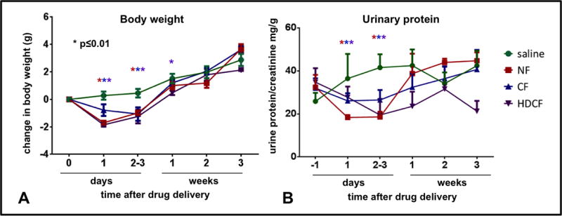
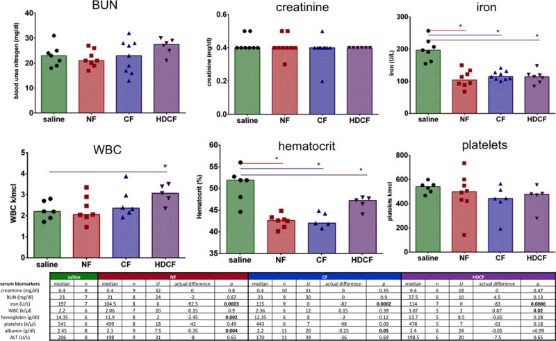
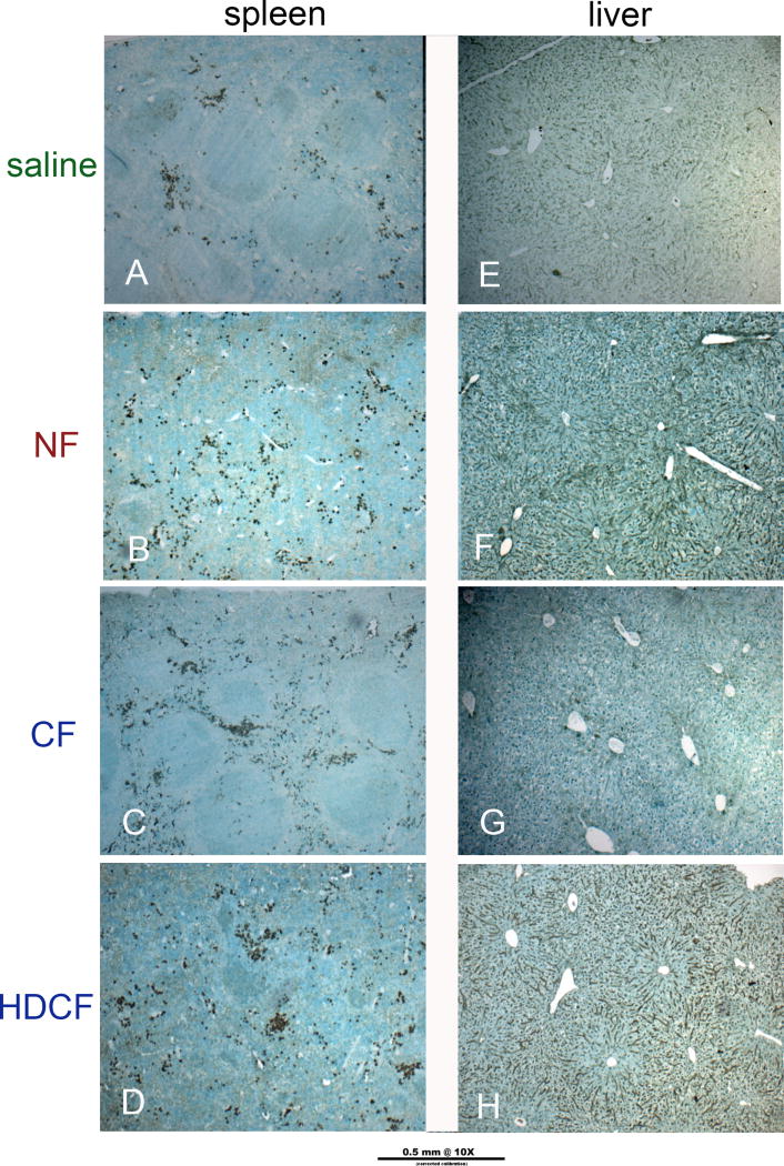
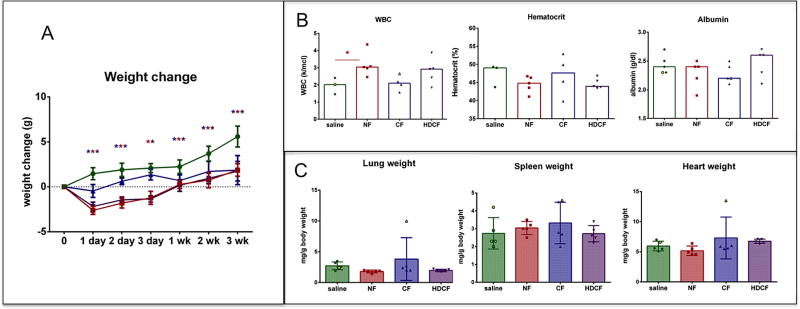
Ferritin is a naturally occurring iron storage protein, proposed as a clinically relevant nanoparticle with applications as a diagnostic and therapeutic agent. Cationic ferritin is a targeted, injectable contrast agent to measure kidney microstructure with MRI. Here, the toxicity of horse spleen ferritin is assessed as a step to clinical translation. Adult male mice received cationic, native and high dose cationic ferritin (CF, NF, or HDCF) or saline and were monitored for 3weeks. Transient weight loss occurred in the ferritin groups with no difference in renal function parameters. Ferritin-injected mice demonstrated a lower serum iron 3weeks after administration. In ferritin-injected animals pre-treated with hydrocortisone, there were no structural or weight differences in the kidneys, liver, lung, heart, or spleen. This study demonstrates a lack of significant detrimental effects of horse-derived ferritin-based nanoparticles at MRI-detectable doses, allowing further exploration of these agents in basic research and clinical diagnostics.

Keywords: Cationic ferritin; Ferritin; Kidney; Magnetic resonance imaging; Nephron number; Toxicity.


Conflict of interest statement

CONFLICTS, DECLARATIONS OF INTEREST

The authors have no financial, consulting or personal relationships with other people or organizations that could influence this work. KMB is the founder of NanoDiagnostics, LLC.

Figures

Figure 1
Figure 1. Intravenous cationic ferritin is detected in perfused, fixed mouse kidney with gradient-recalled echo MRI
Slices from the 3D MRI dataset are shown (19T, GRE-MRI, TE/TR = 8/60 ms, resolution = 34 × 34 × 54 μm). A-Mouse kidney with individual glomeruli labeled due to the accumulation of intravenous CF in the GBM. Glomeruli are visible as punctate dark spots. B- Unlabeled control kidneys are clear of contrast and glomeruli are not visible in MRI.
Figure 2
Figure 2. Body weight and protein excretion of mice receiving CF, NF, HDCF or saline
A-Body weights of mice are depicted as change from baseline (in grams). The CF-injected group (blue) lost significant weight on day 1 compared to the saline group. The NF-injected group lost weight the day after injection but regained weight by 2–3 days and is above baseline and no different than the saline-injected group by 1 week. The HDCF-injected group returned to baseline by 1 week and by week 2 there was no difference between HDCF-injected and the saline group. B-None of the ferritin-injected groups had higher protein excretion than the saline-injected group over the 3 week study period.
Figure 3
Figure 3. Hematologic parameters following ferritin-injections
There was no difference in serum creatinine or BUN. The serum iron level was significantly lower in all ferritin-injected groups. HDCF-injected mice had a higher WBC count. Hematocrit was significantly lower in ferritin-injected mice. There was no difference in the platelet count of ferritin-injected mice.
Figure 4
Figure 4. Iron deposition in the kidney, lung, and spleen
As detected by Perl stain, iron was never seen in kidneys or lungs of the saline-(A, E), NF-(B, F) or CF-(C, G) injected groups. Iron was occasionally detected in the glomeruli of the kidneys (D) or in the interstitium in the lungs (H) of the HDCF-injected group. Iron was detectable in the red pulp of the spleen in higher concentration in NF-(J) and CF-(K) injected mice compared to saline controls. Iron was more densely deposited in the HDCF-injected mice (H-arrows).
Figure 5
Figure 5. Oxidative stress in spleen and liver
4-HNE was used to detect oxidative stress. There was evidence of stress in the spleen co-localized to iron deposition in the red pulp. Although not seen in the CF-injected livers, there was evidence of mild oxidative stress in livers of NF- and HDCF-injected mice which remained present in the steroid treated group (steroid pretreatment group shown).
Figure 6
Figure 6. Hydrocortisone pretreatment group
A-Body weight: Significant weight loss occurred in ferritin-injected mice on day 1 after injection, but by day 2 all groups gained weight. All ferritin groups were back to baseline within one week. B-Hematologic parameters: There was no difference in hematocrit or albumin between any ferritin-injected and saline groups. The NF-injected group had a statistically higher WBC than saline controls. C-Organ Weight: There was no difference in lung, spleen, or heart weight normalized to body weight in any ferritin-injected group compared to saline controls.

References

    1. Aime S, Caravan P. Biodistribution of gadolinium-based contrast agents, including gadolinium deposition. J Magn Reson Imaging. 2009 Dec;30(6):1259–1267. - PMC - PubMed
    1. Pierre VC, Allen MJ, Caravan P. Contrast agents for MRI: 30+ years and where are we going? J Biol Inorg Chem. 2014 Feb;19(2):127–131. - PMC - PubMed
    1. Modo M, Kolosnjaj-Tabi J, Nicholls F, Ling W, Wilhelm C, Debarge O, et al. Considerations for the clinical use of contrast agents for cellular MRI in regenerative medicine. Contrast Media Mol Imaging. 2013 Nov-Dec;8(6):439–455. - PubMed
    1. Cormode DP, Frias JC, Ma Y, Chen W, Skajaa T, Briley-Saebo K, et al. HDL as a contrast agent for medical imaging. Clin Lipidol. 2009 Aug;4(4):493–500. - PMC - PubMed
    1. Cormode DP, Skajaa T, Fayad ZA, Mulder WJ. Nanotechnology in medical imaging: probe design and applications. Arterioscler Thromb Vasc Biol. 2009 Jul;29(7):992–1000. - PMC - PubMed