Review
. 2016 Oct;31(12):1386-93.
doi: 10.1177/0883073816634855. Epub 2016 Apr 12.

Conventional and Advanced Imaging of Diffuse Intrinsic Pontine Glioma

Affiliations
Review

Conventional and Advanced Imaging of Diffuse Intrinsic Pontine Glioma

Jamie Tisnado et al. J Child Neurol. 2016 Oct.

Abstract

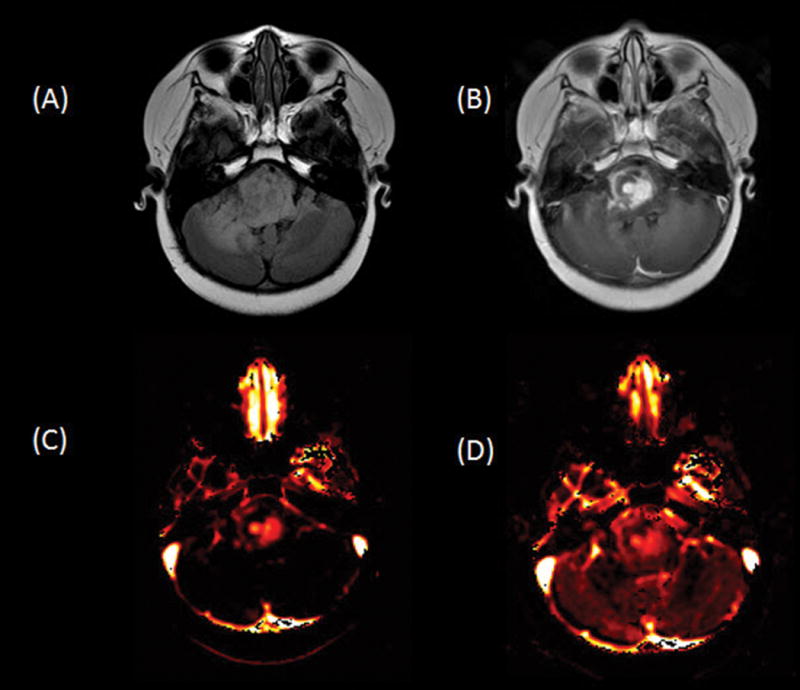
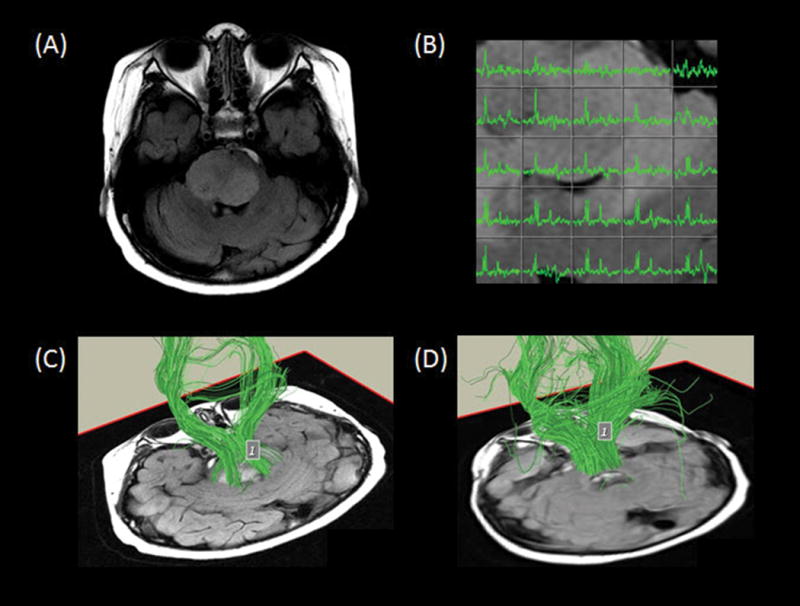
Diffuse intrinsic pontine glioma is the most common brainstem tumor in pediatric patients. This tumor remains one of the most deadly pediatric brain tumors. The diagnosis primarily relies on clinical symptoms and imaging findings. Conventional MRI provides a noninvasive accurate method of diagnosis of these tumors. Advanced MRI techniques are becoming more widely used and studied as additional noninvasive methods to assist clinicians in initial diagnosis and staging, monitoring disease, as well as in surgical and radiation planning. This article will provide an overview of DIPG and describe the typical imaging findings with a focus on advanced imaging techniques.

Keywords: MRI; brainstem; diffuse intrinsic pontine glioma; pediatric; spectroscopy.


Figures

Fig. 1
Fig. 1
Sagittal T1-weighted image shows an expansile hypointense to isointense tumor in the pons, medulla, and cervicomedullary junction with partial effacement of the fourth ventricle and posterior displacement of the cerebellum.
Fig. 2
Fig. 2
Axial fluid-attenuated inversion recovery image shows a large expansile hyperintense tumor in the pons and brachium pontis with partial encasement of the basilar artery anteriorly and partial effacement of the fourth ventricle posteriorly.
Fig. 3
Fig. 3
Axial T2-weighted (A), fluid-attenuated inversion recovery (B), and contrast T1-weighted (C) images demonstrate a hyperintense heterogeneously enhancing tumor centered in the right pons. Note that the exophytic partially cystic anterior portion engulfs the basilar artery flow void.
Fig. 4
Fig. 4
Axial contrast T1-weighted image shows a large ringenhancing component in the left pons and brachium pontis.
Fig. 5
Fig. 5
Axial fluid-attenuated inversion recovery (A) and contrast T1-weighted (B) images show an expansile hyperintense heterogeneously enhancing tumor in the pons and right greater than left cerebellum. Ktrans (C) and plasma volume (D) maps demonstrate heterogeneously increased leakiness and perfusion, respectively, in the areas of enhancing to suggest high-grade disease.
Fig. 6
Fig. 6
Axial fluid-attenuated inversion recovery image (A) demonstrates an expansile hyperintense tumor in the pons, eccentric to the right. Multivoxel spectroscopy (B) reveals increased choline, low N-acetyl-aspartate, and small lipid/lactate peak consistent with high-grade disease. Tractography superimposed on oblique axial T1-weighted images (C, D) demonstrate the descending corticospinal tracts and the transverse pontine fiber tracts traveling through the tumor region.

Similar articles

Cited by

References

    1. Freeman CR, Farmer JP. Pediatric brain stem gliomas: a review. International journal of radiation oncology, biology, physics. 1998;40:265–71. - PubMed
    1. Hargrave D, Bartels U, Bouffet E. Diffuse brainstem glioma in children: critical review of clinical trials. The Lancet Oncology. 2006;7:241–8. - PubMed
    1. Ostrom QT, de Blank PM, Kruchko C, et al. Alex’s Lemonade Stand Foundation Infant and Childhood Primary Brain and Central Nervous System Tumors Diagnosed in the United States in 2007–2011. Neuro-oncology. 2015;16(Suppl 10):x1–x36. - PMC - PubMed
    1. Guillamo JS, Doz F, Delattre JY. Brain stem gliomas. Current opinion in neurology. 2001;14:711–5. - PubMed
    1. Chintagumpala M, Gajjar A. Brain Tumors. Pediatric Clinics of North America. 2015;62:167–78. - PubMed

LinkOut - more resources