. 2016 May;135(5):569-586.
doi: 10.1007/s00439-016-1655-9. Epub 2016 Apr 12.

Pathogenetics of alveolar capillary dysplasia with misalignment of pulmonary veins

Przemyslaw Szafranski  1 Tomasz Gambin  1 Avinash V Dharmadhikari  1   2 Kadir Caner Akdemir  3 Shalini N Jhangiani  1   4 Jennifer Schuette  5 Nihal Godiwala  6 Svetlana A Yatsenko  7   8 Jessica Sebastian  9 Suneeta Madan-Khetarpal  9 Urvashi Surti  7   8   10 Rosanna G Abellar  11 David A Bateman  12 Ashley L Wilson  13 Melinda H Markham  14 Jill Slamon  15 Fernando Santos-Simarro  16   17 María Palomares  16   17 Julián Nevado  16   17 Pablo Lapunzina  16   17 Brian Hon-Yin Chung  18   19 Wai-Lap Wong  18 Yoyo Wing Yiu Chu  18 Gary Tsz Kin Mok  18 Eitan Kerem  20 Joel Reiter  20 Namasivayam Ambalavanan  21   22 Scott A Anderson  23 David R Kelly  24 Joseph Shieh  25 Taryn C Rosenthal  26 Kristin Scheible  27 Laurie Steiner  27 M Anwar Iqbal  28 Margaret L McKinnon  29 Sara Jane Hamilton  29 Kamilla Schlade-Bartusiak  29 Dawn English  29 Glenda Hendson  30 Elizabeth R Roeder  31   32 Thomas S DeNapoli  33 Rebecca Okashah Littlejohn  31 Daynna J Wolff  34 Carol L Wagner  35 Alison Yeung  36 David Francis  36 Elizabeth K Fiorino  37 Morris Edelman  38 Joyce Fox  39 Denise A Hayes  40 Sandra Janssens  41 Elfride De Baere  41 Björn Menten  41 Anne Loccufier  42 Lieve Vanwalleghem  43 Philippe Moerman  44 Yves Sznajer  45 Amy S Lay  46 Jennifer L Kussmann  47 Jasneek Chawla  48   49 Diane J Payton  50   51 Gael E Phillips  50   51 Erwin Brosens  52   53 Dick Tibboel  53 Annelies de Klein  52 Isabelle Maystadt  54 Richard Fisher  55 Neil Sebire  56 Alison Male  57 Maya Chopra  58 Jason Pinner  58 Girvan Malcolm  59 Gregory Peters  60 Susan Arbuckle  61 Melissa Lees  57 Zoe Mead  62 Oliver Quarrell  63 Richard Sayers  63 Martina Owens  64 Charles Shaw-Smith  64 Janet Lioy  65 Eileen McKay  66 Nicole de Leeuw  67 Ilse Feenstra  67 Liesbeth Spruijt  67 Frances Elmslie  68 Timothy Thiruchelvam  69 Carlos A Bacino  1   70 Claire Langston  71 James R Lupski  1   4   70   72 Partha Sen  73 Edwina Popek  71 Paweł Stankiewicz  74   75   76
Affiliations

Pathogenetics of alveolar capillary dysplasia with misalignment of pulmonary veins

Przemyslaw Szafranski et al. Hum Genet. 2016 May.

Abstract

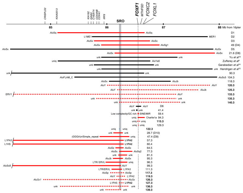
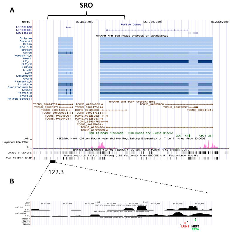
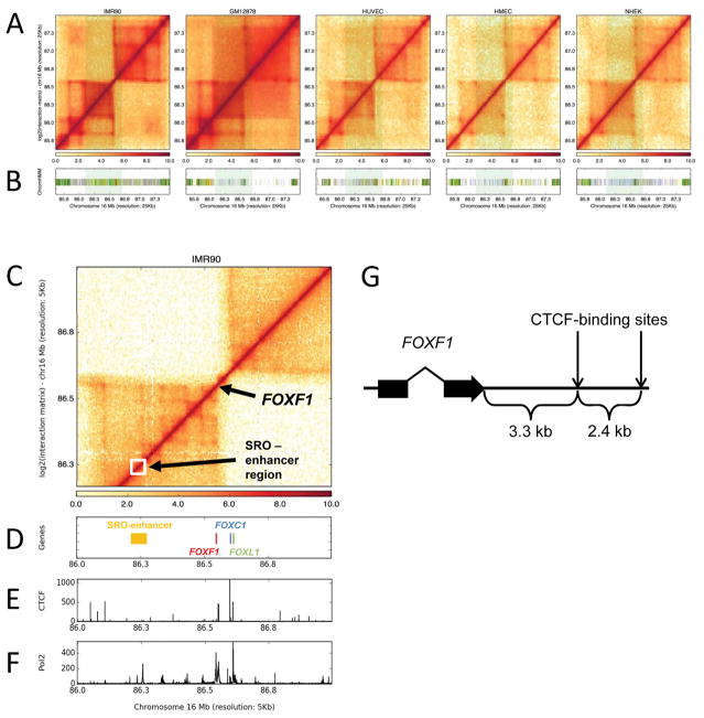
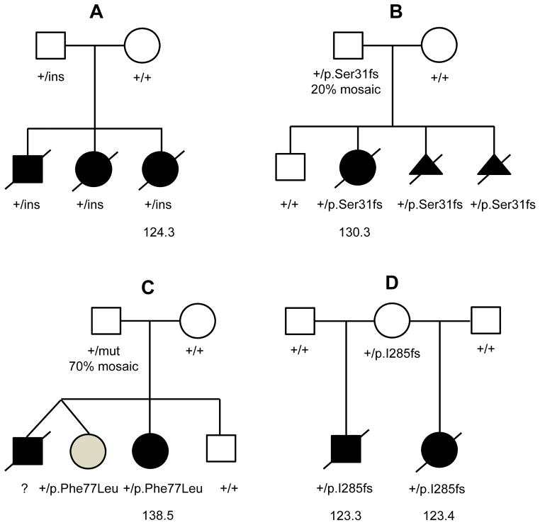
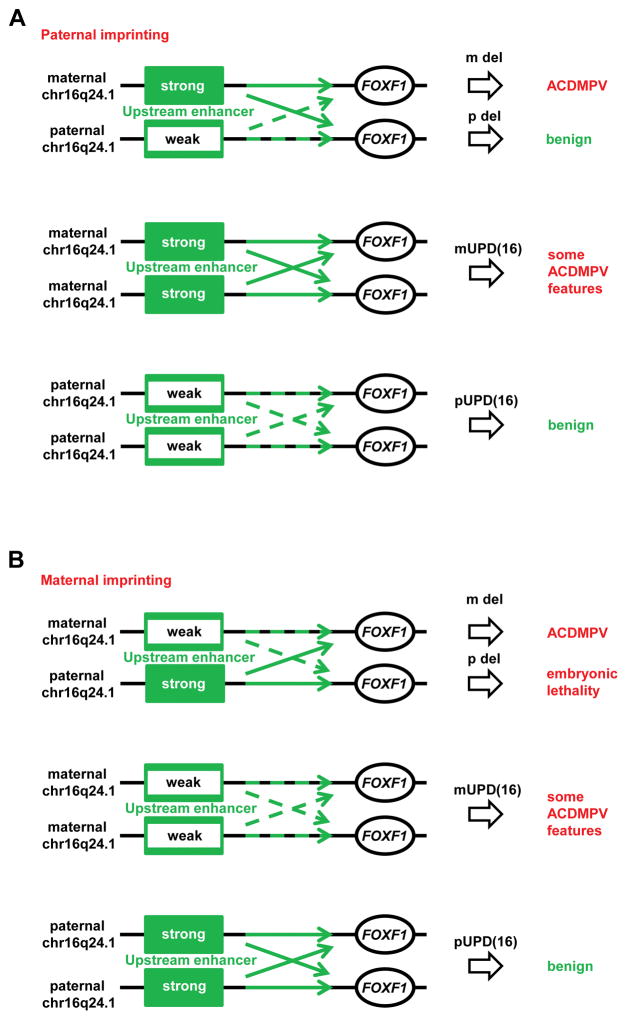
Alveolar capillary dysplasia with misalignment of pulmonary veins (ACDMPV) is a lethal lung developmental disorder caused by heterozygous point mutations or genomic deletion copy-number variants (CNVs) of FOXF1 or its upstream enhancer involving fetal lung-expressed long noncoding RNA genes LINC01081 and LINC01082. Using custom-designed array comparative genomic hybridization, Sanger sequencing, whole exome sequencing (WES), and bioinformatic analyses, we studied 22 new unrelated families (20 postnatal and two prenatal) with clinically diagnosed ACDMPV. We describe novel deletion CNVs at the FOXF1 locus in 13 unrelated ACDMPV patients. Together with the previously reported cases, all 31 genomic deletions in 16q24.1, pathogenic for ACDMPV, for which parental origin was determined, arose de novo with 30 of them occurring on the maternally inherited chromosome 16, strongly implicating genomic imprinting of the FOXF1 locus in human lungs. Surprisingly, we have also identified four ACDMPV families with the pathogenic variants in the FOXF1 locus that arose on paternal chromosome 16. Interestingly, a combination of the severe cardiac defects, including hypoplastic left heart, and single umbilical artery were observed only in children with deletion CNVs involving FOXF1 and its upstream enhancer. Our data demonstrate that genomic imprinting at 16q24.1 plays an important role in variable ACDMPV manifestation likely through long-range regulation of FOXF1 expression, and may be also responsible for key phenotypic features of maternal uniparental disomy 16. Moreover, in one family, WES revealed a de novo missense variant in ESRP1, potentially implicating FGF signaling in the etiology of ACDMPV.


Conflict of interest statement

Conflict of interest No competing interest is declared.

Figures

Fig. 1
Fig. 1
A compilation of chromosome 16q24.1 deletions pathogenic for ACDMPV. Deletions which occurred on maternal chromosome 16, are shown in red, the deletion on paternal chromosome is shown in blue, and deletions, for which parental origin could not be determined, are shown in black. Numbers refer to ACDMPV cases. Locations of deletion breakpoints (BPs) are indicated by names of flanking repetitive elements. Abbreviations: SRO, (smallest deletion overlap - delineating upstream enhancer region); unk, unknown sequence; uniq, unique sequence. Note that most of the deletions are flanked by retrotransposons, mainly by Alu repetitive segments. The LIPA2 element (chr16:86,266,902-86,272,916, GRCh37/hg19) within LINC01081 at three distal deletion breakpoints in patients 57.3, 60.4, and 127.3 is bolded (Supplemental Table S1). Thirteen novel deletions are dashed and their number bolded to distinguished from the published cases.
Fig. 2
Fig. 2
UCSC browser display of the enhancer region upstream to FOXF1. a An ~ 60 kb SRO showing overlapping LINC01081 and LINC01082, CpG islands, H3K27Ac methylation, DNaseI hypersensitivity clusters, and transcription factor ChIP-Seq data. b The ~ 4.1 kb deletion CNV identified in pt 122.3 maps to the FOXF1 upstream enhancer region. Location of the transcription factor and CTCF binding sites, and a diagram of histone modification in the deleted region is shown.
Fig. 3
Fig. 3
Genomic in cis interactions at chromosome 16q24.1. a Hi-C contact maps are visualized as heatmaps for IMR-90, GM12878, HUVEC, NHEK, and HMEC cell lines. b Tiles mark active TSS (red), transcribed (green), enhancer (yellow), low (gray) and heterochromatin (purple) state calls with ChromHMM within the represented locus. Shaded region highlights the active chromatin region in IMR-90 cells whereas the same locus in not enriched for active chromatin states in other cell types, especially on the left-side of TAD boundary. c Zoomed-in view of Hi-C contact map around the FOXF1 locus in IMR-90 cells at 5 kb resolution. d Tiles mark genomic locations of SRO-enhancer region (yellow) and FOXF1 (red), FOXC1 (blue) and FOXL1 (green) genes. e Histogram represents CTCF ChIP-seq enrichment levels within the visualized locus. f Histogram represents RNA PolII ChIP-Seq enrichment levels. g Positions of two strong CTCF-binding sites (chr16:86,551,417–528 and 86,553,812–944; hg19) at the TAD boundary, 3.3 and 5.7 kb downstream of the 3′ end of FOXF1 (chr16: 86,548,070; hg19).
Fig. 4
Fig. 4
Unusual inheritance and manifestation of pathogenic variants in the FOXF1 locus. Pedigrees of ACDMPV families with reportedly healthy parent transmitting FOXF1 pathogenic variant to their affected children. a Family 124 with the complex genomic insertion rearrangement (ins) within the 5′ non-coding part of the FOXF1 1st exon. b Family 130 with the frameshift mutation within the FOXF1 1st exon. c Family 138 with the pathogenic missense mutation in the FOXF1 1st exon transmitted by healthy father, 70% mosaic in peripheral blood. A twin sister (shaded symbol) of the deceased ACDMPV patient has partial anomalous venous return (Reiter et al. (2016). d Family 123 with the frameshift mutation transmitted by healthy mother, likely germline mosaic for the mutation.
Fig. 5
Fig. 5
Proposed model of ACDMPV and UPD(16) etiologies due to (a) paternal or (b) maternal imprinting of the FOXF1 upstream enhancer on chromosome 16q24.1. Epigenetic modification of the enhancer reduces its ability to stimulate FOXF1 transcription. In paternal imprinting model (a), the majority of FOXF1 transcription depends on the strong enhancer located on the maternal chromosome 16. Thus deletion of the enhancer on this chromosome would significantly reduce FOXF1 expression and cause ACDMPV whereas the deletion on the paternal chromosome 16q24.1 would reduce FOXF1 expression less with no clinical consequences. Consequently, maternal UPD(16) would increase FOXF1 expression level (pathogenic) whereas paternal UPD(16) would slightly decrease FOXF1 expression level (benign). In the maternal imprinting model (b), the upstream deletion of the weak enhancer on the maternal chromosome 16 would reduce FOXF1 expression, resulting in ACDMPV. The deletion of the strong enhancer on the paternal chromosome would reduce FOXF1 expression would be embryonic lethal. According to this model, in maternal UPD(16), the level of FOXF1 would be insufficient to prevent development of some ACDMPV features and other anomalies typical for maternal UPD(16). In contrast, point mutations in FOXF1 are found on both parental alleles, suggesting in-trans function of the FOXF1 enhancer, likely using lncRNAs (Szafranski et al. 2013a).

References

    1. Araujo E, Júnior, Palma-Dias R, Martins WP, Reidy K, da Silva Costa F. Congenital heart disease and adverse perinatal outcome in fetuses with confirmed isolated single functioning umbilical artery. J Obstet Gynaecol. 2015;35:85–87. - PubMed
    1. Barlow DP, Bartolomei MS. Genomic imprinting in mammals. Cold Spring Harb Perspect Biol. 2014;6:a018382. - PMC - PubMed
    1. Bartolomei MS, Ferguson-Smith AC. Mammalian genomic imprinting. Cold Spring Harb Perspect Biol. 2011;3:a002592. - PMC - PubMed
    1. Bishop NB, Stankiewicz P, Steinhorn RH. Alveolar capillary dysplasia. Am J Respir Crit Care Med. 2011;184:172–179. - PMC - PubMed
    1. Black BL, Olson EN. Transcriptional control of muscle development by myocyte enhancer factor-2 (MEF2) proteins. Annu Rev Cell Dev Biol. 1998;14:167–196. - PubMed

Publication types

Substances

Supplementary concepts