. 2016 Apr 12:18:87.
doi: 10.1186/s13075-016-0984-3.

Inflammatory properties of inhibitor of DNA binding 1 secreted by synovial fibroblasts in rheumatoid arthritis

Affiliations

Inflammatory properties of inhibitor of DNA binding 1 secreted by synovial fibroblasts in rheumatoid arthritis

Gautam Edhayan et al. Arthritis Res Ther. .

Abstract

Background: Inhibitor of DNA binding 1 (Id1) is a nuclear protein containing a basic helix-loop-helix (bHLH) domain that regulates cell growth by selective binding and prevention of gene transcription. Sources of Id1 production in rheumatoid arthritis synovial tissue (RA ST) and its range of functional effects in RA remain to be clarified.

Methods: We analyzed Id1 produced from synovial fibroblasts and endothelial cells (ECs) with histology and real-time polymerase chain reaction (RT-PCR). Fibroblast supernatants subjected to differential centrifugation to isolate and purify exosomes were measured for Id1 by enzyme-linked immunosorbent assay (ELISA). Western blotting of Id1-stimulated ECs was performed to determine the kinetics of intracellular protein phosphorylation. EC intracellular signaling pathways induced by Id1 were subsequently targeted with silencing RNA (siRNA) for angiogenesis inhibition.

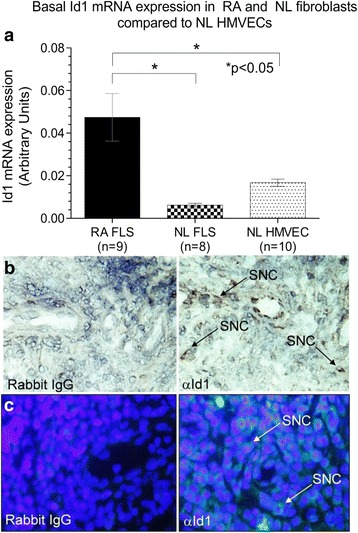
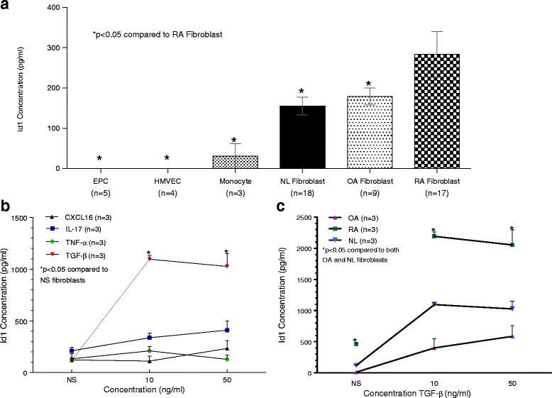
Results: By PCR and histologic analysis, we found that the primary source of Id1 in STs is from activated fibroblasts that correlate with inflammatory scores in human RA ST and in joints from K/BxN serum-induced mice. Normal (NL) and RA synovial fibroblasts increase Id1 production with stimulation by transforming growth factor beta (TGF-β). Most of the Id1 released by RA synovial fibroblasts is contained within exosomes. Endothelial progenitor cells (EPCs) and human dermal microvascular ECs (HMVECs) activate the Jnk signaling pathway in response to Id1, and Jnk siRNA reverses Id1-induced HMVEC vessel formation in Matrigel plugs in vivo.

Conclusions: Id1 is a pleotropic molecule affecting angiogenesis, vasculogenesis, and fibrosis. Our data shows that Id1 is not only an important nuclear protein, but also can be released from fibroblasts via exosomes. The ability of extracellular Id1 to activate signaling pathways expands the role of Id1 in the orchestration of tissue inflammation.

Keywords: Angiogenesis; Fibroblasts; Inflammation; Inhibitor of DNA binding-1 protein (Id1); Rheumatoid arthritis.


Figures

Fig. 1
Fig. 1
Id1 is expressed in ECs and ST fibroblasts. a mRNA was isolated from HMVECs and fibroblasts were isolated from NL and RA ST. mRNA was reverse transcribed into cDNA and underwent PCR for 40 cycles. RA fibroblasts showed significantly elevated Id1 expression compared to NL ST fibroblasts and HMVECs. b Id1 is expressed in RA STs. IHC was performed on RA, OA, and NL ST cryosections. Tissues were blocked and then incubated using a mouse anti-human Id1 (Abcam) primary antibody. After washing, tissues were incubated with a biotinylated anti-mouse secondary antibody (Vector Laboratories). Tissues were washed and subsequently developed with the Vectastain ABC kit (Vector Laboratories). Id1 is found on synovial cells (SNC) in the RA ST. c For immunofluorescence staining, RA, osteoarthritis (OA), and normal (NL) ST sections were fixed in cold acetone for 30 min. The STs were blocked with 5 % donkey serum and 20 % fetal bovine serum (FBS) in PBS at 37 °C for 1 h, and then incubated with rabbit anti-human Id1 antibody (Abcam, 10 μg/ml) or purified nonspecific rabbit IgG for 1 h at 37 °C in blocking buffer. The synovial tissues samples were washed with PBS, and a 1:200 dilution in blocking buffer of fluorescent-conjugated donkey anti-rabbit antibody was added and incubated for an additional 1 h at 37 °C. As shown, we could validate Id1 staining in RA ST similar to what was found using IHC (×400). FLS fibroblast-like synoviocytes, HMVEC human dermal microvascular endothelial cell, Id1 inhibitor of DNA binding 1, IgG immunoglobulin, NL normal, RA rheumatoid arthritis, SNC synovial cell
Fig. 2
Fig. 2
SNC Id1 expression is significantly higher in inflamed ST and in the ankles of K/BxN serum-induced mice. a Id1 expression was visualized on SNCs in ST by IHC. A significantly greater percentage of SNCs were positive for Id1 in RA compared to OA and NL ST. b Percentages of cells expressing Id1 were also evaluated on SNCs from joint tissues taken from K/BxN serum-induced mice. A significantly greater percentage of SNCs positive for Id1 compared to Wt (noninduced mice) was found. c Murine tissues were blocked and then incubated using a rabbit anti-mouse Id1 antibody (CalBioreagents). After washing, tissues were incubated with a biotinylated anti-rabbit secondary antibody (Vector Laboratories). Tissues were washed and subsequently developed with the Vectastain ABC kit (Vector Laboratories). Id1 is found on SNCs near the bone in the K/BxN mouse ankles. Id1 inhibitor of DNA binding 1, IgG immunoglobulin, NL normal, OA osteoarthritis, RA rheumatoid arthritis, SNC synovial cell, ST synovial tissue, Wt wildtype
Fig. 3
Fig. 3
RA ST fibroblasts secrete Id1 and upregulate Id1 expression upon stimulation with TGFβ. a EPCs, HMVECs, monocytes, NL, RA, and OA fibroblasts were plated and serum starved. Cell culture media was replaced and the supernatants were collected 24 h later. The cell culture supernatants were examined for Id1 expression by ELISA (MyBioSource). EPCs, HMVECs as well as all fibroblasts were collected from human tissues, which were digested in a mix of cell culture media supplemented with FBS, collagenase, and hyaluronidase. HMVECs were isolated from skin biopsies while the fibroblasts were isolated from STs of patients with either RA, OA, or from NL patients. HMVECs were isolated and purified using CD31 microbeads (Miltenyi Biotec). All cell lines were between passages 3 and 6. No cytokine stimulation was used. b NL fibroblasts were plated and serum starved under the same conditions as panel a. Cell culture media with the respective cytokine was exchanged and collected 24 h later. CXCL16 (R&D Systems), IL-17 (R&D Systems), TNF-α (Life Technologies) and TGF-β (R&D Systems) were used at 10 ng/mL and 50 ng/mL concentrations. A not stimulated (NS) well was also used with no added cytokine (n = number of experimental replicates). c Synovial fibroblasts were plated under the same conditions as panel a. Cell culture media with TGF-β was exchanged and collected 24 h later. CXCL16 chemokine (C-X-C motif) ligand 16, EPC endothelial progenitor cell, HMVEC human dermal microvascular endothelial cell, Id1 inhibitor of DNA binding 1, IL-17 interleukin 17, NL normal, NS nonstimulated, OA osteoarthritis, RA rheumatoid arthritis, TGF-β transforming growth factor beta, TNF-α tumor necrosis factor alpha
Fig. 4
Fig. 4
Id1 is contained in fibroblast exosomes. Fibroblasts from RA, osteoarthritis (OA), and normal (NL) STs were plated and maintained in CMRL medium supplemented with 20 % FBS, 2 mM L-glutamine, and 1 % penicillin/streptomycin in T175cm2 flasks and were used between passages 4 and 10. Cultures were moved to serum-free media DMEM/F-12 with Peprogrow serum replacement (Peprotech) for 2 days before harvesting. Fibroblast culture supernatants were concentrated to 1 mL (per flask) by centrifugation through an Amicon Ultracel 30 K filter (EMD Millipore, Billerica, MA, USA). Exosomes from cell supernatants were isolated and purified by serial ultracentrifugation. Nanoparticle Tracking Analysis was used to quantify the size and concentration of particles within the exosome fractions. a and b Cells were spun out at 1500 rpm for 5 min. Then the supernatants were cleared of heavier debris by spins at 10,000 × g for 30 min and 30,000 × g for 1 h. Exosomes were then spun down at 110,000 × g for 4–20 h. Exosome pellets were washed in PBS at 110,000 × g for 1.5 h – overnight. Some exosomes were further purified using a density gradient Optiprep (Sigma-Aldrich). Optiprep was diluted with PBS to produce the following layers: 5, 10, 15, 20, 30, 40, and 50 % w/v (densities of 1.031, 1.050, 1.084, 1.110, 1.163, 1.215, and 1.268 g/mL). The expected size of exosomes is between 25 and 135 nm and >70 % of the EVs isolated fall into this range (red area). c Representative diagram showing that we find more exosomes in RA (yellow and blue areas) compared to OA (blue area only) in fibroblast supernatants. d and e Whole and lysed (0.5 % Triton X-100) exosome fractions were then measured for Id1. We found that >80 % of the detected Id1 is contained within RA fibroblast cell supernatant exosomes. We similarly found that OA and RA SF have exosomes containing Id1 (n = number of sample wells measured for exosomes isolated from SF specimens from two separate patients for OA; and three separate patients for RA). All data was pooled in the respective OA and RA SF groups. Id1 inhibitor of DNA binding 1, OA osteoarthritis, RA rheumatoid arthritis, SF synovial fluid
Fig. 5
Fig. 5
Id1 signals through the *pJnk pathway in HMVECs and EPCs. HMVECs and EPCs were cultured in 6-well plates and stimulated at different time intervals with human recombinant Id1. The cell lysate was collected and western blot analysis was performed. The results are shown as fold increase from the nonstimulated (NS), which was arbitrarily set at 1. The upper band represents phosphorylated signaling molecule (*p) and the lower band represents total signaling molecule. Using these bands, the amount of phosphorylated signaling molecule was quantified for each respective blot and results pooled. Upregulation (↑) of *pJnk and *pP38 was statistically significant in EPCs and upregulation of *pJnk was statistically significant in HMVECs. Upregulation of *pJnk plateaued at 5 min in EPCs and later in HMVECs at 30 min. Other signaling molecules were tested but results were not significant (data not shown, n = number of experimental replicates; the delta symbol with slash represents no change. EPC endothelial progenitor cell, HMVEC human dermal microvascular endothelial cell, Id1 inhibitor of DNA binding 1, NS nonstimulated
Fig. 6
Fig. 6
Jnk siRNA lowers HMVEC-mediated angiogenesis in the Matrigel plug assay. HMVECs were transfected with either control or Jnk siRNA designed to inhibit the Jnk signaling pathway. These cells were combined with 10 nM Id1 in Matrigel, which was injected subcutaneously into mice. Five days later, the Matrigel plug was removed, weighed, and homogenized. The hemoglobin assay was run to determine amount of hemoglobin in the plugs as a marker of angiogenesis. Jnk siRNA significantly inhibited angiogenesis in this assay. HMVEC human dermal microvascular endothelial cell, Id1 inhibitor of DNA binding 1, siRNA silencing RNA

References

    1. Gao D, Nolan DJ, Mellick AS, Bambino K, McDonnell K, Mittal V. Endothelial progenitor cells control the angiogenic switch in mouse lung metastasis. Science. 2008;319(5860):195–198. doi: 10.1126/science.1150224. - DOI - PubMed
    1. Mellick AS, Plummer PN, Nolan DJ, Gao D, Bambino K, Hahn M, et al. Using the transcription factor inhibitor of DNA binding 1 to selectively target endothelial progenitor cells offers novel strategies to inhibit tumor angiogenesis and growth. Cancer Res. 2010;70(18):7273–7282. doi: 10.1158/0008-5472.CAN-10-1142. - DOI - PMC - PubMed
    1. Ward SM, Fernando SJ, Hou TY, Duffield GE. The transcriptional repressor ID2 can interact with the canonical clock components CLOCK and BMAL1 and mediate inhibitory effects on mPer1 expression. J Biol Chem. 2010;285(50):38987–39000. doi: 10.1074/jbc.M110.175182. - DOI - PMC - PubMed
    1. Swarbrick A, Akerfeldt MC, Lee CS, Sergio CM, Caldon CE, Hunter LJ, et al. Regulation of cyclin expression and cell cycle progression in breast epithelial cells by the helix-loop-helix protein Id1. Oncogene. 2005;24(3):381–389. doi: 10.1038/sj.onc.1208188. - DOI - PubMed
    1. Kim HJ, Chung H, Yoo YG, Kim H, Lee JY, Lee MO, et al. Inhibitor of DNA binding 1 activates vascular endothelial growth factor through enhancing the stability and activity of hypoxia-inducible factor-1alpha. Mol Cancer Res. 2007;5(4):321–329. doi: 10.1158/1541-7786.MCR-06-0218. - DOI - PubMed

Publication types

MeSH terms

Substances