. 2016 Aug;10(7):966-80.
doi: 10.1016/j.molonc.2016.03.006. Epub 2016 Apr 1.

Improving fascin inhibitors to block tumor cell migration and metastasis

Affiliations

Improving fascin inhibitors to block tumor cell migration and metastasis

Shaoqin Han et al. Mol Oncol. 2016 Aug.

Abstract

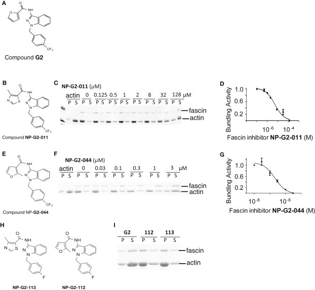
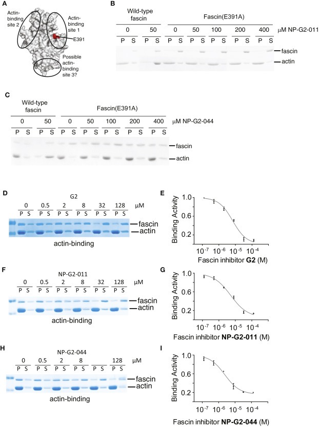
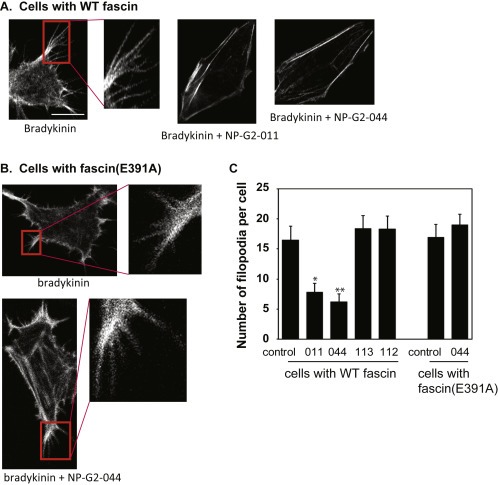
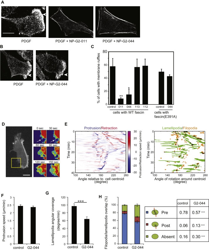
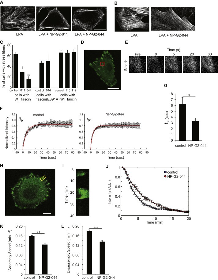
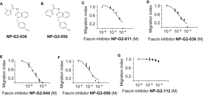
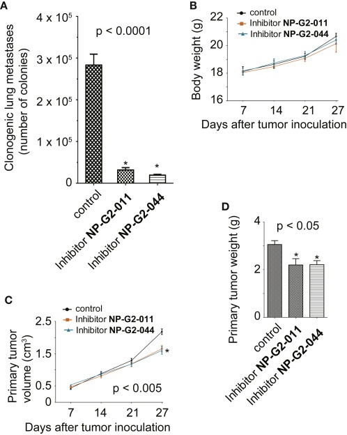
Tumor metastasis is the major cause of mortality of cancer patients, being responsible for ∼90% of all cancer deaths. One of the key steps during tumor metastasis is tumor cell migration which requires actin cytoskeletal reorganization. Among the critical actin cytoskeletal protrusion structures are antenna-like filopodia. Fascin protein is the main actin-bundling protein in filopodia. Here we report the development of fascin-specific small-molecules that inhibit the interaction between fascin and actin. These inhibitors block the in vitro actin-binding and actin-bundling activities of fascin, tumor cell migration and tumor metastasis in mouse models. Mechanistically, these inhibitors likely occupy one of the actin-binding sites, reduce the binding of actin filaments, and thus lead to the inhibition of the bundling activity of fascin. At the cellular level, these inhibitors impair actin cytoskeletal reorganization. Our data indicate that target-specific anti-fascin agents will have great potential for treating metastatic tumors.

Keywords: Fascin; Inhibitors; Metastasis.


Figures

Figure 1
Figure 1
Biological evaluation of analogues of Compound G2. (A) The chemical structure of Compound G2. (B) Chemical structure of Analogue NP‐G2‐011. (C) An example from the actin‐bundling assays shows the decrease of the actin‐bundling function of fascin by Compound NP‐G2‐011. (D) The dose response curve for Compound NP‐G2‐011. (E) Chemical structure of Analogue NP‐G2‐044. (F) An example from the actin‐bundling assays shows the decrease of the actin‐bundling function of fascin by Compound NP‐G2‐044. (G) The dose response curve for Compound NP‐G2‐044. (H and I) Examples of two analogues that did not show inhibitory effect on fascin. Data are representative of three similar experiments.
Figure 2
Figure 2
Inhibitory effect of Compounds G2, NP‐G2‐011 and NP‐G2‐044 on the actin‐binding activity of fascin. (A) Crystal structure of fascin shows the location of the actin‐binding site 1. (B) Actin‐bundling assays for wild‐type fascin and fascin(E391A) in the presence of Compound NP‐G2‐011. (C) Actin‐bundling assays with wild‐type fascin and fascin(E391A) in the presence of Compound NP‐G2‐044. (D) An example from the actin‐binding assays shows the decrease of the actin‐binding function of fascin by Compound G2. (E) The dose response curve for Compound G2. (F) An example from the actin‐binding assays shows the decrease of the actin‐binding function of fascin by Compound NP‐G2‐011. (G) The dose response curve for Compound NP‐G2‐011. (H) An example from the actin‐binding assays shows the decrease of the actin‐binding function of fascin by Compound NP‐G2‐044. (I) The dose response curve for Compound NP‐G2‐044. Data are representative of three similar experiments.
Figure 3
Figure 3
Inhibitory effect of Compounds NP‐G2‐011 and NP‐G2‐044 on filopodial formation. (A) Cells with wild‐type fascin were treated with bradykinin to induce filopodial formation. Addition of Compounds NP‐G2‐011 and NP‐G2‐044 blocked bradykinin‐induced filopodial formation. (B) 4T1 cells expressing fascin(E391A) mutant formed filopodia upon bradykinin treatment. Filopodial formation in these cells was resistant to Compound NP‐G2‐044 inhibition. (C) Quantification of data in A and B. Error bars, mean±SEM. *p < 0.01; **p < 0.005. Scale bar, 10 μm.
Figure 4
Figure 4
Inhibitory effect of Compounds NP‐G2‐011 and NP‐G2‐044 on lamellipodial formation and the transition from filopodia to lamellipodia. (A) Cells with wild‐type fascin were treated with PDGF to induce lamellipodial formation (marked by arrowheads). Addition of Compound NP‐G2‐011 or NP‐G2‐044 blocked PDGF‐induced lamellipodial formation. (B) 4T1 cells expressing fascin(E391A) mutant formed lamellipodia upon PDGF treatment. Lamellipodial formation in these cells was resistant to Compound NP‐G2‐044 inhibition. (C) Quantification of data in A and B. Error bars, mean ± SEM. *p < 0.05; **p < 0.01. Scale bar, 10 μm. (D) Time lapse of a LifeAct‐GFP expressing MDA‐MB‐231 cell showing a filopodium (arrow at 0 and 30 s) extending as a lamellipodium (60–150 s in zoom of yellow box sown at right). (E) Angular projection map of the protrusion and retraction speed of cell membrane extensions along with the identified lamellipodial and filopodial structure map. (F) Average protruding speed of membrane extension with and without NP‐G2‐044 treatment. (G) Quantification of the angular membrane fraction covered by lamellipodia with and without NP‐G2‐044 treatment. (H) Quantification of the lamellipodial and filopodial overlap with and without NP‐G2‐044 treatment. (Pre, presence of a filopodium prior to lamellipodial extension; Post, overlap of a filopodium after the extension of a lamellipodium; Absent, no overlap between filopodia and lamellipodia). Error bars, mean ± s.e.m. Scale bars, 10 μm ***p < 0.01.
Figure 5
Figure 5
Inhibitory effect of Compounds NP‐G2‐011 and NP‐G2‐044 on stress fiber formation and focal adhesion turnover. (A) Cells with wild‐type fascin were treated with LPA to induce actin stress fiber formation. Addition of Compound NP‐G2‐011 or NP‐G2‐044 blocked LPA‐induced stress fiber formation. (B) 4T1 cells expressing fascin(E391A) mutant formed stress fibers upon LPA treatment. Stress fiber formation in these cells was resistant to Compound NP‐G2‐044 inhibition. (C) Quantification of data in A and B. Error bars, mean ± SEM. *p < 0.05; **p < 0.001. Data are representative of three similar experiments. Scale bar, 10 μm. (D) Confocal live cell images of MDA‐MB‐231 cells expressing GFP‐FAK were taken before photobleaching the ROIs (red box) containing focal adhesions. (E) Time lapse sequence of the selected ROIs showing a representative recovery after photobleaching (Pre, pre bleach). (F) Kinetics of recovery of GFP‐FAK after photobleaching of focal adhesions from cells pre‐treated with vehicle (Ctrl) or NP‐G2‐044 are illustrated. The measured fluorescence intensity was normalized and the mean values (18 independent ROIs from different cells for each condition) are shown along with the exponential fit (red line). (G) Corresponding recovery half‐time obtained from FRAP analyses of GFP‐FAK pre‐treated with vehicle (Ctrl) or NP‐G2‐044. (H) Confocal live cell image of a representative MDA‐MB‐231 cell expressing GFP‐FAK and a focal adhesion containing ROI (yellow box). (I) Kymograph of the corresponding focal adhesion over a period of 40 min. Acquisitions were taken every 30 s. (J) Quantitative assessment of focal adhesion disassembly in GFP‐FAK expressing cells pre‐treated with vehicle (Ctrl, blue square) or NP‐G2‐044 (red circle). (K) and (L) Assembly (K) and disassembly (L) rates were obtained by numerical differentiation of the corresponding time‐dependent signal. Error bars, mean ± s.e.m. Scale bars, 10 μm *p < 0.05; **p < 0.01.
Figure 6
Figure 6
Inhibitory effect of analogues of Compound G2 on breast tumor cell migration. (A) The chemical structure of Compound NP‐G2‐036. (B) The chemical structure of Compound NP‐G2‐050. (C) Inhibition of the migration of MDA‐MB‐231 cells by Compound NP‐G2‐011 in response to serum stimulation. (D) Inhibition of the migration of MDA‐MB‐231 cells by Compound NP‐G2‐036 in response to serum stimulation. (E) Inhibition of the migration of MDA‐MB‐231 cells by Compound NP‐G2‐044 in response to serum stimulation. (F) Inhibition of the migration of MDA‐MB‐231 cells by Compound NP‐G2‐050 in response to serum stimulation. (G) Migration of MDA‐MB‐231 cells in the presence of Compound NP‐G2‐112 in response to serum stimulation. Error bars, mean ± s.e.m.
Figure 7
Figure 7
Inhibitory effect of analogue NP‐G2‐011 and NP‐G2‐044 on breast tumor metastasis in mouse models. (A) Inhibition of 4T1 mouse mammary tumor cell metastasis to the lung by NP‐G2‐011 and NP‐G2‐044 in a spontaneous metastasis model (n = 5 for each group). (B) NP‐G2‐011 and NP‐G2‐044 had no effect on the body weights of treated mice. (C) NP‐G2‐011 and NP‐G2‐044 decreased the volume of primary tumors at later stages. (D) NP‐G2‐011 and NP‐G2‐044 decreased the primary tumor weight. Error bars, mean ± s.e.m.

Similar articles

Cited by

References

    1. Adams, J.C. , 2004. Roles of fascin in cell adhesion and motility. Curr. Opin. Cell Biol. 16, 590–596. - PubMed
    1. Aznavoorian, S. , Murphy, A.N. , Stetler-Stevenson, W.G. , Liotta, L.A. , 1993. Molecular aspects of tumor cell invasion and metastasis. Cancer. 71, 1368–1383. - PubMed
    1. Bentley, D. , Toroian-Raymond, A. , 1986. Disoriented pathfinding by pioneer neurone growth cones deprived of filopodia by cytochalasin treatment. Nature. 323, 712–715. - PubMed
    1. Bordeleau, F. , Galarneau, L. , Gilbert, S. , Loranger, A. , Marceau, N. , 2010. Keratin 8/18 modulation of protein kinase C-mediated integrin-dependent adhesion and migration of liver epithelial cells. Mol. Biol. Cell. 21, 1698–1713. - PMC - PubMed
    1. Bryan, J. , Kane, R.E. , 1978. Separation and interaction of the major components of sea urchin actin gel. J. Mol. Biol. 125, 207–224. - PubMed

Publication types

MeSH terms