. 2016 Apr 12:9:216.
doi: 10.1186/s13104-016-1902-0.

An optimized staining technique for the detection of Gram positive and Gram negative bacteria within tissue

Affiliations

An optimized staining technique for the detection of Gram positive and Gram negative bacteria within tissue

Sandra C Becerra et al. BMC Res Notes. .

Abstract

Background: Bacterial infections are a common clinical problem in both acute and chronic wounds. With growing concerns over antibiotic resistance, treatment of bacterial infections should only occur after positive diagnosis. Currently, diagnosis is delayed due to lengthy culturing methods which may also fail to identify the presence of bacteria. While newer costly bacterial identification methods are being explored, a simple and inexpensive diagnostic tool would aid in immediate and accurate treatments for bacterial infections. Histologically, hematoxylin and eosin (H&E) and Gram stains have been employed, but are far from optimal when analyzing tissue samples due to non-specific staining. The goal of the current study was to develop a modification of the Gram stain that enhances the contrast between bacteria and host tissue.

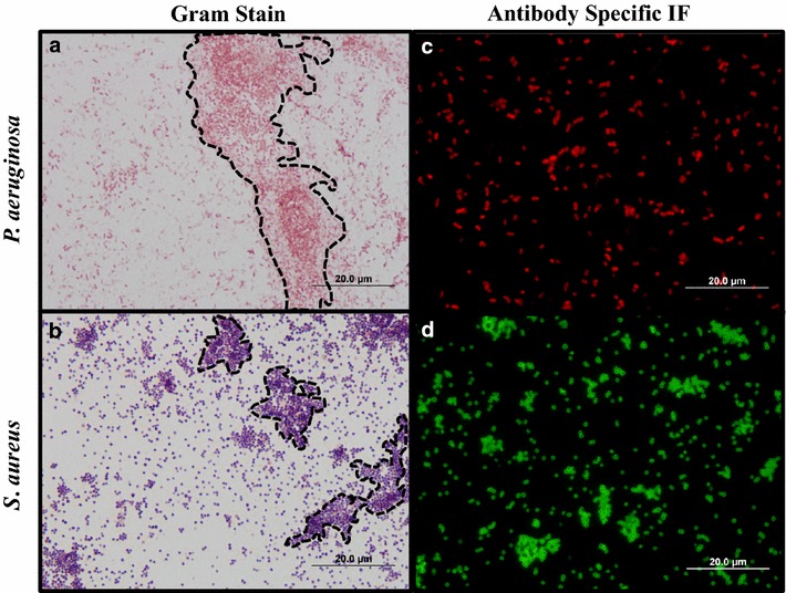
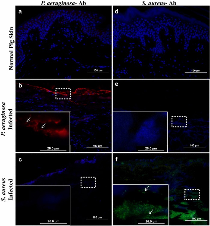
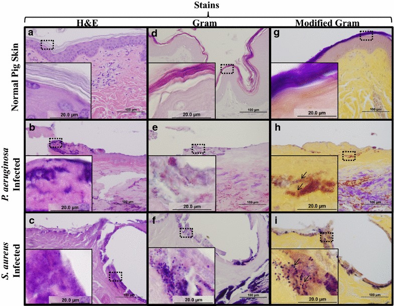
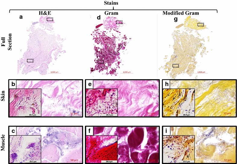
Findings: A modified Gram stain was developed and tested as an alternative to Gram stain that improves the contrast between Gram positive bacteria, Gram negative bacteria and host tissue. Initially, clinically relevant strains of Pseudomonas aeruginosa and Staphylococcus aureus were visualized in vitro and in biopsies of infected, porcine burns using routine Gram stain, and immunohistochemistry techniques involving bacterial strain-specific fluorescent antibodies as validation tools. H&E and Gram stain of serial biopsy sections were then compared to a modification of the Gram stain incorporating a counterstain that highlights collagen found in tissue. The modified Gram stain clearly identified both Gram positive and Gram negative bacteria, and when compared to H&E or Gram stain alone provided excellent contrast between bacteria and non-viable burn eschar. Moreover, when applied to surgical biopsies from patients that underwent burn debridement this technique was able to clearly detect bacterial morphology within host tissue.

Conclusions: We describe a modification of the Gram stain that provides improved contrast of Gram positive and Gram negative microorganisms within host tissue. The samples used in this study demonstrate that this staining technique has laboratory and clinical applicability. This modification only adds minutes to traditional Gram stain with reusable reagents, and results in a cost- and time-efficient technique for identifying bacteria in any clinical biopsy containing connective tissue.

Keywords: Burn; Gram’s stain; Histology; Infection; Pseudomonas aeruginosa; Staphylococcus aureus.


Figures

Fig. 1
Fig. 1
Schematic showing the modified Gram stain procedure. The traditional Gram stain procedure is completed in either case (top box). The major difference in the modified Gram stain procedure occurs during the dehydration process and is the application of alcoholic saffron (lower right box, highlighted in red). The yellow contrast in the connective tissue (collagen) within skin tissue is seen in the low magnification images on the bottom (Scale bar 1 mm)
Fig. 2
Fig. 2
In vitro validation of applied stains. Routine Gram stain shows identification of Gram negative (a P. aeruginosa) and Gram positive (b S. aureus) bacteria in both large aggregates (dotted lines) and individual bacteria within the biofilm. Selected P. aeruginosa (c) and S. aureus (d)-antibodies were used to confirm species identity of cultures used for ex vivo studies
Fig. 3
Fig. 3
Visualization of successful bacterial inoculation ex vivo. Normal pig skin (a, d), P. aeruginosa infected (b, e) and S. aureus infected (c, f) pig skin were subjected to immunohistochemistry with either P. aeruginosa (ac) or S. aureus (df) antibodies. Note that these antibodies allow for specific labeling of bacteria ex vivo as indicated by arrows pointing out unique bacterial morphologies
Fig. 4
Fig. 4
A modified Gram stain improves bacterial detection in tissue sections. Serial sections of normal porcine skin (a, d, g), P. aeruginosa infected (b, e, h) or S. aureus infected (c, f, i) burn wounds were stained with H&E, Gram, and Modified Gram stain. H&E of normal pig skin (a), P. aeruginosa infected (b) and S. aureus infected (c) pig skin illustrate coagulation of tissue, however staining within the burn eschar are not clearly identifiable as bacteria. Gram stain readily differentiates the clusters as Gram positive or Gram negative bacteria (e, f), however there is lack of contrast in the tissue in all samples. This lack of contrast is alleviated in the modified Gram, stain which distinctly enhances the detection of bacterial clusters as Gram negative (h) or Gram positive (i)
Fig. 5
Fig. 5
The modified gram stain applied to clinical surgical biopsies. Serial sections of tissue from a patient that underwent burn debridement were stained with three different stains. Low magnification scans of entire tissue sections show a small amount of dermis overlaying subcutaneous muscle using H&E (a), Gram (b), and Modified Gram (c) stains. Closer inspection of H&E stain in both skin (b) and muscle (c) areas reveal a lack of viable host cells, and hazy purple areas. Closer inspection of the Gram stain in both skin (e) and muscle (f) areas reveal some areas of bacterial morphology that are more apparent where host connective tissue is more sparse. Closer inspection of the modified Gram stain in both skin (h) and muscle (i) illustrates a counterstain that allows for identification of bacterial morphology (arrows) within connective tissue. Magnification insets are ×100

References

    1. Bangert C, Brunner PM, Stingl G. Immune functions of the skin. Clin Dermatol. 2011;29:360–376. doi: 10.1016/j.clindermatol.2011.01.006. - DOI - PubMed
    1. Costerton JW, Stewart PS, Greenberg EP. Bacterial biofilms: a common cause of persistent infections. Science. 1999;284:1318–1322. doi: 10.1126/science.284.5418.1318. - DOI - PubMed
    1. Hall-Stoodley L, Costerton JW, Stoodley P. Bacterial biofilms: from the natural environment to infectious diseases. Nat Rev Microbiol. 2004;2:95–108. doi: 10.1038/nrmicro821. - DOI - PubMed
    1. Ki V, Rotstein C. Bacterial skin and soft tissue infections in adults: a review of their epidemiology, pathogenesis, diagnosis, treatment and site of care. Can J Infect Dis Med Microbiol. 2008;19:173–184. - PMC - PubMed
    1. Swartz MN. Clinical practice. Cellulitis. N Engl J Med. 2004;350:904–912. doi: 10.1056/NEJMcp031807. - DOI - PubMed

Publication types

MeSH terms