Review
. 2016 Apr 12:18:22.
doi: 10.1186/s12968-016-0235-4.

Myocardial arterial spin labeling

Affiliations
Review

Myocardial arterial spin labeling

Frank Kober et al. J Cardiovasc Magn Reson. .

Abstract

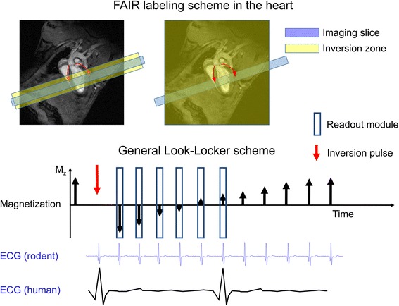
Arterial spin labeling (ASL) is a cardiovascular magnetic resonance (CMR) technique for mapping regional myocardial blood flow. It does not require any contrast agents, is compatible with stress testing, and can be performed repeatedly or even continuously. ASL-CMR has been performed with great success in small-animals, but sensitivity to date has been poor in large animals and humans and remains an active area of research. This review paper summarizes the development of ASL-CMR techniques, current state-of-the-art imaging methods, the latest findings from pre-clinical and clinical studies, and future directions. We also explain how successful developments in brain ASL and small-animal ASL-CMR have helped to inform developments in large animal and human ASL-CMR.

Keywords: Arterial spin labeling; Cardiovascular magnetic resonance; Ischemic heart disease; Myocardial perfusion.


Figures

Fig. 1
Fig. 1
ASL is a subtraction technique. Images are acquired (top) with and (bottom) without a preceding RF pulse that labels inflowing blood. The difference between these two images can be made directly proportional to myocardial blood flow in units of mL-blood/g-tissue/min. (Figure courtesy of Eric C. Wong, MD, PhD, University of California, San Diego)
Fig. 2
Fig. 2
The FAIR labeling scheme (upper part) consists in two inversion recovery measurements. In the first measurement the labeling zone is only around the imaging slice, and in the second measurement the inversion is global. FAIR is the most commonly used ASL technique in the heart. The timing of the inversion and that of the readout modules should be ECG-gated and occur in the same cardiac phase. The Look-Locker inversion-recovery readout (lower part) has been used mainly (although not exclusively) in the rodent heart, where the rapid heart rate permits dense sampling of the magnetization recovery curve. Due to the lower heart rate in humans, only one readout at a specific inversion time is generally performed
Fig. 3
Fig. 3
Look-Locker FAIR recovery signal behavior in myocardial and chest muscle regions in a mouse at 4.7 T (magnitude data, symbols represent the average of pixel signals in each ROI after global or slice-selective inversion, continuous line is an exponential fit for T1gl and T1sl). The signal difference due to perfusion is nearly absent in the very weakly perfused chest muscle, whereas myocardium shows a clear difference in relaxation. Data: CRMBM Marseille
Fig. 4
Fig. 4
Myocardial perfusion and cardiac function under continuous isoproterenol administration in rats. Both perfusion and function are strongly increased by this inotropic agent shortly after infusion start (day 1). The strong contractility is sustained over seven days while perfusion diminishes to lower values leading to morphological alterations at longer term (adapted from Desrois et al. [65])
Fig. 5
Fig. 5
ECG-gated FAIR Pulse Sequence Diagram with single inversion time readout mainly (but not exclusively) used in the human heart. Labeling and imaging are placed in the same cardiac phase to ensure the same volume of myocardium is excited. Imaging is preceded by a fat saturation and an initial preparation consisting of a catalyzation ramp to reduce transient oscillations
Fig. 6
Fig. 6
Acquisition timing diagram for spASL and FAIR ASL-CMR. In spASL, labeling and imaging occur every heartbeat during end-systole and mid-diastole, respectively. In FAIR, labeling and imaging occur in consecutive heartbeats at mid-diastole. An additional 6 sec wait is required to allow for T1 recovery of the label before the next acquisition. spASL has approximately 5x higher scan efficiency than FAIR, but is also more sensitive to gating errors and heart rate variability
Fig. 7
Fig. 7
ASL-CMR in patients with single-vessel CAD. Patient with total LAD occlusion shows reduced perfusion reserve by (a) ASL consistent with (b) angiography (red arrows). Patient with total RCA occlusion shows reduced perfusion reserve by (c) ASL consistent with (d) angiography (green arrows). The myocardial perfusion reserve (MPR) color scale represents 0.0 – 2.0 ml/g/min. (Data from Zun Z, et al. JACC: Cardiovasc Imag 2011, 4:1253–1261)
Fig. 8
Fig. 8
ASL-CMR pilot study in patients with suspected CAD demonstrated that ASL detected clinically relevant increases in MBF associated with vasodilation in (a) normal myocardium, and was able to differentiate those from (b) segments that were most ischemic based on coronary angiography. (Data from Zun Z, et al. JACC: Cardiovasc Imag 2011, 4:1253–1261)
Fig. 9
Fig. 9
ASL-CMR in a pig with 4-week old septal infarct. a first-pass CMR image from peak myocardial enhancement. b Late Gadolinium Enhanced image and c ASL MBF map at rest. The color scale ranges from 0-4 ml/g/min. The infarcted myocardium (blue arrows) is thinned, and shows low MBF. Normal myocardium (yellow arrows) shows MBF of approximately 1.5 ml/g/min. (Data courtesy of Nilesh Ghugre, Sunnybrook Research Institute, University of Toronto, Toronto, Canada)

References

    1. Calamante F, Thomas DL, Pell GS, Wiersma J, Turner R. Measuring cerebral blood flow using magnetic resonance imaging techniques. J Cereb Blood Flow Metab. 1999;19:701–35. doi: 10.1097/00004647-199907000-00001. - DOI - PubMed
    1. Wolf RL, Detre JA. Clinical neuroimaging using arterial spin-labeled perfusion magnetic resonance imaging. Neurother J Am Soc Exp Neurother. 2007;4:346–59. doi: 10.1016/j.nurt.2007.04.005. - DOI - PMC - PubMed
    1. Klocke FJ, Baird MG, Lorell BH, Bateman TM, Messer JV, Berman DS, O’Gara PT, Carabello BA, Russell RO, Cerqueira MD, St John Sutton MG, DeMaria AN, Udelson JE, Kennedy JW, Verani MS,Williams KA, Antman EM, Smith SC, Alpert JS, Gregoratos G, Anderson JL, Hiratzka LF, Faxon DP,Hunt SA, Fuster V, Jacobs AK, Gibbons RJ, Russell RO, American College of Cardiology, American Heart Association, et al. ACC/AHA/ASNC guidelines for the clinical use of cardiac radionuclide imaging--executive summary: a report of the American College of Cardiology/American Heart Association Task Force on Practice Guidelines (ACC/AHA/ASNC Committee to Revise the 1995 Guidelines for the Clinical Use of Cardiac Radionuclide Imaging). J Am Coll Cardiol. 2003;42:1318–33. - PubMed
    1. Ragosta M, Bishop AH, Lipson LC, Watson DD, Gimple LW, Sarembock IJ, Powers ER. Comparison between angiography and fractional flow reserve versus single-photon emission computed tomographic myocardial perfusion imaging for determining lesion significance in patients with multivessel coronary disease. Am J Cardiol. 2007;99:896–902. doi: 10.1016/j.amjcard.2006.11.035. - DOI - PubMed
    1. Gerber BL, Raman SV, Nayak K, Epstein FH, Ferreira P, Axel L, Kraitchman DL. Myocardial first-pass perfusion cardiovascular magnetic resonance: history, theory, and current state of the art. J Cardiovasc Magn Reson. 2008;10:18. doi: 10.1186/1532-429X-10-18. - DOI - PMC - PubMed

Publication types