

Mapping polyclonal antibody responses to bacterial infection using next generation phage display
- PMID: 27072017
- PMCID: PMC4829855
- DOI: 10.1038/srep24232
Mapping polyclonal antibody responses to bacterial infection using next generation phage display
Abstract
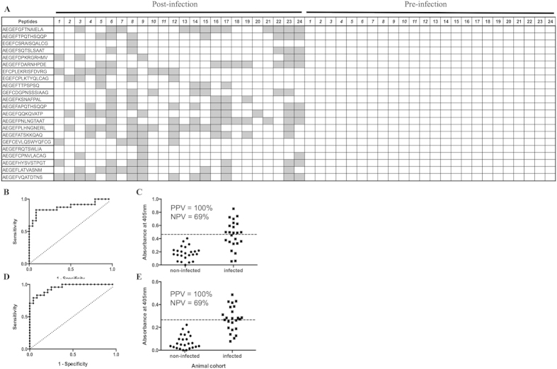
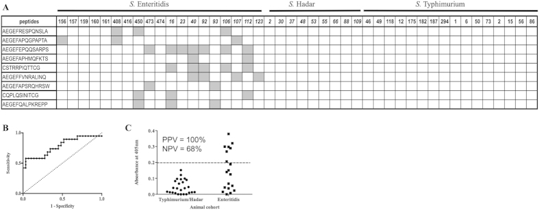
Mapping polyclonal antibody responses to infectious diseases to identify individual epitopes has the potential to underpin the development of novel serological assays and vaccines. Here, phage-peptide library panning coupled with screening using next generation sequencing was used to map antibody responses to bacterial infections. In the first instance, pigs experimentally infected with Salmonella enterica serovar Typhimurium was investigated. IgG samples from twelve infected pigs were probed in parallel and phage binding compared to that with equivalent IgG from pre-infected animals. Seventy-seven peptide mimotopes were enriched specifically against sera from multiple infected animals. Twenty-seven of these peptides were tested in ELISA and twenty-two were highly discriminatory for sera taken from pigs post-infection (P < 0.05) indicating that these peptides are mimicking epitopes from the bacteria. In order to further test this methodology, it was applied to differentiate antibody responses in poultry to infections with distinct serovars of Salmonella enterica. Twenty-seven peptides were identified as being enriched specifically against IgY from multiple animals infected with S. Enteritidis compared to those infected with S. Hadar. Nine of fifteen peptides tested in ELISA were highly discriminatory for IgY following S. Enteritidis infection (p < 0.05) compared to infections with S. Hadar or S. Typhimurium.
Figures

Similar articles
-
Mapping B-cell responses to Salmonella enterica serovars Typhimurium and Enteritidis in chickens for the discrimination of infected from vaccinated animals.Sci Rep. 2016 Aug 11;6:31186. doi: 10.1038/srep31186. Sci Rep. 2016. PMID: 27510219 Free PMC article.
-
[Agglutination of hen egg-yolk immunoglobulins (IgY) against Salmonella enterica, serovar enteritidis].Rev Argent Microbiol. 1998 Apr-Jun;30(2):84-92. Rev Argent Microbiol. 1998. PMID: 9744035 Spanish.
-
A proteomic approach to the development of DIVA ELISA distinguishing pigs infected with Salmonella Typhimurium and pigs vaccinated with a Salmonella Typhimurium-based inactivated vaccine.BMC Vet Res. 2016 Nov 11;12(1):252. doi: 10.1186/s12917-016-0879-1. BMC Vet Res. 2016. PMID: 27835998 Free PMC article.
-
High throughput functional epitope mapping: revisiting phage display platform to scan target antigen surface.MAbs. 2014;6(6):1368-76. doi: 10.4161/mabs.36144. MAbs. 2014. PMID: 25484050 Free PMC article. Review.
-
Massively-multiplexed epitope mapping techniques for viral antigen discovery.Front Immunol. 2023 Sep 25;14:1192385. doi: 10.3389/fimmu.2023.1192385. eCollection 2023. Front Immunol. 2023. PMID: 37818363 Free PMC article. Review.
Cited by
-
Depth of Sequencing Plays a Determining Role in the Characterization of Phage Display Peptide Libraries by NGS.Int J Mol Sci. 2023 Mar 11;24(6):5396. doi: 10.3390/ijms24065396. Int J Mol Sci. 2023. PMID: 36982469 Free PMC article.
-
Isolation of antigen-specific, disulphide-rich knob domain peptides from bovine antibodies.PLoS Biol. 2020 Sep 4;18(9):e3000821. doi: 10.1371/journal.pbio.3000821. eCollection 2020 Sep. PLoS Biol. 2020. PMID: 32886672 Free PMC article.
-
A Simple Whole-Plasmid PCR Method to Construct High-Diversity Synthetic Phage Display Libraries.Mol Biotechnol. 2022 Jul;64(7):791-803. doi: 10.1007/s12033-021-00442-4. Epub 2022 Feb 2. Mol Biotechnol. 2022. PMID: 35107752 Free PMC article.
-
Mapping B-cell responses to Salmonella enterica serovars Typhimurium and Enteritidis in chickens for the discrimination of infected from vaccinated animals.Sci Rep. 2016 Aug 11;6:31186. doi: 10.1038/srep31186. Sci Rep. 2016. PMID: 27510219 Free PMC article.
-
Mapping Polyclonal Antibody Responses to Infection Using Next-Generation Phage Display.Methods Mol Biol. 2023;2702:467-487. doi: 10.1007/978-1-0716-3381-6_25. Methods Mol Biol. 2023. PMID: 37679636
References
-
- Stalhammar-Carlemalm M., Waldermarsson J., Johnsson E., Areschoug T. & Lindahl G. Nonimmunodominant regions are effective as building blocks in streptococcal fusion protein vaccine. Cell Host Microbe 2, 1780–1887 (2007). - PubMed
-
- Tjalsma H., Schaeps R. M. J. & Swinkels D. Immunoproteomics: from biomarker discovery to diagnostic applications. Proteomics Clin. Appl. 2, 167–180 (2008). - PubMed
-
- Luzzago A., Felici F., Tramontano A., Pessi A. & Cortese R. Mimicking of discontinuous epitopes by phage displayed peptides, I. Epitope mapping of human H ferritin using a phage library of constrained peptides. Gene 128, 51–57 (1993). - PubMed
Publication types
MeSH terms
Substances
LinkOut - more resources
Full Text Sources
Other Literature Sources
Molecular Biology Databases
