

Reliability of standard pupillometry practice in neurocritical care: an observational, double-blinded study
- PMID: 27072310
- PMCID: PMC4828754
- DOI: 10.1186/s13054-016-1239-z
Reliability of standard pupillometry practice in neurocritical care: an observational, double-blinded study
Abstract
Background: In critical care units, pupil examination is an important clinical parameter for patient monitoring. Current practice is to use a penlight to observe the pupillary light reflex. The result seems to be a subjective measurement, with low precision and reproducibility. Several quantitative pupillometer devices are now available, although their use is primarily restricted to the research setting. To assess whether adoption of these technologies would benefit the clinic, we compared automated quantitative pupillometry with the standard clinical pupillary examination currently used for brain-injured patients.
Methods: In order to determine inter-observer agreement of the device, we performed repetitive measurements in 200 healthy volunteers ranging in age from 21 to 58 years, providing a total of 400 paired (alternative right eye, left eye) measurements under a wide variety of ambient light condition with NeuroLight Algiscan pupillometer. During another period, we conducted a prospective, observational, double-blinded study in two neurocritical care units. Patients admitted to these units after an acute brain injury were included. Initially, nursing staff measured pupil size, anisocoria and pupillary light reflex. A blinded physician subsequently performed measurement using an automated pupillometer.
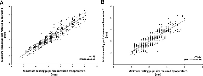
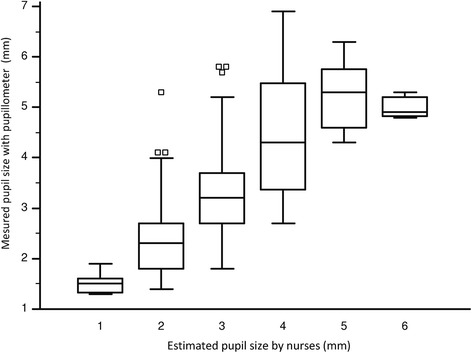
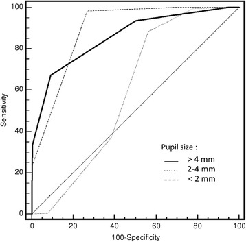
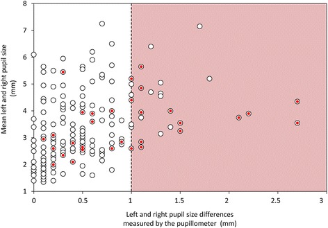
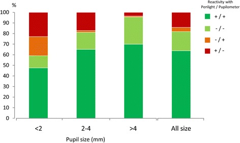
Results: In 200 healthy volunteers, intra-class correlation coefficient for maximum resting pupil size was 0.95 (IC: 0.93-0.97) and for minimum pupil size after light stimulation 0.87 (0.83-0.89). We found only 3-pupil asymmetry (≥ 1 mm) in these volunteers (1.5% of the population) with a clear pupil asymmetry during clinical inspection. The mean pupil light reactivity was 40 ± 7%. In 59 patients, 406 pupillary measurements were prospectively performed. Concordance between measurements for pupil size collected using the pupillometer, versus subjective assessment, was poor (Spearmen's rho = 0.75, IC: 0.70-0.79; P < 0.001). Nursing staff failed to diagnose half of the cases (15/30) of anisocoria detected using the pupillometer device. A global rate of discordance of 18% (72/406) was found between the two techniques when assessing the pupillary light reflex. For measurements with small pupils (diameters <2 mm) the error rate was 39% (24/61).
Conclusion: Standard practice in pupillary monitoring yields inaccurate data. Automated quantitative pupillometry is a more reliable method with which to collect pupillary measurements at the bedside.
Keywords: Anisocoria; Neurocritical Care; Neurological examination; Pupillary light reflex; Pupillary reactivity; Pupillary size; Pupillometer.
Figures
References
-
- Chestnut RM, Ghajar J, Maas AI, Marion DW, Servadei F, Teasdale GM. Part 2: early indicators of prognosis in severe traumatic brain injury. J Neurotrauma. 2000;17:557-627.
-
- Marmarou A, Lu J, Butcher I, McHugh GS, Murray GD, Steyerberg EW, et al. Prognositc value of the Glsasgow Coma Scale and pupill reactivity in traumatic brain injury assessed pre-hospital and on enrollment; an IMPACT analysis. J Neurotrauma. 2007;24:270–280. doi: 10.1089/neu.2006.0029. - DOI - PubMed
-
- Morris GF, Juul N, Marshall SB, Benedict B, Marshall LF. Neurological deterioration as a potential alternative endpoint in human clinical trials of experimental pharmacological agents for treatment of severe traumatic brain injuries. Executive Committee of the International Selfotel Trial. Neurosurgery. 1998;43(6):1369–1372. - PubMed
Publication types
MeSH terms
LinkOut - more resources
Full Text Sources
Other Literature Sources
Medical
