

Liquid-based three-dimensional tumor models for cancer research and drug discovery
- PMID: 27072562
- PMCID: PMC4950350
- DOI: 10.1177/1535370216643772
Liquid-based three-dimensional tumor models for cancer research and drug discovery
Abstract
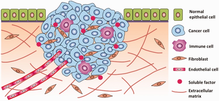
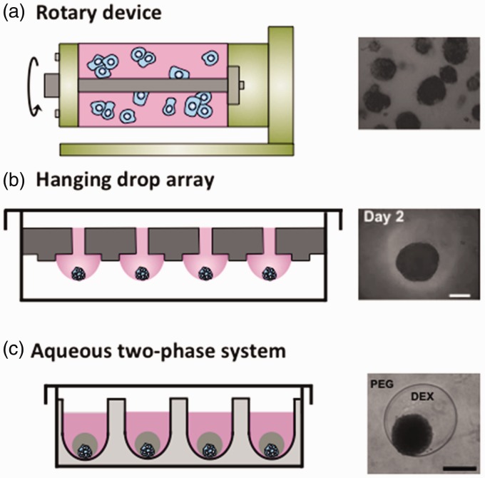
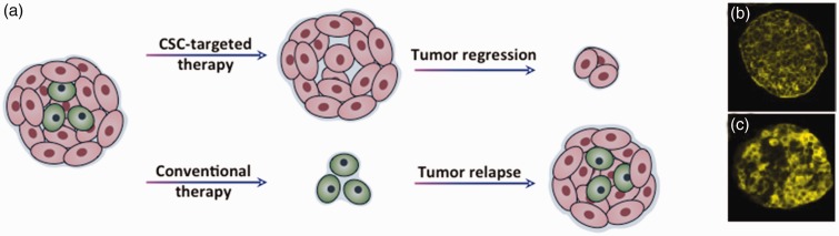
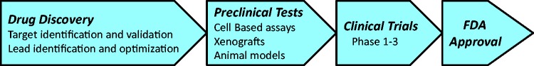
Tumors are three-dimensional tissues where close contacts between cancer cells, intercellular interactions between cancer and stromal cells, adhesion of cancer cells to the extracellular matrix, and signaling of soluble factors modulate functions of cancer cells and their response to therapeutics. Three-dimensional cultures of cancer cells overcome limitations of traditionally used monolayer cultures and recreate essential characteristics of tumors such as spatial gradients of oxygen, growth factors, and metabolites and presence of necrotic, hypoxic, quiescent, and proliferative cells. As such, three-dimensional tumor models provide a valuable tool for cancer research and oncology drug discovery. Here, we describe different tumor models and primarily focus on a model known as tumor spheroid. We summarize different technologies of spheroid formation, and discuss the use of spheroids to address the influence of stromal fibroblasts and immune cells on cancer cells in tumor microenvironment, study cancer stem cells, and facilitate compound screening in the drug discovery process. We review major techniques for quantification of cellular responses to drugs and discuss challenges ahead to enable broad utility of tumor spheroids in research laboratories, integrate spheroid models into drug development and discovery pipeline, and use primary tumor cells for drug screening studies to realize personalized cancer treatment.
Keywords: 3D tumor models; anti-cancer drug discovery; cancer cell spheroids; cancer stem cells; co-culture spheroids; tumor microenvironment.
© 2016 by the Society for Experimental Biology and Medicine.
Figures

Similar articles
-
A Novel Stromal Fibroblast-Modulated 3D Tumor Spheroid Model for Studying Tumor-Stroma Interaction and Drug Discovery.J Vis Exp. 2020 Feb 28;(156). doi: 10.3791/60660. J Vis Exp. 2020. PMID: 32176195
-
3D-3 Tumor Models in Drug Discovery for Analysis of Immune Cell Infiltration.Methods Mol Biol. 2019;1953:151-162. doi: 10.1007/978-1-4939-9145-7_10. Methods Mol Biol. 2019. PMID: 30912021
-
Establishment and Analysis of a 3D Co-Culture Spheroid Model of Pancreatic Adenocarcinoma for Application in Drug Discovery.Methods Mol Biol. 2019;1953:163-179. doi: 10.1007/978-1-4939-9145-7_11. Methods Mol Biol. 2019. PMID: 30912022
-
Three-Dimensional Cell Cultures as an In Vitro Tool for Prostate Cancer Modeling and Drug Discovery.Int J Mol Sci. 2020 Sep 16;21(18):6806. doi: 10.3390/ijms21186806. Int J Mol Sci. 2020. PMID: 32948069 Free PMC article. Review.
-
Taking a Full Snapshot of Cancer Biology: Deciphering the Tumor Microenvironment for Effective Cancer Therapy in the Oncology Clinic.OMICS. 2020 Apr;24(4):175-179. doi: 10.1089/omi.2020.0019. Epub 2020 Mar 13. OMICS. 2020. PMID: 32176591 Review.
Cited by
-
Modeling adaptive drug resistance of colorectal cancer and therapeutic interventions with tumor spheroids.Exp Biol Med (Maywood). 2021 Nov;246(22):2372-2380. doi: 10.1177/15353702211014185. Epub 2021 Jun 8. Exp Biol Med (Maywood). 2021. PMID: 34102903 Free PMC article. Review.
-
Cyclical Treatment of Colorectal Tumor Spheroids Induces Resistance to MEK Inhibitors.Transl Oncol. 2019 Mar;12(3):404-416. doi: 10.1016/j.tranon.2018.11.009. Epub 2018 Dec 14. Transl Oncol. 2019. PMID: 30550927 Free PMC article.
-
3D hanging spheroid plate for high-throughput CAR T cell cytotoxicity assay.J Nanobiotechnology. 2022 Jan 10;20(1):30. doi: 10.1186/s12951-021-01213-8. J Nanobiotechnology. 2022. PMID: 35012567 Free PMC article.
-
iRhom2 regulates ERBB signalling to promote KRAS-driven tumour growth of lung cancer cells.J Cell Sci. 2022 Sep 1;135(17):jcs259949. doi: 10.1242/jcs.259949. Epub 2022 Sep 8. J Cell Sci. 2022. PMID: 35971826 Free PMC article.
-
Image Analysis of 3D Conjunctival Melanoma Cell Cultures Following Electrochemotherapy.Biomedicines. 2020 Jun 13;8(6):158. doi: 10.3390/biomedicines8060158. Biomedicines. 2020. PMID: 32545782 Free PMC article.
References
-
- Choe C, Shin Y-S, Kim S-H, Jeon M-J, Choi S-J, Lee J, Kim J. Tumor–stromal interactions with direct cell contacts enhance motility of non-small cell lung cancer cells through the hedgehog signaling pathway. Anticancer Res 2013; 33: 3715–23. - PubMed
-
- Nakamura T, Kato Y, Fuji H, Horiuchi T, Chiba Y, Tanaka K. E-cadherin-dependent intercellular adhesion enhances chemoresistance. Int J Mol Med 2003; 12: 693–700. - PubMed
-
- Green SK, Francia G, Isidoro C, Kerbel RS. Antiadhesive antibodies targeting E-cadherin sensitize multicellular tumor spheroids to chemotherapy in vitro. Mol Cancer Ther 2004; 3: 149–59. - PubMed
Publication types
MeSH terms
Substances
Grants and funding
LinkOut - more resources
Full Text Sources
Other Literature Sources
Research Materials