. 2016 Sep 7;11(9):1664-1674.
doi: 10.2215/CJN.13791215. Epub 2016 Apr 12.

The Players: Cells Involved in Glomerular Disease

Affiliations

The Players: Cells Involved in Glomerular Disease

A Richard Kitching et al. Clin J Am Soc Nephrol. .

Abstract

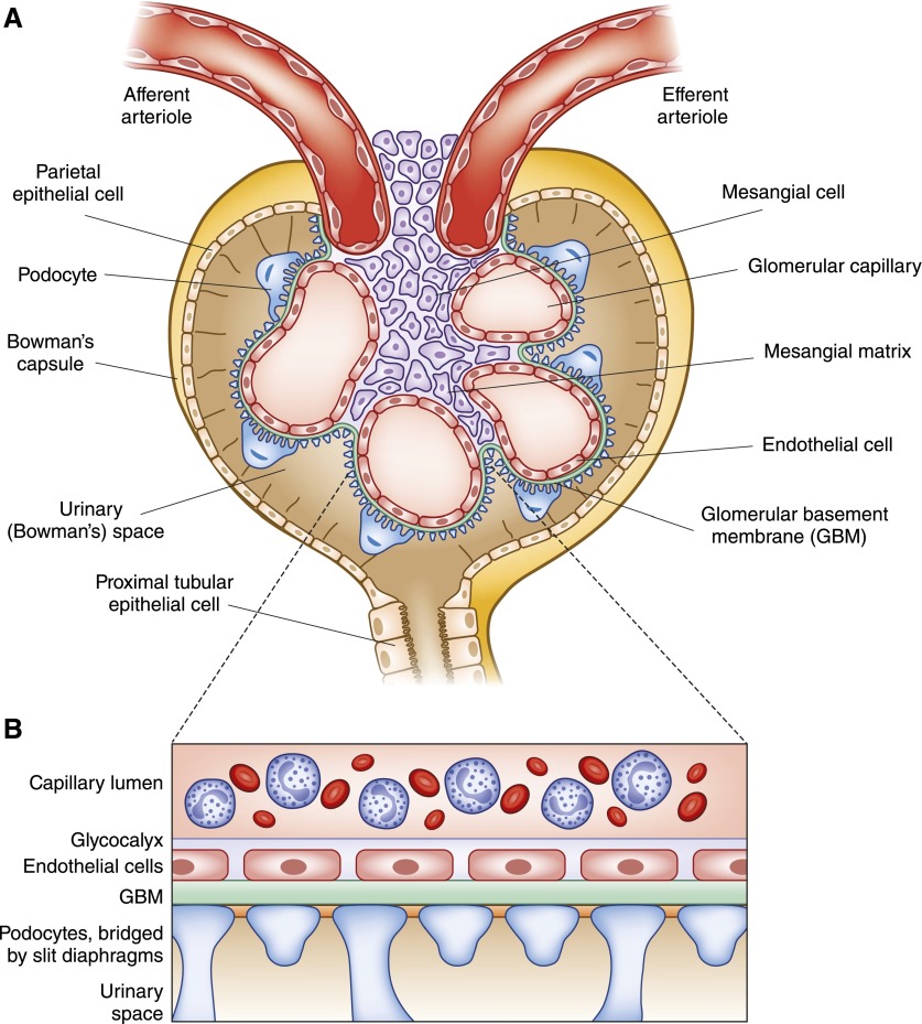
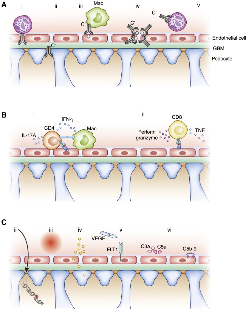
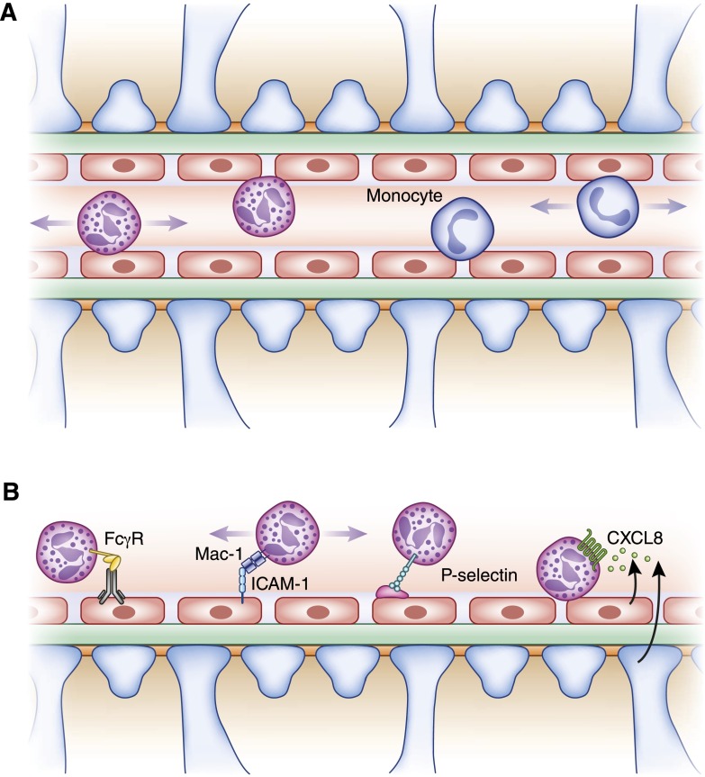
Glomerular diseases are common and important. They can arise from systemic inflammatory or metabolic diseases that affect the kidney. Alternately, they are caused primarily by local glomerular abnormalities, including genetic diseases. Both intrinsic glomerular cells and leukocytes are critical to the healthy glomerulus and to glomerular dysregulation in disease. Mesangial cells, endothelial cells, podocytes, and parietal epithelial cells within the glomerulus all play unique and specialized roles. Although a specific disease often primarily affects a particular cell type, the close proximity, and interdependent functions and interactions between cells mean that even diseases affecting one cell type usually indirectly influence others. In addition to those cells intrinsic to the glomerulus, leukocytes patrol the glomerulus in health and mediate injury in disease. Distinct leukocyte types and subsets are present, with some being involved in different ways in an individual glomerular disease. Cells of the innate and adaptive immune systems are important, directing systemic immune and inflammatory responses, locally mediating injury, and potentially dampening inflammation and facilitating repair. The advent of new genetic and molecular techniques, and new disease models means that we better understand both the basic biology of the glomerulus and the pathogenesis of glomerular disease. This understanding should lead to better diagnostic techniques, biomarkers, and predictors of prognosis, disease severity, and relapse. With this knowledge comes the promise of better therapies in the future, directed toward halting pathways of injury and fibrosis, or interrupting the underlying pathophysiology of the individual diseases that lead to significant and progressive glomerular disease.

Keywords: Glomerulus; Immunology and pathology; Inflammation; Leukocytes; Podocytes; Prognosis; endothelial cells; fibrosis; kidney; mesangial cells.


Figures

Figure 1.
Figure 1.
Basic structure of the glomerulus and the glomerular filtration barrier. (A) Each glomerulus is composed of an afferent arteriole, which supplies the glomerular capillaries, and an efferent arteriole, into which they drain. Mesangial cells and mesangial matrix provide structural support for the glomerular capillaries, lined by specialized fenestrated endothelium, and then the glomerular basement membrane. On the urinary side of the glomerular basement membrane are podocytes, with foot processes that wrap around the glomerular capillaries. The urinary space is lined by a cup-like layer of parietal epithelial cells which adhere to the basement membrane of Bowman’s capsule. (B) The glomerular filtration barrier is a specialized molecular sieve, with properties that aid filtration of small solutes from the blood to the urine, while limiting the passage of macromolecules such as albumin.
Figure 2.
Figure 2.
Simplified diagrammatic representation of a selection of mechanisms of glomerular injury. (A) Antibody-mediated glomerular injury. From left to right, (i) neutrophils (shown) and macrophages induce injury after anti-α3(IV)NC1 autoantibodies bind to the GBM in anti-GBM GN; (ii) in membranous glomerulopathy autoantibodies against PLA2R1 (and other antigens) on podocytes are deposited subepithelially, with the involvement of complement; (iii) antibodies can bind to antigens lodged in the glomerulus (grey dots) with recruitment of macrophages (shown) and neutrophils, and the activation of complement; (iv) circulating immune complexes can be deposited in glomeruli, activate complement, and recruit leukocytes; (v) ANCA, (with complement) activates neutrophils and enables their recruitment to the glomerulus. Not shown, but important, is IgA deposition in mesangial areas. (B) Cell-mediated immune mechanisms. (i) Effector CD4+ cells (often Th1 or Th17 type) recognize antigens that can be intrinsic to or planted in the glomeruli. This occurs via their T cell receptor recognizing MHC class II peptide complexes (several cell types could possibly be involved in this process). Activated T cells produce cytokines (IL-17A and IFN-γ as examples) that have direct effects on intrinsic kidney cells and activate, together with costimulatory molecules (e.g., CD154/CD40), innate leukocytes such as macrophages. Not shown are interactions between intrinsic renal cells and T cells that include costimulation and cytokines. (ii) CD8+ cells can recognize antigenic peptides with MHC class I on intrinsic cells and secrete cytokines or induce cell death. (C) Metabolic, vascular, and other mechanisms of injury. Podocyte and foot process injury and dysfunction occurs due to (i) genetic abnormalities of slit diaphragm proteins and (ii) in minimal change disease and FSGS due to circulating permeability factors. Metabolic factors such as (iii) systemic and intraglomerular hypertension and (iv) hyperglycemia and its consequences are common, and affect both the cells and the structural components of the glomerulus. Both glomerular endothelial cell and podocyte injury are important consequences of preeclampsia, involved a number of mediators including soluble fms-like tyrosine kinase-1. C3 glomerulopathy, as well as some types of atypical hemolytic uremic syndrome (vi), can be induced by autoantibodies to, or genetic abnormalities in, complement regulatory proteins, resulting in complement activation. α3(IV)NC1, the non-collagenous domain of the α3 chain of type IV collagen; FLT1, fms-like tyrosine kinase-1; GBM, glomerular basement membrane; Mac, macrophage; M-type PLA2R1, phospholipase A2 receptor 1; Th, T helper; VEGF, vascular endothelial growth factor.
Figure 3.
Figure 3.
Leukocytes in glomeruli of patients with rapidly progressive GN. High powered photomicrographs as illustrative examples of leukocytes within glomeruli in ANCA-associated GN. In all panels cell nuclei are stained with 4′,6-diamidino-2-phenylindole (blue). (A) CD45+ cells (green, CD45 is a common leukocyte marker) in the glomerulus, (B) CD3+ T cells (green), (C) CD68+ macrophages (green), and (D) myeloperoxidase (red) expressing leukocytes (neutrophils or macrophages) in a segmental glomerular lesion with local loss of CD34 (green), an endothelial cell lesion. Nephrin (marking podocytes) is in white. Magnification: (A), (C ) and (D) x400; (B) x600. Photomicrographs from Ms. Kim O’Sullivan.
Figure 4.
Figure 4.
Leukocyte recruitment and behavior in the glomerulus. (A) In health, neutrophils and monocytes (and potentially other leukocytes) patrol the glomerulus. While some are static, the majority migrate bidirectionally within glomerular capillaries. (B) In acute disease, leukocytes can be recruited and retained in glomeruli via a number of molecular processes. Examples are given with reference to the neutrophil (from left to right). Neutrophils can be recruited via direct FcγR-Fc interactions. Adhesion molecules participate in recruitment, migration, and retention, with, for example, Mac-1 (CD11b/CD18) on neutrophils slowing migration and inducing retention. P-selectins is not constitutively expressed by glomerular endothelial cells, but in some situations P-selectin can participate in forming bridges with recruited platelets (pink oval) to recruit neutrophils. Lastly, chemokines, for example CXCL8 (IL-8) secreted by endothelial cells, podocytes, mesangial cells (not shown), or other leukocytes (not shown), attract leukocytes expressing appropriate chemokine receptors down a concentration gradient. Other mechanisms, for example complement (not shown), can also attract leukocytes to the glomerulus. FcγR, Fcγ receptor; ICAM-1, intercellular adhesion molecule 1.

References

    1. Shankland SJ, Smeets B, Pippin JW, Moeller MJ: The emergence of the glomerular parietal epithelial cell. Nat Rev Nephrol 10: 158–173, 2014 - PubMed
    1. Puelles VG, Douglas-Denton RN, Cullen-McEwen LA, Li J, Hughson MD, Hoy WE, Kerr PG, Bertram JF: Podocyte Number in Children and Adults: Associations with Glomerular Size and Numbers of Other Glomerular Resident Cells. J Am Soc Nephrol 26: 2277–2288, 2015 - PMC - PubMed
    1. Schlöndorff D, Banas B: The mesangial cell revisited: no cell is an island. J Am Soc Nephrol 20: 1179–1187, 2009 - PubMed
    1. Saleem MA: One hundred ways to kill a podocyte. Nephrol Dial Transplant 30: 1266–1271, 2015 - PubMed
    1. Abboud HE: Mesangial cell biology. Exp Cell Res 318: 979–985, 2012 - PubMed

Publication types