. 2016:2016:2974605.
doi: 10.1155/2016/2974605. Epub 2016 Mar 17.

Reduced Numbers and Impaired Function of Regulatory T Cells in Peripheral Blood of Ischemic Stroke Patients

Affiliations

Reduced Numbers and Impaired Function of Regulatory T Cells in Peripheral Blood of Ischemic Stroke Patients

Johanna Ruhnau et al. Mediators Inflamm. 2016.

Abstract

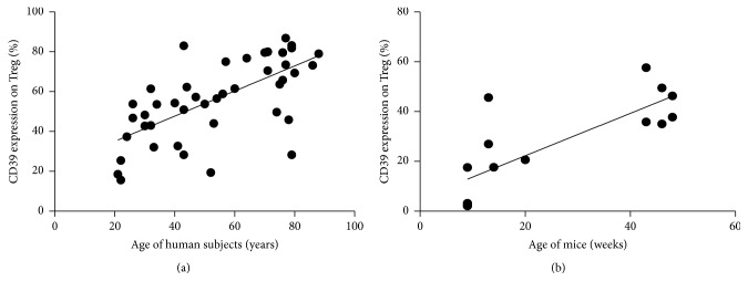
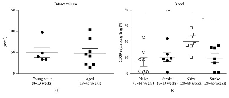
Background and purpose: Regulatory T cells (Tregs) have been suggested to modulate stroke-induced immune responses. However, analyses of Tregs in patients and in experimental stroke have yielded contradictory findings. We performed the current study to assess the regulation and function of Tregs in peripheral blood of stroke patients. Age dependent expression of CD39 on Tregs was quantified in mice and men.

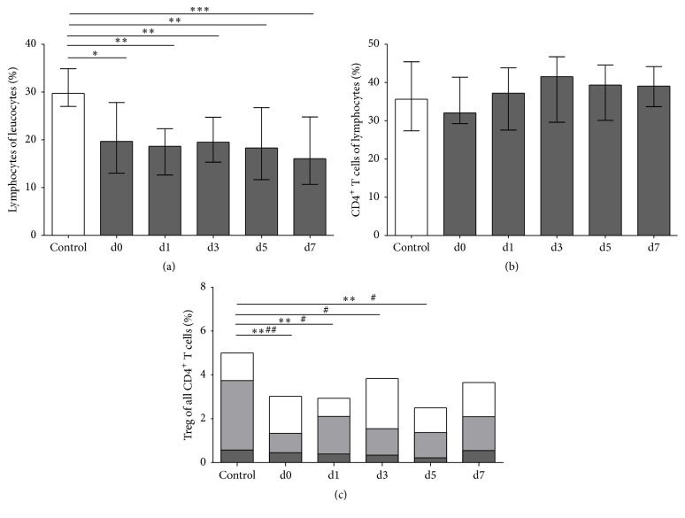
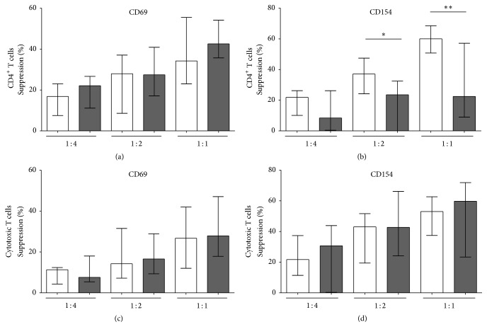
Methods: Total FoxP3(+) Tregs and CD39(+)FoxP3(+) Tregs were quantified by flow cytometry in controls and stroke patients on admission and on days 1, 3, 5, and 7 thereafter. Treg function was assessed by quantifying the inhibition of activation-induced expression of CD69 and CD154 on T effector cells (Teffs).

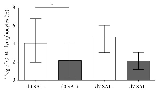
Results: Total Tregs accounted for 5.0% of CD4(+) T cells in controls and <2.8% in stroke patients on admission. They remained below control values until day 7. CD39(+) Tregs were most strongly reduced in stroke patients. On day 3 the Treg-mediated inhibition of CD154 upregulation on CD4(+) Teff was impaired in stroke patients. CD39 expression on Treg increased with age in peripheral blood of mice and men.

Conclusion: We demonstrate a loss of active FoxP3(+)CD39(+) Tregs from stroke patient's peripheral blood. The suppressive Treg function of remaining Tregs is impaired after stroke.


Figures

Figure 1
Figure 1
CD39 expression on Treg correlates positively with age in man and mice. (a) Human, peripheral blood: the percentage of CD39 expression was determined on CD4+CD25+CD49dFoxP3+ Treg and correlated to age for a total of 45 healthy controls (age from 21 to 88 years). Pearson r = 0.6612, p < 0.0001, and R squared = 0.4372. (b) Mice, blood: the percentage of CD39 expression was determined on CD4+CD25+CD49dFoxP3+ Treg and correlated to age for a total of 15 naive animals (age from 8 to 48 weeks). Pearson r = 0.7895, p < 0.0005, and R squared = 0.6233.
Figure 2
Figure 2
T helper cells and Tregs and their subpopulations. Cytometric analysis of nonstroke controls (white bars) and stroke patients (dark grey bars) was performed for (a) lymphocytes (n control,d0,1,3,5,7 = 15, 35, 35, 35, 32, 30, resp.), shown as the percentage of leukocytes. p < 0.05, ∗∗ p < 0.01, and ∗∗∗ p < 0.001. (b) CD4+ T helper cells (n control,d0,1,3,5,7 = 15, 35, 35, 35, 32, 29, resp.), shown as the percentage of lymphocytes. (c) Total Tregs (n control,d0,1,3,5,7 = 15, 35, 35, 34, 32, 27, resp.) combined with their subpopulations. Naive Tregs (n control,d0,1,3,5,7 = 15, 34, 35, 33, 31, 27, resp.) are shown by dark grey bars and activated Tregs (n control,d0,1,3,5,7 = 14, 35, 34, 33, 32, 27, resp.) by light grey bars. Patient samples were obtained on admission (d0), the next morning (d1), and on days 3, 5, and 7 (d3, d5, and d7, resp.). ∗∗ p < 0.01 for Tregs in stroke patients versus controls; # p < 0.05 and ## p < 0.01 for activated Tregs in patients versus controls. Medians and interquartile ranges (a, b) are provided.
Figure 3
Figure 3
Age dependency of infarct volume and CD39 expression on Treg after experimental stroke in mice. (a) The infarct volume as assessed by MRI in young adult and aged mice was compared on day 1 after transient middle cerebral artery occlusion. (b) The CD39 expression on CD4+CD25+CD49dFoxP3+ Treg in blood was compared between naive and stroked young adult and naive and stroked aged mice on day 3 after transient middle cerebral artery occlusion. p < 0.05, ∗∗ p < 0.01. Means and SEM are provided.
Figure 4
Figure 4
Suppressive activity of Tregs. Treg function was evaluated by measuring the CD4+CD25+CD127dim/− Treg-mediated inhibition of CD69 (a, c) and CD154 (b, d) induction on T effector cells (Teff) after anti-CD3/anti-CD28 stimulation. Suppression is shown for T helper cells (a, b) and cytotoxic T cells (c, d) in control subjects (white bars) versus stroke patients (dark grey bars) on day 3 with different Treg : PBMC concentrations (Treg : PBMC = 1 : 4; 1 : 2; and 1 : 1). n control,stroke  on  day  3 = 11 and 10, respectively. p < 0.05 and ∗∗ p < 0.01 for stroke patients versus controls. Medians and interquartile ranges are provided.
Figure 5
Figure 5
Comparison of SAI− and SAI+ patients. On the day of admission (d0) and day 7 after stroke (d7) the percentage of Treg of CD4+ T helper cells was compared between patients without poststroke infections SAI− (white bars; n d0,d7 = 14, 12, resp.) and patients with poststroke infections SAI+ (grey bars; n d0,d7 = 7, 7, resp.). p < 0.05. Means and SD are provided.

References

    1. Prass K., Meisel C., Höflich C., et al. Stroke-induced immunodeficiency promotes spontaneous bacterial infections and is mediated by sympathetic activation reversal by poststroke T helper cell type 1–like immunostimulation. The Journal of Experimental Medicine. 2003;198(5):725–736. doi: 10.1084/jem.20021098. - DOI - PMC - PubMed
    1. Vogelgesang A., Dressel A. Immunological consequences of ischemic stroke: immunosuppression and autoimmunity. Journal of Neuroimmunology. 2011;231(1-2):105–110. doi: 10.1016/j.jneuroim.2010.09.023. - DOI - PubMed
    1. Vogelgesang A., Grunwald U., Langner S., et al. Analysis of lymphocyte subsets in patients with stroke and their influence on infection after stroke. Stroke. 2007;39(1):237–241. doi: 10.1161/strokeaha.107.493635. - DOI - PubMed
    1. Chamorro A., Amaro S., Vargas M., et al. Catecholamines, infection, and death in acute ischemic stroke. Journal of the Neurological Sciences. 2007;252(1):29–35. doi: 10.1016/j.jns.2006.10.001. - DOI - PubMed
    1. Hug A., Liesz A., Muerle B., et al. Reduced efficacy of circulating costimulatory cells after focal cerebral ischemia. Stroke. 2011;42(12):3580–3586. doi: 10.1161/strokeaha.111.620948. - DOI - PubMed

Publication types