. 2016 Feb;11(2):298-304.
doi: 10.4103/1673-5374.177739.

Human neural stem cells promote proliferation of endogenous neural stem cells and enhance angiogenesis in ischemic rat brain

Affiliations

Human neural stem cells promote proliferation of endogenous neural stem cells and enhance angiogenesis in ischemic rat brain

Sun Ryu et al. Neural Regen Res. 2016 Feb.

Abstract

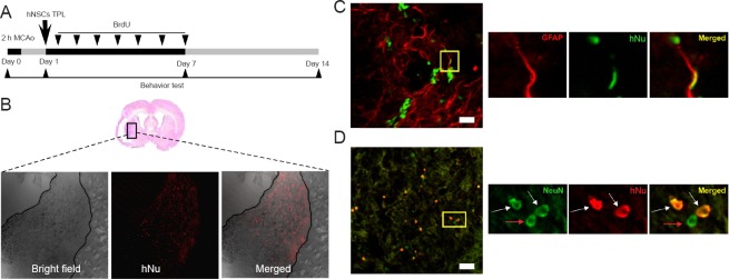
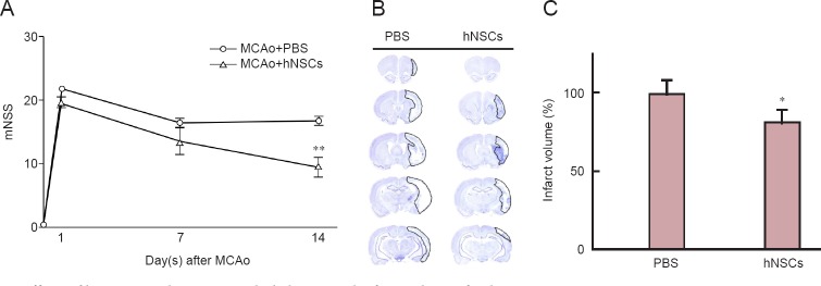
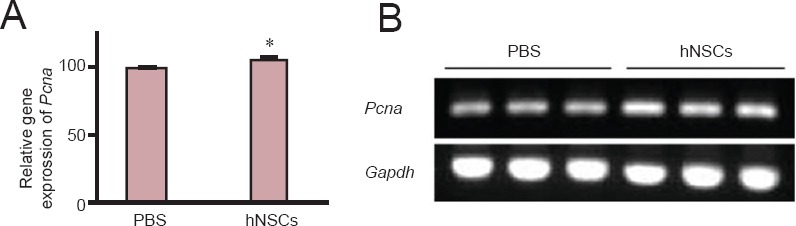
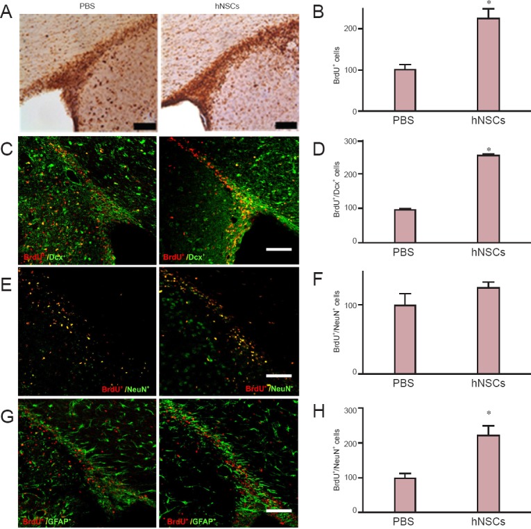
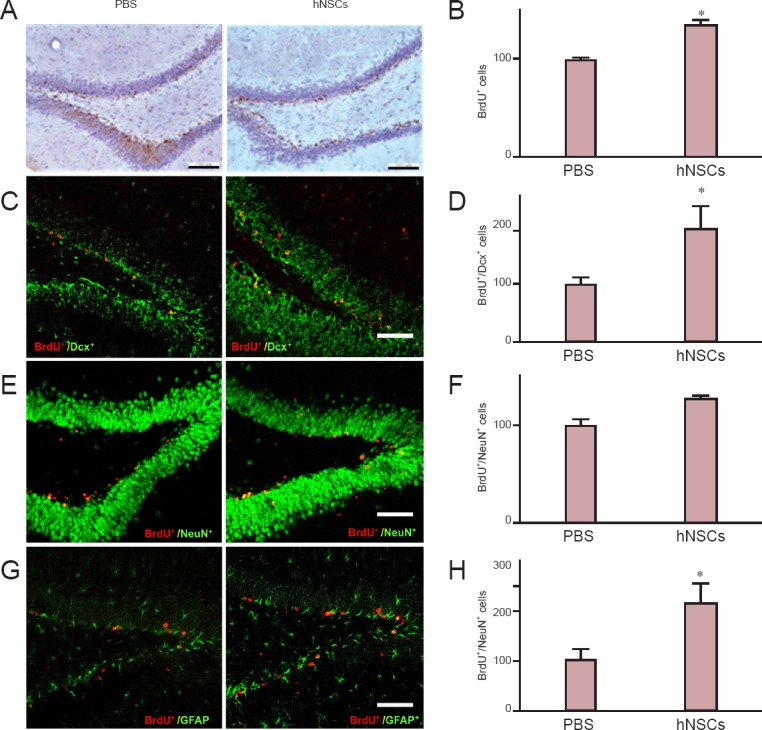
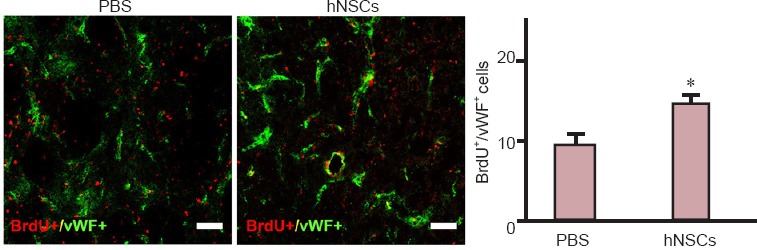
Transplantation of human neural stem cells into the dentate gyrus or ventricle of rodents has been reportedly to enhance neurogenesis. In this study, we examined endogenous stem cell proliferation and angiogenesis in the ischemic rat brain after the transplantation of human neural stem cells. Focal cerebral ischemia in the rat brain was induced by middle cerebral artery occlusion. Human neural stem cells were transplanted into the subventricular zone. The behavioral performance of human neural stem cells-treated ischemic rats was significantly improved and cerebral infarct volumes were reduced compared to those in untreated animals. Numerous transplanted human neural stem cells were alive and preferentially localized to the ipsilateral ischemic hemisphere. Furthermore, 5-bromo-2'-deoxyuridine-labeled endogenous neural stem cells were observed in the subventricular zone and hippocampus, where they differentiated into cells immunoreactive for the neural markers doublecortin, neuronal nuclear antigen NeuN, and astrocyte marker glial fibrillary acidic protein in human neural stem cells-treated rats, but not in the untreated ischemic animals. The number of 5-bromo-2'-deoxyuridine-positive ⁄ anti-von Willebrand factor-positive proliferating endothelial cells was higher in the ischemic boundary zone of human neural stem cells-treated rats than in controls. Finally, transplantation of human neural stem cells in the brains of rats with focal cerebral ischemia promoted the proliferation of endogenous neural stem cells and their differentiation into mature neural-like cells, and enhanced angiogenesis. This study provides valuable insights into the effect of human neural stem cell transplantation on focal cerebral ischemia, which can be applied to the development of an effective therapy for stroke.

Keywords: angiogenesis; behavioral analysis; differentiation; endogenous neurogenesis; focal cerebral ischemia; human neural stem cells; infarct size; middle cerebral artery occlusion; nerve regeneration; neural regeneration; rats; transplantation.


Conflict of interest statement

Conflicts of interest: None declared.

Figures

Figure 1
Figure 1
Transplanted hNSCs exhibited directional migration and differentiation in the ischemic lesion of rats. Survival and differentiation pattern of hNSCs transplanted into the ipsilateral subventricular zone of the rat brain subjected to ischemic stroke. (A) Experimental design of the present study. Ischemic stroke was induced by MCAo, and hNSCs were transplanted into the ipsilateral ventricle of the brain at 1 day post-stroke. The following day, daily intraperitoneal injections of BrdU were started and continued for 7 days. Behavioral testing was performed at the indicated intervals. (B–D) Immunohistochemical staining for hNu (red) to identify transplanted hNSCs in the ischemic penumbra, 50× magnification (B), hNu (green) + GFAP (red) (C), and hNu (red) + NeuN (green) (D) in the ischemic striatum 14 days after stroke. White arrows: Double-labeled cells; red arrow: single-labeled cells. Scale bars: 100 μm. hNSCs: Human neural stem cells; hNu: human-specific nuclei; NeuN: neuronal nuclei antigen; GFAP: glial fibrillary acidic protein; MCAo: occlusion of the middle cerebral artery.
Figure 2
Figure 2
Effects of hNSC transplantation on the behavior and infarct volume of ischemic rats. (A) Intraventricularly transplanted hNSCs improved behavioral recovery after MCAo. Behavioral tests were performed using the mNSS at days 1, 7, and 14 after hNSC transplantation. Lower mNSS scores represent better neurological function. (B) Transplanted hNSCs reduced infarct volume at day 14 after MCAo. Images show Nissl staining of coronal serial sections of the brain at day 14 after MCAo in the PBS-injected (PBS) and hNSCs-transplanted (hNSCs) rats. (C) Quantification of the infarct volume shown in B. The data are expressed as the mean ± SD (n = 7); data were analyzed using two-sample t-test. *P < 0.05, **P < 0.01, vs. PBS-injected rats. hNSCs: Human neural stem cells; PBS: phosphate buffered saline; MCAo: occlusion of the middle cerebral artery; mNSS: modified Neurological Severity Score.
Figure 3
Figure 3
Transplanted hNSCs increased mRNA expression of Pcna in the hippocampus of ischemic rats. (A) Quantification of mRNA expression of PCNA in the hippocampus. (B) Gel visualization of the PCR products. GAPDH was used as an internal control. The data are presented as the mean ± SD of three independent experiments (n = 3). Data were analyzed using two-sample t-test. *P < 0.05, vs. PBS injection. hNSCs: Human neural stem cells; PCNA: proliferation cell nuclear antigen.
Figure 4
Figure 4
Transplanted hNSCs promoted proliferation of endogenous NSCs in the SVZ of cerebral ischemic rats. Transplanted hNSCs enhanced proliferation and differentiation of endogenous neural stem cells in the SVZ of ischemic rats. Images show immunostaining for BrdU (A), BrdU (red)/Dcx (green) (C), BrdU (red)/NeuN (green) (E), and BrdU (red)/GFAP (green) (G) double-positive cells in the SVZ of hNSCs-transplanted (hNSCs) and control (PBS) rats at day 14 after middle cerebral artery occlusion. Bar graphs show quantification of BrdU+ (B), BrdU+/Dcx+ (D), BrdU+/NeuN+ (F), and BrdU+/GFAP+ (H) cells. The data are expressed as the mean ± SD (n = 7) and analyzed using two sample t-test. *P < 0.05, vs. PBS injection. Scale bars: 100 μm. hNSCs: Human neural stem cells; SVZ: subventricular zone; PBS: phosphate buffered saline; BrdU: 5-bromo-2′-deoxyuridine; NeuN: neuronal nuclei antigen; Dcx: doublecortin; GFAP: glial fibrillary acidic protein.
Figure 5
Figure 5
Transplanted hNSCs promoted proliferation of endogenous NSCs in the DG of the hoppocampus in ischemic rats. Transplanted hNSCs enhanced proliferation and differentiation of endogenous neural stem cells in the DG of ischemic rats. Images show immunostaining for BrdU (A), BrdU (red)/Dcx (green) (C), BrdU (red)/NeuN (green) (E), and BrdU (red)/GFAP (green) (G) double-positive cells in the DG of hNSCs-transplanted (hNSCs) and control (PBS) rats at day 14 after MCAo. Bar graphs show quantification of BrdU+ (B), BrdU+/Dcx+ (D), BrdU+/NeuN+ (F), and BrdU+/GFAP+ (H) cells. The data are expressed as the mean ± SD (n = 7) and analyzed using two-sample t-test. *P < 0.05, vs. PBS injection. Scale bars: 100 μm. hNSCs: Human neural stem cells; DG: dentate gyrus; PBS: phosphate buffered saline; BrdU: 5-bromo-2’-deoxyuridine; NeuN: neuronal nuclei antigen; Dcx: doublecortin; GFAP: glial fibrillary acidic protein.
Figure 6
Figure 6
Transplanted hNSCs promoted focal angiogenesis in the ischemic penumbra in ischemic rats. (A) Images show immunohistochemistry staining for vWF (green) and BrdU (red) in the ischemic penumbra at day 14 after ischemia. (B) Bar graph shows quantification of BrdU+/vWF+ cells in hNSCs-transplanted (hNSCs) and control (PBS) rats. The data are expressed as the mean ± SD (n = 7). Data were analyzed using two-sample t-test. *P < 0.05, vs. PBS injection. hNSCs: Human neural stem cells; PBS: phosphate buffered saline; BrdU: 5-bromo-2′-deoxyuridine; vWF: von Willebrand factor.

References

    1. Chen J, Li Y, Wang L, Zhang Z, Lu D, Lu M, Chopp M. Therapeutic benefit of intravenous administration of bone marrow stromal cells after cerebral ischemia in rats. Stroke. 2001;32:1005–1119. - PubMed
    1. Chu K, Kim M, Park KI, Jeong SW, Park HK, Jung KH, Lee ST, Kang L, Lee K, Park DK, Kim SU, Roh JK. Human neural stem cells improve sensorimotor deficits in the adult rat brain with experimental focal ischemia. Brain Res. 2004;1016:145–153. - PubMed
    1. Ergul A, Alhusban A, Fagan SC. Angiogenesis: a harmonized target for recovery after stroke. Stroke. 2012;43:2270. - PMC - PubMed
    1. Go AS, Mozaffarian D, Roger VL, Benjamin EJ, Berry JD, Blaha MJ, Dai S, Ford ES, Fox CS, Franco S, Fullerton HJ, Gillespie C, Hailpern SM, Heit JA, Howard VJ, Huffman MD, Judd SE, Kissela BM, Kittner SJ, Lackland DT, et al. Heart disease and stroke statistics-2014 update: a report from the American Heart Association. Circulation. 2014;129:e28–292. - PMC - PubMed
    1. Hacke W, Kaste M, Bluhmki E, Brozman M, Davalos A, Guidetti D, Larrue V, Lees K, Medeghri Z, Machnig T, Schneider D, Kummer R, Wahlgren N, Toni D. Thrombolysis with alteplase 3 to 4. 5 hours after acute ischemic stroke. N Engl J Med. 2008;359:1317–1329. - PubMed