. 2016 Apr 13;11(4):e0153308.
doi: 10.1371/journal.pone.0153308. eCollection 2016.

Induction of Autophagy in the Striatum and Hypothalamus of Mice after 835 MHz Radiofrequency Exposure

Affiliations

Induction of Autophagy in the Striatum and Hypothalamus of Mice after 835 MHz Radiofrequency Exposure

Ju Hwan Kim et al. PLoS One. .

Abstract

The extensive use of wireless mobile phones and associated communication devices has led to increasing public concern about potential biological health-related effects of the exposure to electromagnetic fields (EMFs). EMFs emitted by a mobile phone have been suggested to influence neuronal functions in the brain and affect behavior. However, the affects and phenotype of EMFs exposure are unclear. We applied radiofrequency (RF) of 835 MHz at a specific absorption rate (SAR) of 4.0 W/kg for 5 hours/day for 4 and 12 weeks to clarify the biological effects on mouse brain. Interestingly, microarray data indicated that a variety of autophagic related genes showed fold-change within small range after 835 MHz RF exposure. qRT-PCR revealed significant up-regulation of the autophagic genes Atg5, LC3A and LC3B in the striatum and hypothalamus after a 12-week RF. In parallel, protein expression of LC3B-II was also increased in both brain regions. Autophagosomes were observed in the striatum and hypothalamus of RF-exposed mice, based on neuronal transmission electron microscopy. Taken together, the results indicate that RF exposure of the brain can induce autophagy in neuronal tissues, providing insight into the protective mechanism or adaptation to RF stress.


Conflict of interest statement

Competing Interests: The authors have declared that no competing interests exist.

Figures

Fig 1
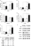
Fig 1. Expression level of autophagy genes in the striatum of mice after 835MHz radiofrequency (RF) exposure.
Striatal RNA extracted control and RF-exposed mice were analysed for the expression of autophagy genes by semiquantitative reverse-transcription PCR and quantitative real-time PCR. (A-E) Quantification of Atg4a, Atg5, Beclin1, LC3A and LC3B mRNA by qRT-PCR and (F) Syto60-stained agarose gel showing differential expression of autophagy genes by sqRT-PCR. The expression values of the striatum of RF-exposed mice were normalized to those of the sham-exposed mice. The relative mRNA levels of Atg4a, Atg5, Becn1, LC3A and LC3B was calculated by normalizing to expression of GAPDH by the 2-ΔΔCt method (n = 5). Table (G) shows the average fold-change. Each bar represents the mean ± SEM of three independent experiments. Statistical significance was evaluated using a t-test: *P<0.05, **P<0.01, *** P< 0.00.
Fig 2
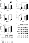
Fig 2. Expression of autophagy genes in the hypothalamus of mice following 835MHz RF exposure.
Hypothalamic RNA extracted from control and RF-exposed mice were analysed for the expression of autophagy genes by semiquantitative RT-PCR and quantitative RT-PCR. (A-E) Quantification of Atg4a, Atg5, Beclin1, LC3A and LC3B mRNA by qRT-PCR and (F) expression level of autophagy genes by sqRT-PCR. The expression values of the hypothalamus of RF-exposed mice were normalized to those of the sham-exposed mice. The relative mRNA levels of Atg4a, Atg5, Beclin1, LC3A and LC3B was calculated by normalizing to expression of GAPDH by the 2-ΔΔCt method (n = 5). Table (G) shows the average fold change. Each bar represents the mean ± SEM of three independent experiments. Statistical significance was evaluated using two tailed t-test: *P<0.05, **P<0.01, *** P< 0.001, **** P< 0.0001.
Fig 3
Fig 3. Western blot analysis of autophagy-related proteins in the striatum and hypothalamus of RF-EMF exposed mice.
Total protein extracted from mice stratum and hypothalamus was subjected to 15% sodium dodecyl sulfate-polyacrylamide gel electrophoresis and resolved proteins were immunoblotted with antibody against LC3B (A) and Beclin1 (B). The intensity of the bands was quantified by densitometry. The protein level of LC3B-II or Beclin1 was normalized relative to LC3B-I or α-tubulin, respectively. Each bar shows mean of three independent experiments with SEM. Statistical significance was evaluated using two tailed t-test (*P<0.05, *** P< 0.001, **** P< 0.0001).
Fig 4
Fig 4. Comparison of intracellular organelles in neuron of hypothalamus and striatum between control and radiofrequency-exposed mice.
(A-D). Representative TEM micrographs of neuronal cell body in hypothalamus and striatum were acquired from control mice and radiofrequency-exposed (4 weeks) mice, respectively. Comparative images showing structural indifference of intracellular organelles between control mice and radiofrequency-exposed mice in neuronal cell body of hypothalamus (A vs. B) and striatum (C vs. D). (E-H) Representative TEM micrographs of neuronal cell body in hypothalamus and striatum were acquired from control mice and radiofrequency-exposed (12 weeks) mice, respectively. In radiofrequency-exposed mice during 12 weeks, many autophagy (AU in F and H) were observed in neuronal cell body of hypothalamus (F) and striatum (H) in difference with that of sham control (E and G). However, most of the mitochondria (M), rough endoplasmic reticulum (RER), and Golgi apparatus (Ga) were maintained the structural integrity as similar with that of control mice. Abbreviations are: Nu, nucleus; M, mitochondria; RER, rough endoplasmic reticulum; Ga, Golgi apparatus and AU, autophagy.

Similar articles

Cited by

References

    1. Christ A, Gosselin MC, Christopoulou M, Kuhn S, Kuster N. Age-dependent tissue-specific exposure of cell phone users. Physics in medicine and biology. 2010;55(7):1767–83. 10.1088/0031-9155/55/7/001 . - DOI - PubMed
    1. Dubreuil D, Jay T, Edeline J. Does head-only exposure to GSM-900 electromagnetic fields affect. Behav Brain Res. 2002;129:203–10. - PubMed
    1. Leszczynski D, Joenväärä S, Reivinen J, Kuokka R. Non-thermal activation of the hsp27/p38MAPK stress pathway by mobile phone radiation in human endothelial cells: Molecular mechanism for cancer- and blood-brain barrier-related effects. Differentiation. 2002;70(2–3):120–9. . - PubMed
    1. Nittby H, Brun A, Eberhardt J, Malmgren L, Persson BR, Salford LG. Increased blood-brain barrier permeability in mammalian brain 7 days after exposure to the radiation from a GSM-900 mobile phone. Pathophysiology: the official journal of the International Society for Pathophysiology / ISP. 2009;16(2–3):103–12. 10.1016/j.pathophys.2009.01.001 . - DOI - PubMed
    1. Salford LG, Brun AE, Eberhardt JL, Malmgren L, Persson BRR. Nerve Cell Damage in Mammalian Brain after Exposure to Microwaves from GSM Mobile Phones. Environmental Health Perspectives. 2003;111(7):881–3. 10.1289/ehp.6039 - DOI - PMC - PubMed

Publication types