. 2016 Jun;79(6):959-69.
doi: 10.1002/ana.24654. Epub 2016 Apr 30.

Mutation-specific effects on thin filament length in thin filament myopathy

Affiliations

Mutation-specific effects on thin filament length in thin filament myopathy

Josine M de Winter et al. Ann Neurol. 2016 Jun.

Abstract

Objective: Thin filament myopathies are among the most common nondystrophic congenital muscular disorders, and are caused by mutations in genes encoding proteins that are associated with the skeletal muscle thin filament. Mechanisms underlying muscle weakness are poorly understood, but might involve the length of the thin filament, an important determinant of force generation.

Methods: We investigated the sarcomere length-dependence of force, a functional assay that provides insights into the contractile strength of muscle fibers as well as the length of the thin filaments, in muscle fibers from 51 patients with thin filament myopathy caused by mutations in NEB, ACTA1, TPM2, TPM3, TNNT1, KBTBD13, KLHL40, and KLHL41.

Results: Lower force generation was observed in muscle fibers from patients of all genotypes. In a subset of patients who harbor mutations in NEB and ACTA1, the lower force was associated with downward shifted force-sarcomere length relations, indicative of shorter thin filaments. Confocal microscopy confirmed shorter thin filaments in muscle fibers of these patients. A conditional Neb knockout mouse model, which recapitulates thin filament myopathy, revealed a compensatory mechanism; the lower force generation that was associated with shorter thin filaments was compensated for by increasing the number of sarcomeres in series. This allowed muscle fibers to operate at a shorter sarcomere length and maintain optimal thin-thick filament overlap.

Interpretation: These findings might provide a novel direction for the development of therapeutic strategies for thin filament myopathy patients with shortened thin filament lengths. Ann Neurol 2016;79:959-969.


Figures

FIGURE 1
FIGURE 1
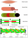
Schematic of the skeletal muscle thin filament. The thin filament is an essential structure in the sarcomere, the smallest contractile unit in skeletal muscle. The actin-based backbone of the thin filament is decorated with proteins that are involved in the regulation of thin filament length and in muscle activation. Tropomyosin and the troponin complex decorate the thin filament along its entire length and are important players in thin filament activation. Actin, nebulin, and leiomodin-3 are involved in both specifying the length of the thin filament and thin filament activation.,, Both cofilin-2 and the kelch proteins—KBTBD13, KLHL40, and KLHL41—are associated with thin filament proteins: cofilin-2 by regulating actin dynamics, KLHL40 and KLHL41 by interacting with nebulin, and KLHL40 by stabilizing leiomodin-3. The role of KBTBD13 has not yet been elucidated, but as it is required for the formation of a functional cullin-3 ubiquitin ligase complex, and as mutations in KBTBD13 result in thin filament myopathy, it is hypothesized to be involved in stabilizing thin filament proteins.
FIGURE 2
FIGURE 2
The force–sarcomere length relation in muscle fibers from thin filament myopathy patients and control (CTRL) subjects. (A) The shape of the force–sarcomere length relation is determined by the amount of overlap between the thick and thin filament. Force at incremental sarcomere lengths is fitted using a 2nd order polynomial. This yields 3 parameters describing the force–sarcomere length relation: the sarcomere length at which maximum force is generated (SLopt), the sarcomere length at which 50% of maximum force is generated (SL50), and the sarcomere length at which the fit crosses the x-axis (SLx). Data shown are from control subjects. The fits represent the mean of individual subjects per genotype. (B, C) NEB (n = 9) and ACTA1 (n = 14) muscle fibers exhibit a downward shift of the descending limb of the force–sarcomere length relation compared to controls. Note that in patients with mutations in NEB (B), this results in a doubling of the force deficit (right y-axis) across the in vivo sarcomere length range (shaded area). (D–I) Muscle fibers from patients with mutations in TPM3 (n = 12), TPM2 (n = 3), TNNT1 (n = 1), KBTBD13 (n = 10), KLHL40 (n = 1), and KLHL41 (n = 1) exhibit a preserved force–sarcomere length relation. Note that the protein products of TPM3 and TPM2 are predominantly expressed in type I fibers and that these fibers exhibited normal force–sarcomere length relations. t tests were performed between genotype and CTRL; p < 0.05 was considered statistically significant (indicated by asterisk). For each biopsy, 3 to 9 muscle fibers were measured.
FIGURE 3
FIGURE 3
Confocal microscopy confirms shorter thin filament lengths in NEB and ACTA1 patients. (A–C) Within the NEB and ACTA1 cohorts, fibers of some patients exhibited force–sarcomere length relation characteristics that were within the range of control (CTRL) values (ie, NEB-1, ACTA1-6), whereas other patients (ie, NEB-2, and ACTA1-1) had values that were markedly below this range (normal range for sarcomere length at which 50% of maximum force is generated [SL50] is in gray). (D–G) Fluorescein isothiocyanate–labeled phalloidin (green) was used to measure thin filament length, and antibodies against both slow and fast myosin heavy chain isoforms (red) were used to measure thick filament length. Thick filament length is normal in muscle fibers from CTRL, NEB, and ACTA1 biopsies, whereas thin filaments are shorter in fibers that revealed a downward shift of the descending limb of the force–sarcomere length relation (NEB-2 and ACTA1-1; n = 4 for CTRL, >5 images per genotype). t tests were performed between genotype and CTRL; p < 0.05 was considered statistically significant.
FIGURE 4
FIGURE 4
A mouse model for nemaline myopathy compensates for shorter thin filaments by adding more sarcomeres in series. (A) Soleus muscle fibers from a mouse model that phenocopies human nemaline myopathy (conditional nebulin knockout, cNeb−/−) revealed a downward shift of the descending limb of the force–sarcomere length relation compared to muscle fibers from cNeb+/+ mice (n = 4 mice/group). (B, C) Confocal microscopy revealed shorter thin filaments in cNeb−/− mice. (D) cNeb−/− mice compensated for the force deficit caused by shorter thin filaments by increasing the number of sarcomeres in series (n = 6 mice/group). (E) At optimal length for force generation, intact soleus muscle of cNeb−/− mice operated at a shorter sarcomere length (SL) than cNeb+/+ mice (n = 20 mice/group). (F) The addition of sarcomeres in series increases the amount of overlap between the thin and thick filaments and enables the fibers with shorter thin filaments to operate at a length closer to their optimal length. t tests were performed between cNeb+/+ and cNeb−/− mice, p < 0.05 was considered statistically significant (indicated by asterisk). SLopt 5 SL at which maximum force is generated; SL50 = SL at which 50% of maximum force is generated; SLx = SL at which the fit crosses the x-axis.

References

    1. Colombo I, Scoto M, Manzur AY, et al. Congenital myopathies: natural history of a large pediatric cohort. Neurology. 2015;84:28–35. - PMC - PubMed
    1. Sanoudou D, Beggs AH. Clinical and genetic heterogeneity in nemaline myopathy—a disease of skeletal muscle thin filaments. Trends Mol Med. 2001;7:362–368. - PubMed
    1. Sambuughin N, Yau KS, Olivé M, et al. Dominant mutations in KBTBD13, a member of the BTB/kelch family, cause nemaline myopathy with cores. Am J Hum Genet. 2010;87:842–847. - PMC - PubMed
    1. Ravenscroft G, Miyatake S, Lehtokari V-L, et al. Mutations in KLHL40 are a frequent cause of severe autosomal-recessive nemaline myopathy. Am J Hum Genet. 2013;93:6–18. - PMC - PubMed
    1. Gupta VA, Ravenscroft G, Shaheen R, et al. Identification of KLHL41 mutations implicates BTB-kelch-mediated ubiquitination as an alternate pathway to myofibrillar disruption in nemaline myopathy. Am J Hum Genet. 2013;93:1108–1117. - PMC - PubMed