

Implementation of full/half bowtie filter models in a commercial treatment planning system for kilovoltage cone-beam CT dose estimations
- PMID: 27074480
- PMCID: PMC5874958
- DOI: 10.1120/jacmp.v17i2.5988
Implementation of full/half bowtie filter models in a commercial treatment planning system for kilovoltage cone-beam CT dose estimations
Abstract
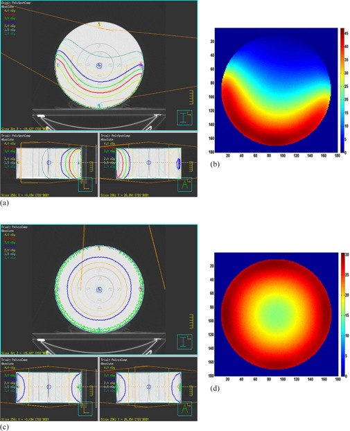
The purpose of this study was to implement full/half bowtie filter models in a com-mercial treatment planning system (TPS) to calculate kilovoltage (kV) cone-beam CT (CBCT) doses of Varian On-Board Imager (OBI) kV X-ray imaging system. The full/half bowtie filter models were created as compensators in Pinnacle TPS using MATLAB software. The physical profiles of both bowtie filters were imported and hard-coded in the MATLAB system. Pinnacle scripts were written to import bowtie filter models into Pinnacle treatment plans. Bowtie filter-free kV X-ray beam models were commissioned and the bowtie filter models were validated by analyzing the lateral and percent-depth-dose (PDD) profiles of anterior/posterior X-ray beams in water phantoms. A CT dose index (CTDI) phantom was employed to calculate CTDI and weighted CTDI values for pelvis and pelvis-spotlight CBCT protocols. A five-year-old pediatric anthropomorphic phantom was utilized to evaluate absorbed and effective doses (ED) for standard and low-dose head CBCT protocols. The CBCT dose calculation results were compared to ion chamber (IC) and Monte Carlo (MC) data for the CTDI phantom and MOSFET and MC results for the pediatric phantom, respectively. The differences of lateral and PDD profiles between TPS calculations and IC measurements were within 6%. The CTDI and weighted CTDI values of the TPS were respectively within 0.25 cGy and 0.08 cGy compared to IC measurements. The absorbed doses ranged from 0 to 7.22 cGy for the standard dose CBCT and 0 to 1.56 cGy for the low-dose CBCT. The ED values were found to be 36-38 mSv and 7-8 mSv for the standard and low-dose CBCT protocols, respectively. This study demonstrated that the established full/half bowtie filter beam models can produce reasonable dose calculation results. Further study is to be performed to evaluate the models in clinical situations.
Figures


References
-
- Islam MK, Purdie TG, Norrlinger BD, et al. Patient dose from kilovoltage cone beam computed tomography imaging in radiation therapy. Med Phys. 2006;33(6):1573–82. - PubMed
-
- Kan MWK, Leung LHT, Wong W, Lam N. Radiation dose from cone beam computed tomography for image‐guided radiation therapy. Int J Radiat Oncol Biol Phys. 2008;70(1):272–79. - PubMed
-
- Song WY, Kamath S, Ozawa S, et al. A dose comparison study between XVI and OBI CBCT systems. Med Phys. 2008;35(2):480–86. - PubMed
-
- Alaei P, Gerbi BJ, Geise RA. Generation and use of photon energy deposition kernels for diagnostic quality x rays. Med Phys. 1999;26(8):1687–97. - PubMed
-
- Alaei P, Gerbi BJ, Geise RA. Evaluation of a model‐based treatment planning system for dose computations in the kilovoltage energy range. Med. Phys. 2000;27(12):2821–26. - PubMed
MeSH terms
LinkOut - more resources
Full Text Sources
Other Literature Sources
