. 2016 Apr 12;110(7):1499-1509.
doi: 10.1016/j.bpj.2016.02.037.

Effect of Oxidative Damage on the Stability and Dimerization of Superoxide Dismutase 1

Affiliations

Effect of Oxidative Damage on the Stability and Dimerization of Superoxide Dismutase 1

Drazen Petrov et al. Biophys J. .

Abstract

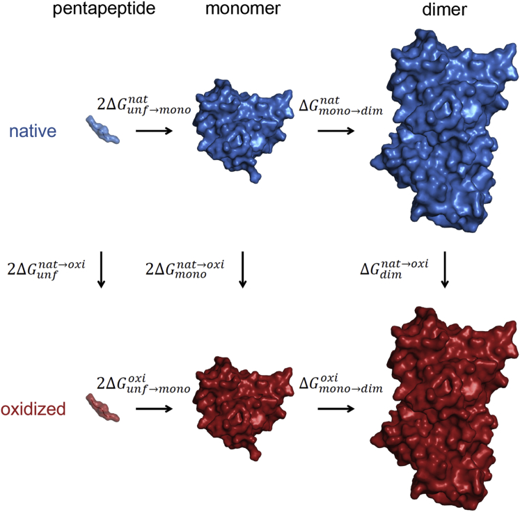
During their life cycle, proteins are subject to different modifications involving reactive oxygen species. Such oxidative damage to proteins may lead to the formation of insoluble aggregates and cytotoxicity and is associated with age-related disorders including neurodegenerative diseases, cancer, and diabetes. Superoxide dismutase 1 (SOD1), a key antioxidant enzyme in human cells, is particularly susceptible to such modifications. Moreover, this homodimeric metalloenzyme has been directly linked to both familial and sporadic amyotrophic lateral sclerosis (ALS), a devastating, late-onset motor neuronal disease, with more than 150 ALS-related mutations in the SOD1 gene. Importantly, oxidatively damaged SOD1 aggregates have been observed in both familial and sporadic forms of the disease. However, the molecular mechanisms as well as potential implications of oxidative stress in SOD1-induced cytotoxicity remain elusive. In this study, we examine the effects of oxidative modification on SOD1 monomer and homodimer stability, the key molecular properties related to SOD1 aggregation. We use molecular dynamics simulations in combination with thermodynamic integration to study microscopic-level site-specific effects of oxidative "mutations" at the dimer interface, including lysine, arginine, proline and threonine carbonylation, and cysteine oxidation. Our results show that oxidative damage of even single residues at the interface may drastically destabilize the SOD1 homodimer, with several modifications exhibiting a comparable effect to that of the most drastic ALS-causing mutations known. Additionally, we show that the SOD1 monomer stability decreases upon oxidative stress, which may lead to partial local unfolding and consequently to increased aggregation propensity. Importantly, these results suggest that oxidative stress may play a key role in development of ALS, with the mutations in the SOD1 gene being an additional factor.


Figures

Figure 1
Figure 1
Summary of the studied oxidative modifications: 1) lysine to aminoadipic semialdehye (carbonylation), 2) proline and arginine to glutamic semialdehyde (carbonylation), 3) threonine to 2-amino-3-ketobutyric acid (carbonylation), and 4) cysteine to cysteic acid (oxidation) modifications. Note that the chemical structures are shown in their neutral forms, whereas in simulations the protonation states of all residues correspond to the most abundant state in solution at neutral pH. To see this figure in color, go online.
Figure 2
Figure 2
Thermodynamic cycle. The relative free-energy difference between the native and the oxidized SOD1 dimer/monomer stability was determined by evaluating the changes in the free energy of an alchemical modification from a native residue of interest to its oxidized form (ΔGdimnatoxi for an alchemical modification in the dimer, ΔGmononatoxi for an alchemical modification in the monomer, and ΔGunfnatoxi for an alchemical modification in the pentapeptide, i.e., unfolded state), according to the above thermodynamic cycle and Eqs. 1 and 2. To see this figure in color, go online.
Figure 3
Figure 3
Typical <∂H/∂λ> curves. Ensemble average are derivative of the system Hamiltonian with respect to λ shown as a function of λ: (A) LYS9, (B) THR54, and (C) ARG115. Multiple points at a given λ come from independent simulations, with the average curves shown in solid lines.
Figure 4
Figure 4
Local unfolding as a consequence of oxidative damage of CYS6. While the affected residue is buried in its native form (A), it flips to the protein surface and becomes solvent exposed upon oxidative modification, additionally destabilizing local β-sheet structure (B). Inset: A close-up picture of the affected residue is shown. To see this figure in color, go online.
Figure 5
Figure 5
Impact on SOD1 dimer stability of oxidative damage of residues at the homodimer interface. (A) Changes in free energy of homodimer formation with error bars calculated by block averaging and standard propagation of error are shown. Location of the studied interface residues and effects of their oxidative modifications on the SOD1 dimer are shown in the context of (B) one of the monomers (view at the interface) and (C) SOD1 dimer. The color code for the protein structure: interface (white) and rest of the protein (gray). To see this figure in color, go online.
Figure 6
Figure 6
Impact on SOD1 monomer stability of oxidative damage of residues at the homodimer interface. (A) Changes in free energy of folding with error bars calculated by block averaging and propagation of error are shown. (B) Location of the studied interface residues and effects of their oxidative modifications on the SOD1 monomer (view at the interface) are shown. The color code for the protein structure: interface (white) and rest of the protein (gray). To see this figure in color, go online.
Figure 7
Figure 7
Microscopic analysis of the effects of oxidative damage on residues at the SOD1-homodimer interface: (A) root-mean-square fluctuations averaged over all atoms of a given amino acid; (B) root-mean-square deviation from the native structure; (C) interaction energy between a given residue and its surrounding as defined by the force field and simulation parameters used; (D) number of hydrogen bonds formed by a given residue; and (E) number of favorable (positive-negative) charge-charge interactions between a given residue and its surrounding in the native state, within 0.3 nm (first bar) and 0.6 nm (second bar). The bars in A through D represent averages over native and oxidized end-state simulations, whereas error bars in all panels show the standard deviation. To see this figure in color, go online.

References

    1. Levine R.L., Stadtman E.R. Oxidative modification of proteins during aging. Exp. Gerontol. 2001;36:1495–1502. - PubMed
    1. Nyström T. Role of oxidative carbonylation in protein quality control and senescence. EMBO J. 2005;24:1311–1317. - PMC - PubMed
    1. Dalle-Donne I., Rossi R., Milzani A. Biomarkers of oxidative damage in human disease. Clin. Chem. 2006;52:601–623. - PubMed
    1. Davies K.J. Degradation of oxidized proteins by the 20S proteasome. Biochimie. 2001;83:301–310. - PubMed
    1. Grune T., Jung T., Davies K.J. Decreased proteolysis caused by protein aggregates, inclusion bodies, plaques, lipofuscin, ceroid, and ‘aggresomes’ during oxidative stress, aging, and disease. Int. J. Biochem. Cell Biol. 2004;36:2519–2530. - PubMed

Publication types

Substances

LinkOut - more resources