. 2016 Apr 12;110(7):1615-1624.
doi: 10.1016/j.bpj.2016.03.003.

Emergent Global Contractile Force in Cardiac Tissues

Affiliations

Emergent Global Contractile Force in Cardiac Tissues

Meghan B Knight et al. Biophys J. .

Abstract

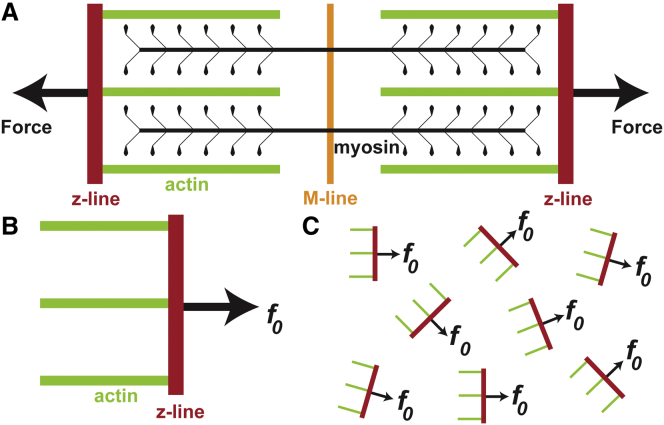
The heart is a complex organ whose structure and function are intricately linked at multiple length scales. Although several advancements have been achieved in the field of cardiac tissue engineering, current in vitro cardiac tissues do not fully replicate the structure or function necessary for effective cardiac therapy and cardiotoxicity studies. This is partially due to a deficiency in current understandings of cardiac tissue organization's potential downstream effects, such as changes in gene expression levels. We developed a novel (to our knowledge) in vitro tool that can be used to decouple and quantify the contribution of organization and associated downstream effects to tissue function. To do so, cardiac tissue monolayers were designed into a parquet pattern to be organized anisotropically on a local scale, within a parquet tile, and with any desired organization on a global scale. We hypothesized that if the downstream effects were muted, the relationship between developed force and tissue organization could be modeled as a sum of force vectors. With the in vitro experimental platforms of parquet tissues and heart-on-a-chip devices, we were able to prove this hypothesis for both systolic and diastolic stresses. Thus, insight was gained into the relationship between the generated stress and global myofibril organization. Furthermore, it was demonstrated that the developed quantitative tool could be used to estimate the changes in stress production due to downstream effects decoupled from tissue architecture. This has the potential to elucidate properties coupled to tissue architecture, which change force production and pumping function in the diseased heart or stem cell-derived tissues.


Figures

Figure 1
Figure 1
The basic model approximation of a sarcomere. (A) A sarcomere is a dipole in which force is produced perpendicular to the z-line and parallel to actin the fibrils. (B) For the model, the sarcomere is simplified to have a single force vector. (C) Within a cardiac tissue, each sarcomere will produce an average force f0 parallel to its actin fibrils. To see this figure in color, go online.
Figure 2
Figure 2
Locally organized globally disorganized parquet tissues. (A and B) Isotropic FN. (C and D) The parquet FN pattern. (E and F) The globally aligned anisotropic FN pattern. (A, C, and E) Local scale (96 × 96 μm); (B, D, and F) global scale (1 × 1 mm). (GJ) Immunostain images of actin fibrils (green), sarcomeric z-disks (red), and nuclei (blue) for isotropic tissue (G), globally aligned anisotropic tissue (H), tissues within a parquet tile (I), and tissue from a border region of multiple parquet tiles (J) like the one indicated with the white rectangle in (D). (K) Actin fibril OOP on the global and local scale for each tissue type. Error bars represent the standard deviation of the data. Significance was tested within each group (global, local), and was p < 0.05 unless labeled “No. Sig.” (Table S1). (L) Log twofold change for globally aligned anisotropic (N = 3) and parquet (N = 5) samples normalized to isotropic (N = 3) samples (Table S2). Scale bars, 10 μm (A, C, and E); 100 μm (B, D, and F); 25 μm (GJ). To see this figure in color, go online.
Figure 3
Figure 3
Contractility experiments using heart-on-a-chip assays. (A and B) Sample video frames for the three tissue types showing diastole and systole, respectively. Pink outlines show original film length, orange bars track films’ x-projections, and yellow dashed lines provide a visual cue for the change in contraction between diastole and systole (scale bars, 1 mm). (C) Example stress traces of raw data for anisotropic (top, purple line), parquet (middle, blue line), and isotropic (bottom, red line) tissue from a contractility experiment. (D) Schematic of stresses measured from each stress trace. (E) Comparison of stresses for anisotropic (N = 28), parquet (N = 22), and isotropic (N = 27) tissue versus the basic model prediction (2nd, green bar). Significance was assumed if p < 0.05 (Table S3). Within systolic and diastolic stress, pairwise comparisons were significant unless labeled with “No. Sig.” Within active stress, pairwise statistical significance is indicated by (). To see this figure in color, go online.
Figure 4
Figure 4
Experiment versus model for varying global organization. (A) Various parquet tiles used for tissue design. (B) FN pattern OOP and global actin OOP from all parquet tissues (Table S4). Error bars: standard deviation. (C) Systolic and diastolic stresses as a function of OOP predicted by the model based on calculated parameter σ0 (thick, green line). The mean systolic stress for each parquet tissue type (light, blue circles) falls within the 95% confidence limit of the model (light brown, shading). The mean systolic and diastolic stresses of the isotropic tissues (dark, red circle) falls outside the confidence limits of the model. Error bars (black) represent standard error of the mean (mean ± SD for all stresses in Table S5). To see this figure in color, go online.

References

    1. Gregorio C.C., Antin P.B. To the heart of myofibril assembly. Trends Cell Biol. 2000;10:355–362. - PubMed
    1. Helm P., Beg M.F., Winslow R.L. Measuring and mapping cardiac fiber and laminar architecture using diffusion tensor MR imaging. Ann. N. Y. Acad. Sci. 2005;1047:296–307. - PubMed
    1. Ehler E. Springer; New York: 2015. Cardiac Cytoarchitecture: How to Maintain a Working Heart.
    1. Helm P.A., Younes L., Winslow R.L. Evidence of structural remodeling in the dysynchronous failing heart. Circ. Res. 2006;98:125–132. - PubMed
    1. Ahmad F., Seidman J.G., Seidman C.E. The genetic basis for cardiac remodeling. Annu. Rev. Genomics Hum. Genet. 2005;6:185–216. - PubMed

Publication types