. 2016 Aug;41(9):2399-410.
doi: 10.1038/npp.2016.52. Epub 2016 Apr 14.

Cocaine-Induced Synaptic Alterations in Thalamus to Nucleus Accumbens Projection

Affiliations

Cocaine-Induced Synaptic Alterations in Thalamus to Nucleus Accumbens Projection

Peter A Neumann et al. Neuropsychopharmacology. 2016 Aug.

Abstract

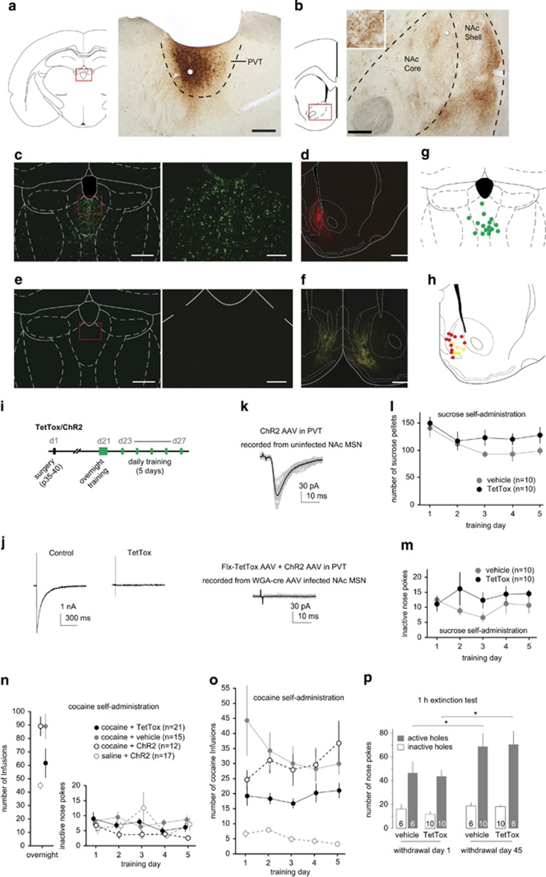
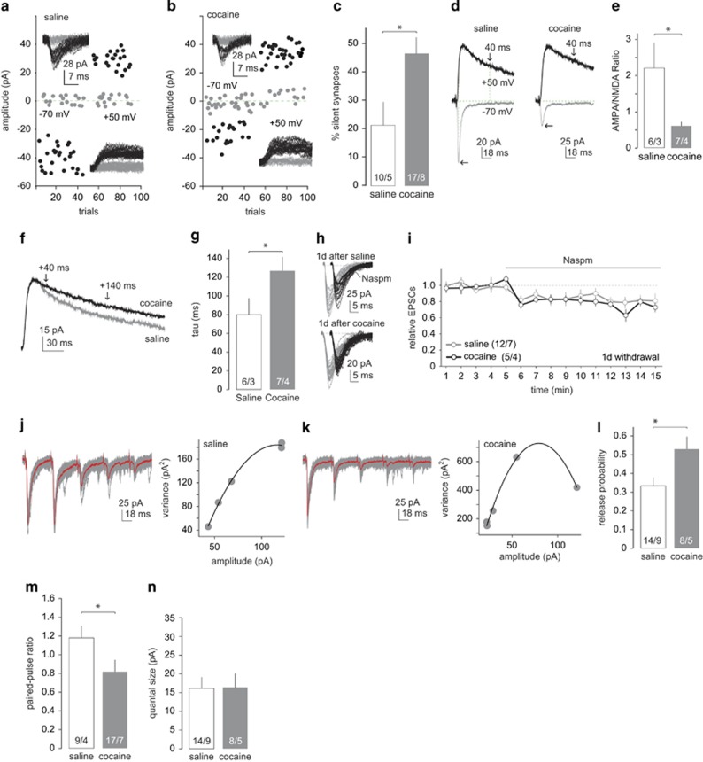
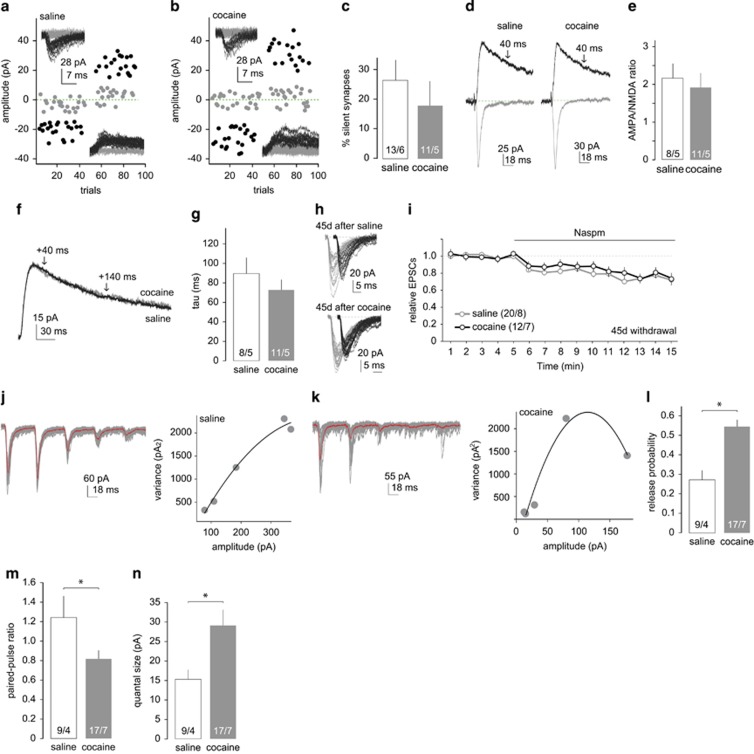
Exposure to cocaine induces addiction-associated behaviors partially through remodeling neurocircuits in the nucleus accumbens (NAc). The paraventricular nucleus of thalamus (PVT), which projects to the NAc monosynaptically, is activated by cocaine exposure and has been implicated in several cocaine-induced emotional and motivational states. Here we show that disrupting synaptic transmission of select PVT neurons with tetanus toxin activated via retrograde trans-synaptic transport of cre from NAc efferents decreased cocaine self-administration in rats. This projection underwent complex adaptations after self-administration of cocaine (0.75 mg/kg/infusion; 2 h/d × 5 d, 1d overnight training). Specifically, 1d after cocaine self-administration, we observed increased levels of AMPA receptor (AMPAR)-silent glutamatergic synapses in this projection, accompanied by a decreased ratio of AMPAR-to-NMDA receptor (NMDAR)-mediated EPSCs. Furthermore, the decay kinetics of NMDAR EPSCs was significantly prolonged, suggesting insertion of new GluN2B-containing NMDARs to PVT-to-NAc synapses. After 45-d withdrawal, silent synapses within this projection returned to the basal levels, accompanied by a return of the AMPAR/NMDAR ratio and NMDAR decay kinetics to the basal levels. In amygdala and infralimbic prefrontal cortical projections to the NAc, a portion of cocaine-generated silent synapses becomes unsilenced by recruiting calcium-permeable AMPARs (CP-AMPARs) after drug withdrawal. However, the sensitivity of PVT-to-NAc synapses to CP-AMPAR-selective antagonists was not changed after withdrawal, suggesting that CP-AMPAR trafficking is not involved in the evolution of cocaine-generated silent synapses within this projection. Meanwhile, the release probability of PVT-to-NAc synapses was increased after short- and long-term cocaine withdrawal. These results reveal complex and profound alterations at PVT-to-NAc synapses after cocaine exposure and withdrawal.


Figures

Figure 1
Figure 1
Disrupting synaptic transmission in PVT neurons that project to the NAc shell decreases cocaine self-administration. (a) Diagram (left) and image (right) showing BDA tracer injection to the PVT. (b) Diagram (left) and image (shell) showing the NAc shell containing neural fibers arising from BDA-traced PVT neurons. The square region, shown at higher magnification in the insert, illustrates axon varicosities. Scale bar in panel a represents 250 μm for panels a and b, and 25 μm for inset. (c) Image of an example coronal slice showing EGFP-TetTox expression in PVT neurons after intra-PVT injection of Flx-TetTox-expressing AAV and intra-NAc shell injection of WGA-cre-expressing AAV. Scale bar, left 500 μm, right 150 μm. (d) Example of image showing mCherry fluorescence indicating the expression of WGA-cre in the NAc shell. Scale bar 500 μm. (e) Example of image showing lack of EGFP-TetTox expression in the PVT after intra-PVT injection of Flx-TetTox-expressing AAV and intra-NAc shell injection of control (without WGA-cre component) YFP-expressing AAV. Scale bar, left 500 μm, right 150 μm. (f) Example of image showing the expression of YFP after intra-NAc shell injection of YFP-expressing control AAV vehicle (without WGA-cre). Scale bar 500 μm. (g) Diagram showing the injection sites (green dots) of Flx-TetTox-expressing AAV in rats for self-administration tests. (h) Diagram showing the injection sites of WGA-cre AAVs (red) and YFP AAV vehicle controls (yellow) in the NAc. (i) Experimental timeline showing when rats received stereotaxic injections of ChR2- and TetTox-expressing AAVs and underwent self-administration training. (j) Example of postsynaptic responses evoked from an uninfected, control presynaptic neuron (left) and from a presynaptic neuron expressing TetTox (right) in hippocampal cultures. (k) Upper: Example of optogenetically evoked traces in a control NAc MSN from a rat with intra-PVT expression of ChR2 alone; lower: Example of optogenetically evoked traces in a WGA-cre-expressing NAc MSN from a rat with intra-PVT expression of ChR2 and Flx-TetTox together. The black trace is the average of individual traces in each condition. (l) Rats with intra-PVT injection of TetToxAAV and intra-NAc injection of WGA-cre AAV exhibited normal sucrose self-administration. (m) Rats with intra-PVT injections of Flx-TetTox AAV and intra-NAc injections of WGA-cre AAV also had equivalent inactive nose poke behavior. (n) After an overnight session of cocaine/saline self-administration training (left), cocaine-exposed rats exhibited equally low levels of nosepokes to inactive holes during subsequent 2 h/d × 5 d training (right). (o) Rats with intra-PVT injection of Flx-TetTox-expressing AAV and intra-NAc injection of WGA-cre AAV exhibited decreased cocaine self-administration compared with the control and vehicle groups. (p) Summary showing that, after prolonged withdrawal from cocaine self-administration, rats with intra-PVT injection of Flx-TetTox-expressing AAV and intra-NAc injection of WGA-cre AAV exhibited similar incubation of cue-induced cocaine craving as rats without TetTox-cre manipulation. n values labeled in key and bars. *p<0.01.
Figure 2
Figure 2
Alterations in PVT-to-NAc synaptic transmission 1–2 d after cocaine self-administration. (a, b) Example of minimal stimulation EPSCs and their trials from PVT-to-NAC projections 1–2 d after saline (a) and cocaine (b) self-administration. (c) Summary showing increased levels of silent synapses in the PVT-to-NAc projection 1–2 d after cocaine self-administration. (d) Example of EPSCs evoked at −70 mV and +50 mV from PVT-to-NAc synapses 1–2 d after saline or cocaine self-administration. (e) Summary showing decreased AMPAR/NMDAR ratios at PVT-to-NAc synapses. (f) Normalized EPSCs from example of MSNs 1–2 d after saline or cocaine self-administration. Arrows indicate the range for measuring decay kinetics of NMDAR-mediated EPSCs. (g) Summary showing that the decay kinetics of NMDAR-mediated EPSCs was prolonged 1–2 d after cocaine self-administration. (h, i) Example of traces (h) and summary (i) showing similar Naspm-induced inhibition of the peak amplitude of EPSCs at PVT-to-NAc synapses 1–2 d after saline or cocaine self-administration. (j, k) Example of EPSCs and their variance–mean hyperbolic fittings at PVT-to-NAc synapses upon 5-pulse 20 Hz stimulation 1–2 d after saline (j) or cocaine (k) self-administration. (l) Calculated probability of presynaptic release at PVT-to-NAc synapses 1–2 d after saline and cocaine self-administration. (m) Summary showing decreased paired-pulse ratio following saline and cocaine self-administration. (n) Summary showing equivalent quantal sizes following saline and cocaine self-administration. n/m (cell/animal) values labeled in key and bars. *p<0.05.
Figure 3
Figure 3
Alterations in PVT-to-NAc synaptic transmission 45 d after cocaine self-administration. (a, b) Example minimal stimulation EPSCs and their trials from PVT-to-NAc projections 45 d after saline (a) and cocaine (b) self-administration. (c) Summary showing similar levels of silent synapses in the PVT-to-NAc projection 45 d after cocaine or saline self-administration. (d) Example of EPSCs evoked at −70 mV and +50 mV from PVT-to-NAc synapses 45 d after saline or cocaine self-administration. (e) Summary showing similar AMPAR/NMDAR ratios at PVT-to-NAc synapses 45 d after saline or cocaine self-administration. (f) Normalized EPSCs from example of MSNs 45 d after saline or cocaine self-administration. Arrows indicate the range for measuring decay kinetics of NMDAR-mediated EPSCs. (g) Summary showing similar decay kinetics of NMDAR-mediated EPSCs 45 d after saline or cocaine self-administration. (h, i) Example of traces (h) and summary (i) showing similar Naspm-induced inhibition of the peak amplitude of EPSC at PVT-to-NAc synapses 45 d after saline or cocaine self-administration. (j, k) Example of EPSCs and their variance–mean hyperbolic fittings at PVT-to-NAc synapses upon 5-pulse 20 Hz stimulation 45 d after saline (j) or cocaine (k) self-administration. (l) Calculated probability of presynaptic release at PVT-to-NAc synapses 45 d after saline and cocaine self-administration. (m) Summary showing decreased paired-pulse ratio following cocaine self-administration. (n) Summary showing increased quantal size following 45 d withdrawal from cocaine self-administration. n/m (cell/animal) values labeled in key and bars. *p<0.05.

References

    1. Auer S, Sturzebecher AS, Juttner R, Santos-Torres J, Hanack C, Frahm S et al (2010). Silencing neurotransmission with membrane-tethered toxins. Nat Methods 7: 229–236. - PubMed
    1. Berendse HW, Groenewegen HJ (1990). Organization of the thalamostriatal projections in the rat, with special emphasis on the ventral striatum. J Comp Neurol 299: 187–228. - PubMed
    1. Braz JM, Rico B, Basbaum AI (2002). Transneuronal tracing of diverse CNS circuits by Cre-mediated induction of wheat germ agglutinin in transgenic mice. Proc Natl Acad Sci USA 99: 15148–15153. - PMC - PubMed
    1. Britt JP, Benaliouad F, McDevitt RA, Stuber GD, Wise RA, Bonci A (2012). Synaptic and behavioral profile of multiple glutamatergic inputs to the nucleus accumbens. Neuron 76: 790–803. - PMC - PubMed
    1. Brown EE, Robertson GS, Fibiger HC (1992). Evidence for conditional neuronal activation following exposure to a cocaine-paired environment: role of forebrain limbic structures. J Neurosci 12: 4112–4121. - PMC - PubMed

Publication types