. 2016 Apr 13:14:30.
doi: 10.1186/s12915-016-0250-9.

Muscle cell identity requires Pax7-mediated lineage-specific DNA demethylation

Affiliations

Muscle cell identity requires Pax7-mediated lineage-specific DNA demethylation

Elvira Carrió et al. BMC Biol. .

Abstract

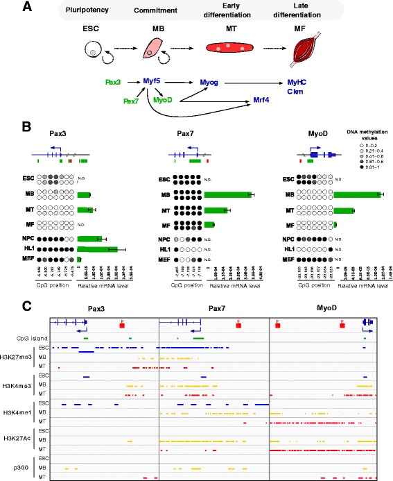
Background: Skeletal muscle stem cells enable the formation, growth, maintenance, and regeneration of skeletal muscle throughout life. The regeneration process is compromised in several pathological conditions, and muscle progenitors derived from pluripotent stem cells have been suggested as a potential therapeutic source for tissue replacement. DNA methylation is an important epigenetic mechanism in the setting and maintenance of cellular identity, but its role in stem cell determination towards the myogenic lineage is unknown. Here we addressed the DNA methylation dynamics of the major genes orchestrating the myogenic determination and differentiation programs in embryonic stem (ES) cells, their Pax7-induced myogenic derivatives, and muscle stem cells in proliferating and differentiating conditions.

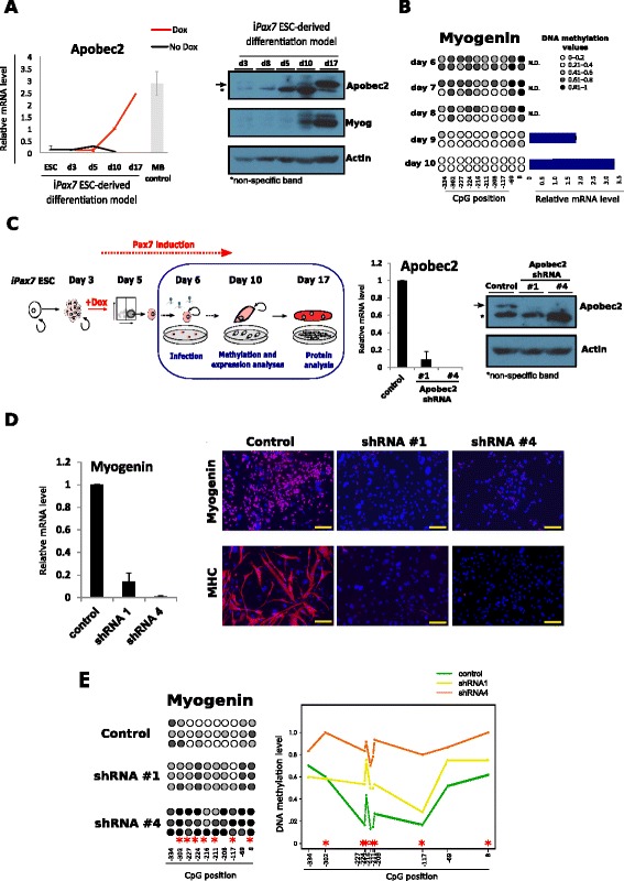
Results: Our data showed a common muscle-specific DNA demethylation signature required to acquire and maintain the muscle-cell identity. This specific-DNA demethylation is Pax7-mediated, and it is a prime event in muscle stem cells gene activation. Notably, downregulation of the demethylation-related enzyme Apobec2 in ES-derived myogenic precursors reduced myogenin-associated DNA demethylation and dramatically impaired the expression of differentiation markers and, ultimately, muscle differentiation.

Conclusions: Our results underscore DNA demethylation as a key mechanism driving myogenesis and identify specific Pax7-mediated DNA demethylation signatures to acquire and maintain the muscle-cell identity. Additionally, we provide a panel of epigenetic markers for the efficient and safe generation of ES- and induced pluripotent stem cell (iPS)-derived myogenic progenitors for therapeutic applications.

Keywords: Apobec2; Cellular identity; DNA methylation; Epimarkers; Myogenesis; Pax7-induced ESCs.


Figures

Fig. 1
Fig. 1
Epigenetic profile of myogenic genes harbouring a CpG island-promoter during myogenic differentiation. a Diagram of the myogenic differentiation model and the main genes driving myogenesis. CpG-rich and CpG-poor promoter genes are indicated in green and in blue, respectively. b Scheme of the Pax3, Pax7 and MyoD genes and their associated CGIs (green) and enhancer regions (red). DNA methylation and expression of each gene were analysed in two biological duplicates of ESCs, myoblasts (MB), myotubes (MT), myofibres (MF), and NPC, HL1 and MEF cell lines. Each circle represents a CpG dinucleotide, and its position relative to the TSS is indicated below. The colour gradient represents the level of methylation indicated in the legend. Bar charts show gene expression values measured by qRT-PCR and normalized by 18S expression (n = 2, mean ± SD). N.D. means non detectable. c Histone marks distribution and p300 binding in ESC (blue), MB (yellow) and MT (red) obtained from ENCODE Project and Dynlatch’s lab [38]. CGIs CpG islands, ESCs embryonic stem cells, NPCs neuronal precursor cells, HL1 cardiomyocytes, MEFs mouse embryonic fibroblasts, TSS transcription start site
Fig. 2
Fig. 2
Epigenetic profile of muscle-specific CpG-poor promoter genes during myogenesis. a Scheme of the Myog, Mrf4, Ckm and Myh1, 4 and 8 loci. Regions analysed by sodium bisulphite sequencing are indicated in red. Two biological replicates were performed for ESCs, myoblasts (MB), myotubes (MT) and myofibres (MF) and in NPC, HL1 and MEF cell lines. Each circle represents a CpG dinucleotide and its position relative to the TSS is indicated below. The colour gradient represents the level of methylation indicated in the legend. Bar charts show gene expression values measured by qRT-PCR and normalized by 18S expression (n = 2, mean ± SD). N.D. means non detectable. b Histone marks distribution and p300 binding in ESC (blue), MB (yellow) and MT (red) obtained from ENCODE Project and Dynlatch’s lab [38]. ESCs embryonic stem cells, NPCs neuronal precursor cells, HL1 cardiomyocytes, MEFs mouse embryonic fibroblasts, TSS transcription start site
Fig. 3
Fig. 3
Pax7-engineered ESCs efficiently acquired the myogenic DNA methylation profile. a Experimental scheme of the differentiation protocol to generate ES-derived myogenic progenitors using iPax7 mES cells (left). Pax7 induction assessed by qRT-PCR (right). b Expression profiles of markers of pluripotency (Pou5f1 and Dppa4), muscle lineage commitment (Pax3, Myf5 and MyoD) and muscle differentiation (Myog, Ckm and Myh8) were measured by qRT-PCR at the successive time points of iPax7 ESC-derived myogenic model and normalized by Gapdh expression (n = 3, mean ± SD). c DNA methylation dynamics of Pax3, Myf5 and MyoD enhancers and CpG-poor promoters of muscle-specific genes (Myf5 Myog, Ckm and Myh8) and pluripotency genes (Pou5f1 and Dppa4) during Pax7-induced myogenesis (+Dox shown in red and -Dox shown in black). The results of three biological replicates are shown. Each circle represents a CpG dinucleotide and the colour gradient represents the level of methylation indicated in the legend and assessed by sodium bisulphite sequencing. Demethylation waves occurring during the Pax7-induced myogenesis are denoted by red dotted boxes. ESCs embryonic stem cells, Dox doxycycline; iPax7 inducible Pax7
Fig. 4
Fig. 4
Impaired myogenic progression and differentiation and reduced DNA demethylation in Apobec2 knockdown myogenic precursors. a Time course of Apobec2 expression during differentiation of iPax7 ES cells in presence or absence of Pax7, which was induced by adding doxycycline (Dox) to the culture media. Apobec2 expression was measured by qRT-PCR (left), and by western blotting (WB) (right). Apobec2 mRNA expression was normalized by Gapdh expression (n = 3, mean ± SD) and as a reference the expression level in primary myoblasts (MB control) was measured (n = 2, mean ± SD). Myog and actin WB were used as controls for myogenic differentiation and equal loading, respectively. b DNA methylation and gene expression analyses of Myogenin were performed in a daily time course after Dox induction in the inducible myogenic model (n = 2). Each circle represents a CpG dinucleotide and its distance to the gene TSS is indicated below. The colour gradient represents the methylation level indicated in the legend. N.D. means non detectable. c Scheme of the experimental approach followed to knockdown Apobec2 in the inducible Pax7 model (left). Gene expression and protein level to evaluate Apobec2 knockdown efficiency using two independent clones were compared to control vector (right) (n = 3, mean ± SD; representative Apobec2 WB with the corresponding actin loading control). d Myogenin expression was measured in Pax7-induced myogenic precursors infected with Apobec2 shRNAs 4 days upon lentiviral transduction (right) (n = 3, mean ± SD). Representative images of Pax7-induced myogenic precursors infected with Apobec2 shRNAs under differentiation conditions stained with Myogenin and MHC antibodies. Scale bar: 100 μm. e DNA methylation analysis by sodium bisulphite sequencing of the Myogenin promoter in Pax7 ES-derived myogenic precursors transduced with Apobec2 shRNA1 and shRNA4 in three biological replicates. Each circle represents a CpG dinucleotide and its distance to the gene TSS is indicated below. The colour gradient represents the level of methylation indicated in the legend. Statistical significance with respect to the control samples is indicated with * for p-value < 0.05 applying the Kruskal-Wallis test. ESC embryonic stem cell, TSS transcription start site, shRNA short hairpin RNA
Fig. 5
Fig. 5
Model for the epigenetic control of muscle-specific gene expression during myogenesis. Diagram proposing a simplified model where myogenic transcription factors would activate muscle genes by modulating both the recruitment, direct or indirect, of DNA demethylases to reduce DNA methylation levels and chromatin compaction, and histone modifiers/chromatin remodelers to activate transcription of muscle genes. In proliferating myoblasts, the differentiation genes would be kept in a silenced/poised transcriptional state by PRC2 and HDACs complexes, whereas in myotubes they would be expressed upon the recruitment of chromatin activating complexes. Abbreviations: DDMs DNA demethylases, HATs histone acetyltransferases, HDACs histone deacetylases, HMTs histone methyltransferases, PRC2 Polycomb repressive complex 2, SWI/SNF SWI/SNF ATP-dependent chromatin remodelling complex, YY1 Yin and Yang 1

References

    1. Mohn F, Schübeler D. Genetics and epigenetics: stability and plasticity during cellular differentiation. Trends in Genetics. 2009;25(3):129–36. doi: 10.1016/j.tig.2008.12.005. - DOI - PubMed
    1. Berdasco M, Esteller M. DNA methylation in stem cell renewal and multipotency. Stem Cell Res Ther. 2011;2(5):42. doi: 10.1186/scrt83. - DOI - PMC - PubMed
    1. Cedar H, Bergman Y. Programming of DNA methylation patterns. Annu Rev Biochem. 2012;2012(81):97–117. doi: 10.1146/annurev-biochem-052610-091920. - DOI - PubMed
    1. Smith ZD, Meissner A. DNA methylation: roles in mammalian development. Nat Rev Genet. 2013;14(3):204–20. doi: 10.1038/nrg3354. - DOI - PubMed
    1. Jones PA. Functions of DNA methylation: islands, start sites, gene bodies and beyond. Nat Rev Genet. 2012;13(7):484–92. doi: 10.1038/nrg3230. - DOI - PubMed

Publication types

Substances