. 2016 Jun;44(4):502-35.
doi: 10.1177/0192623316631844. Epub 2016 Apr 12.

Proceedings of the 2015 National Toxicology Program Satellite Symposium

Affiliations

Proceedings of the 2015 National Toxicology Program Satellite Symposium

Susan A Elmore et al. Toxicol Pathol. 2016 Jun.

Abstract

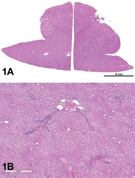
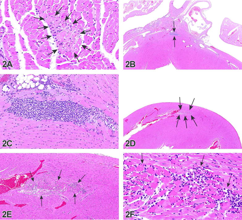
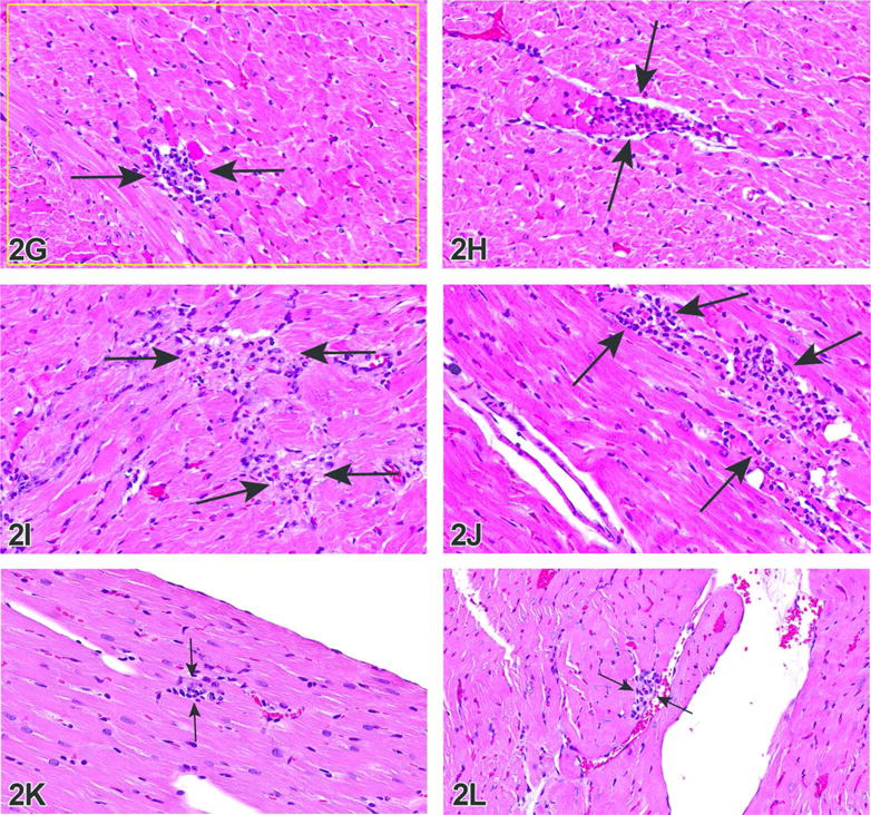
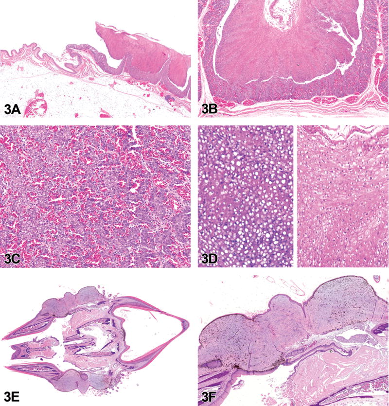
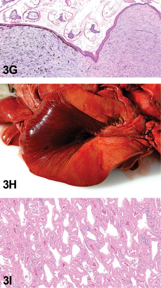
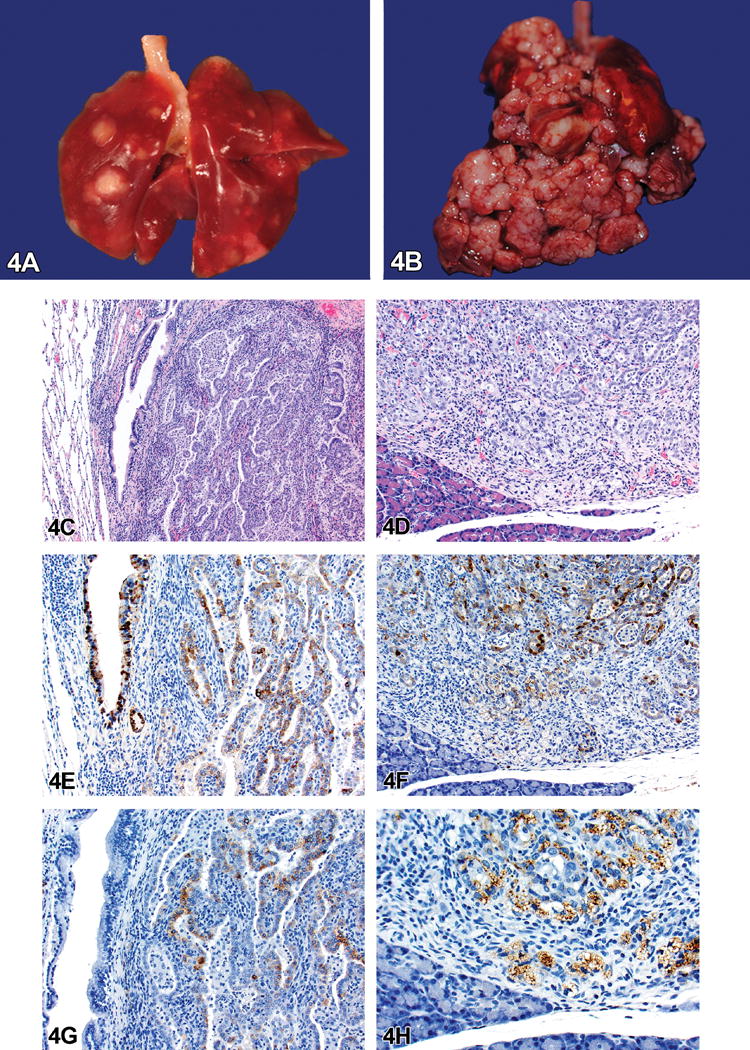
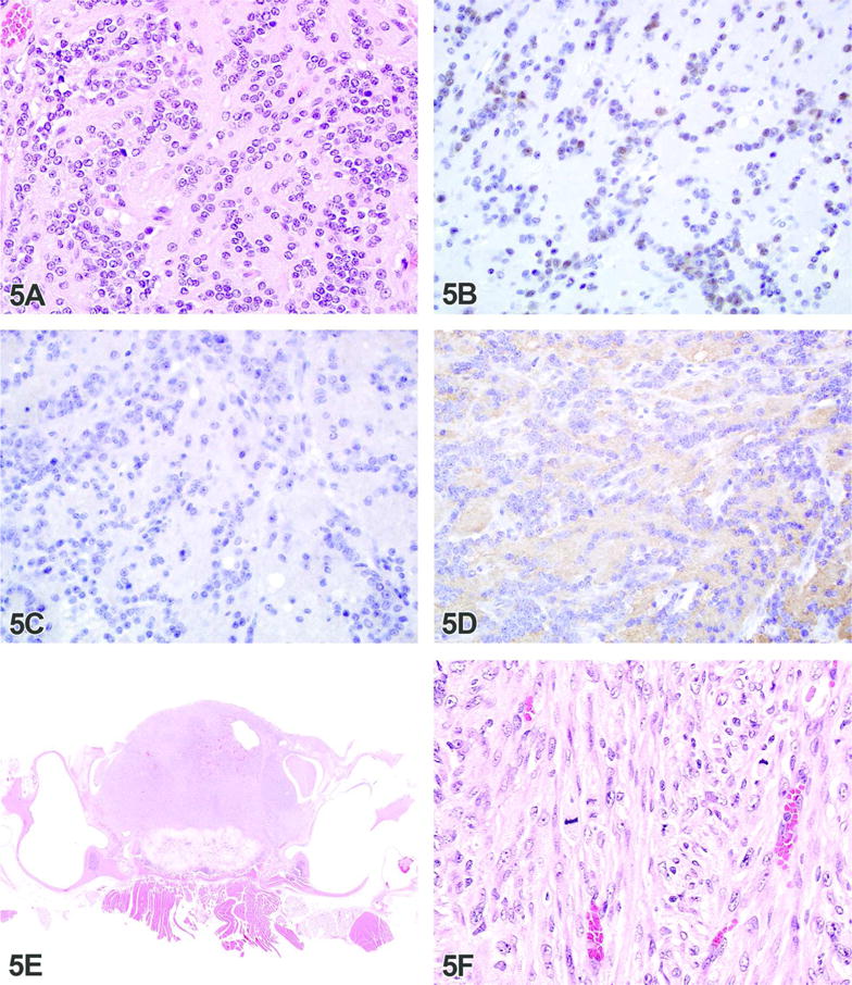
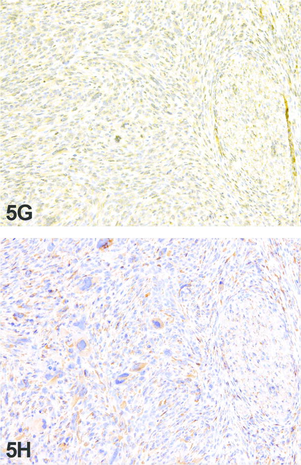
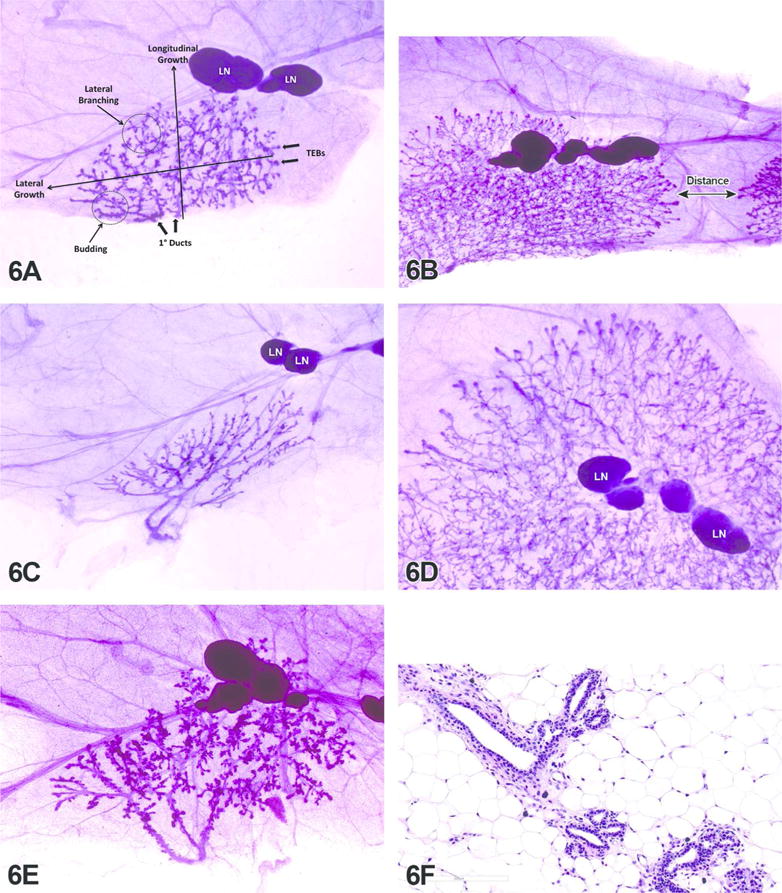
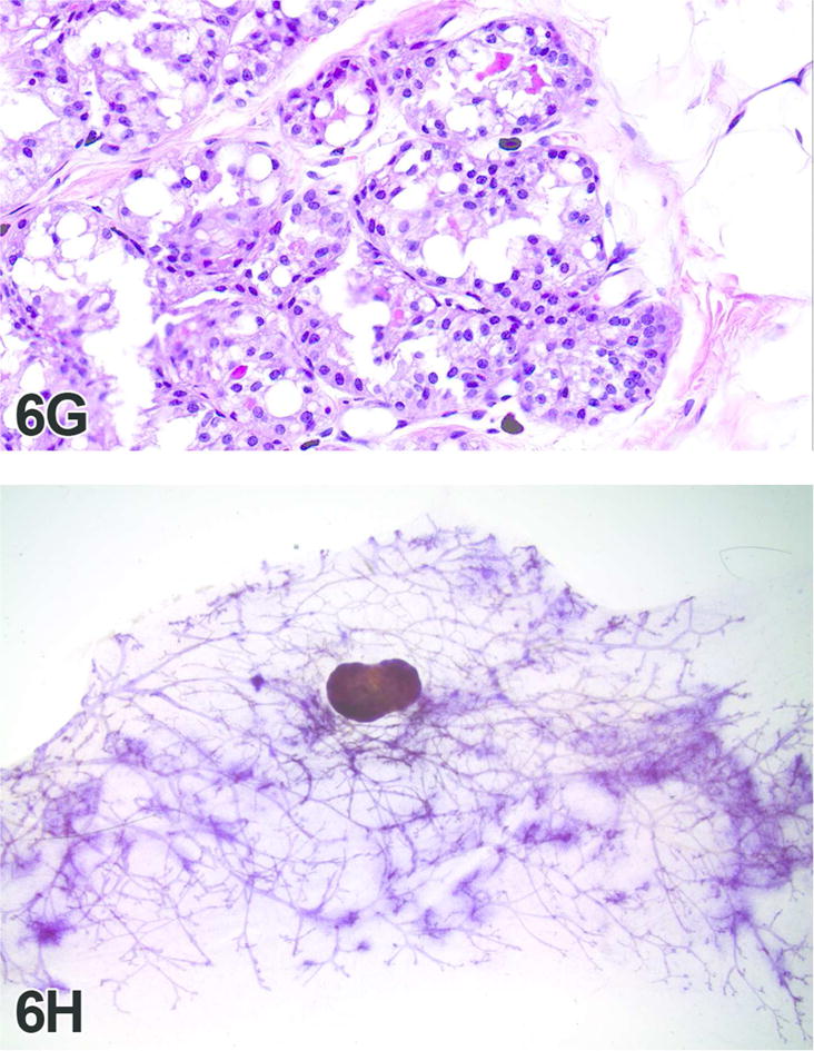
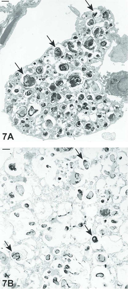
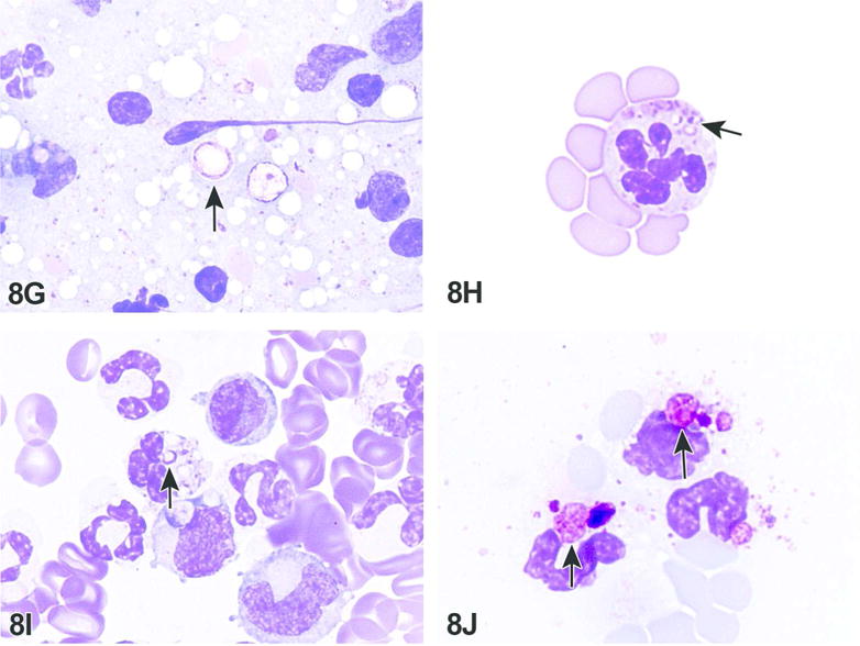
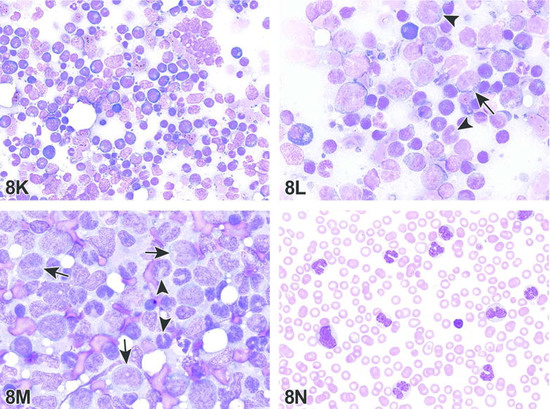
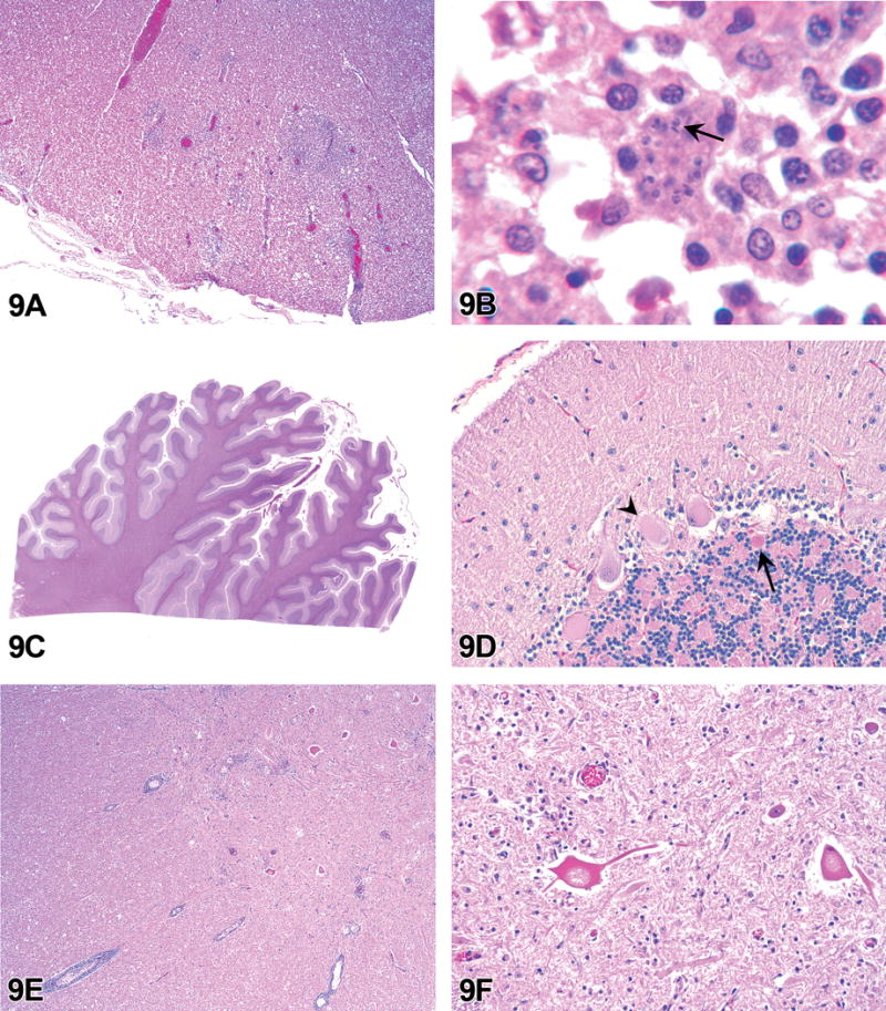
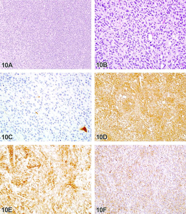
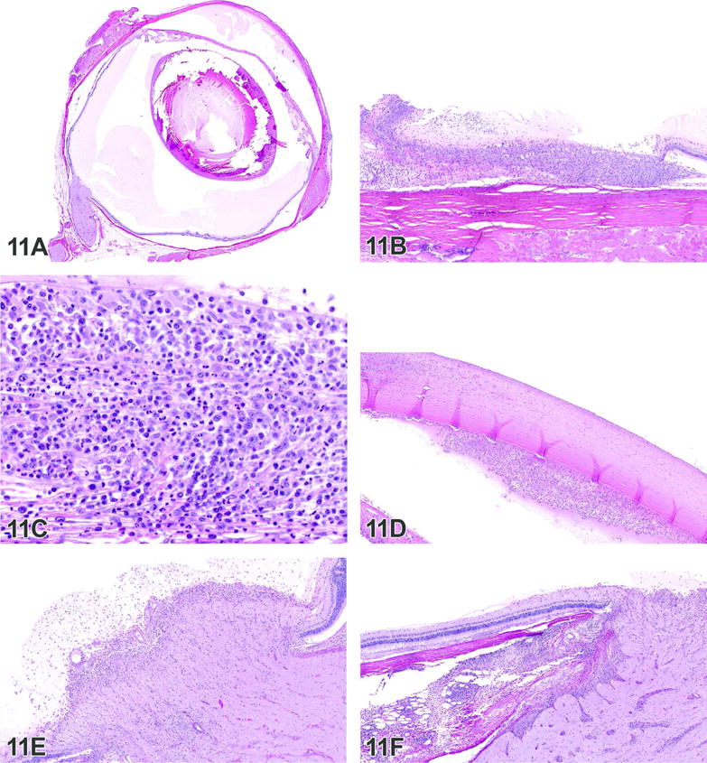
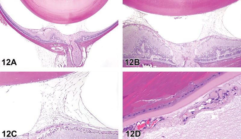
The 2015 Annual National Toxicology Program Satellite Symposium, entitled "Pathology Potpourri" was held in Minneapolis, Minnesota, at the American College of Veterinary Pathologists/American Society for Veterinary Clinical Pathology/Society of Toxicologic Pathology combined meeting. The goal of this symposium is to present and discuss diagnostic pathology challenges or nomenclature issues. Because of the combined meeting, both laboratory and domestic animal cases were presented. This article presents summaries of the speakers' talks, including challenging diagnostic cases or nomenclature issues that were presented, along with select images that were used for audience voting and discussion. Some lesions and topics covered during the symposium included hepatocellular lesions, a proposed harmonized diagnostic approach to rat cardiomyopathy, crop milk in a bird, avian feeding accoutrement, heat exchanger in a tuna, metastasis of a tobacco carcinogen-induced pulmonary carcinoma, neurocytoma in a rat, pituicytoma in a rat, rodent mammary gland whole mounts, dog and rat alveolar macrophage ultrastructure, dog and rat pulmonary phospholipidosis, alveolar macrophage aggregation in a dog, degenerating yeast in a cat liver aspirate, myeloid leukemia in lymph node aspirates from a dog, Trypanosoma cruzi in a dog, solanum toxicity in a cow, bovine astrovirus, malignant microglial tumor, and nomenclature challenges from the Special Senses International Harmonization of Nomenclature and Diagnostic Criteria Organ Working Group.

Keywords: INHAND; NTP Satellite Symposium; cardiomyopathy; crop milk; diagnostic neuropathology; focal nodular hyperplasia; histoplasmosis; leukemia +/− cytology; mammary gland whole mounts; neurocytoma; ocular inflammation; oral ornamentation; persistent fetal vasculature; persistent hyperplastic primary vitreous; persistent hyperplastic tunica vasculosa lentis; pulmonary metastatic tumor; pulmonary ultrastructure; tuna heat exchanger.


Conflict of interest statement

The author(s) declared no potential conflicts of interest with respect to the research, authorship, and/or publication of this article.

Figures

Figure 1
Figure 1
(A–B). Benign nodular lesion of hepatocytes in the dog. Photomicrograph of a cross section of a liver mass on the left liver lobe of a dog (A) and a higher magnification photomicrograph of the mass in figure 1A (B), demonstrating portal tracts and distorted lobules accentuated by minimal portal bridging inflammation, fibrosis, and biliary hyperplasia. H&E.
Figure 2
Figure 2
(A–F). Heart lesions representing the spectrum of morphologies comprising cardiomyopathy in control male SD rats. Case 1 (A)-high magnification of a predominantly necrotic lesion within the left ventricle. Case 2 (B)-low magnification of a predominantly cellular lesion within the base of the heart and (C) high magnification. Case 3 (D)-low magnification of a mixed necrotic and cellular lesion within the apex of the heart, (E) mid magnification and (F) high magnification. H&E. (G–L). Case 4 (G, H, I, J)-high magnifications of 4 lesions within the same heart (apex, right ventricle, left ventricle and septum). Case 5 (K)-high magnification of an “equivocal” lesion within the right ventricle. Case 6 (L)-high magnification of clear but very small lesion within the left ventricle. H&E.
Figure 2
Figure 2
(A–F). Heart lesions representing the spectrum of morphologies comprising cardiomyopathy in control male SD rats. Case 1 (A)-high magnification of a predominantly necrotic lesion within the left ventricle. Case 2 (B)-low magnification of a predominantly cellular lesion within the base of the heart and (C) high magnification. Case 3 (D)-low magnification of a mixed necrotic and cellular lesion within the apex of the heart, (E) mid magnification and (F) high magnification. H&E. (G–L). Case 4 (G, H, I, J)-high magnifications of 4 lesions within the same heart (apex, right ventricle, left ventricle and septum). Case 5 (K)-high magnification of an “equivocal” lesion within the right ventricle. Case 6 (L)-high magnification of clear but very small lesion within the left ventricle. H&E.
Figure 3
Figure 3
(A–F). Various tissues from a Rock dove, a Gouldian finch and a tuna. (A) Esophagus (left) extending into crop (right), Rock dove (pigeon). There is massive hyperplasia (A) of the surface epithelium. (B) The basilar layers of the epithelial surface have massive interdigitated infoldings of a (C) highly vascularized subepithelial stroma. (D) The surface is a thick layer of epithelial cells that contain heavy concentrations of lipid (clear vacuoles) and protein (eosinophilia, hyperkeratosis). (E) The oral maxillary and mandibular surfaces of a young (nestling) Gouldian finch. (F) There are symmetric fibrous nodules at the commissures of the beak, one side of which is shown. The nodules are composed of organized collagen fibers, two of which (outer, grossly blue) have melanocytes interspersed with the stroma. H&E. (G–I). The central nodule (G) is grossly yellow, with arrayed collagen similar to a fibrous tapetum. Scattered light through the fibrous arrays produce the blue and yellow colors seen grossly (http://www.aqua.org/explore/animals/gouldian-finch, accessed 11/30/15). Gross image (H) of the liver from a tuna. The visceral rete (I) is formed by massive branching of the celiac artery into parallel arteries that interdigitate with veins. The arteries and veins are closely apposed to cool cardiac arterial blood and warm visceral venous blood. Histology images are H&E.
Figure 3
Figure 3
(A–F). Various tissues from a Rock dove, a Gouldian finch and a tuna. (A) Esophagus (left) extending into crop (right), Rock dove (pigeon). There is massive hyperplasia (A) of the surface epithelium. (B) The basilar layers of the epithelial surface have massive interdigitated infoldings of a (C) highly vascularized subepithelial stroma. (D) The surface is a thick layer of epithelial cells that contain heavy concentrations of lipid (clear vacuoles) and protein (eosinophilia, hyperkeratosis). (E) The oral maxillary and mandibular surfaces of a young (nestling) Gouldian finch. (F) There are symmetric fibrous nodules at the commissures of the beak, one side of which is shown. The nodules are composed of organized collagen fibers, two of which (outer, grossly blue) have melanocytes interspersed with the stroma. H&E. (G–I). The central nodule (G) is grossly yellow, with arrayed collagen similar to a fibrous tapetum. Scattered light through the fibrous arrays produce the blue and yellow colors seen grossly (http://www.aqua.org/explore/animals/gouldian-finch, accessed 11/30/15). Gross image (H) of the liver from a tuna. The visceral rete (I) is formed by massive branching of the celiac artery into parallel arteries that interdigitate with veins. The arteries and veins are closely apposed to cool cardiac arterial blood and warm visceral venous blood. Histology images are H&E.
Figure 4
Figure 4
(A–B). Alveolar/bronchiolar carcinoma with metastasis to the pancreas in male F344 rats from a 2-year carcinogenicity bioassay. Gross images of multifocal lung tumors from a rat treated with (R)-NNAL (A*), lung tumors with extrapulmonary masses invading the mediastinum and thoracic cavity (B*). (C–H). Histomicrographs of alveolar/bronchiolar carcinoma from a rat treated with racemic NNAL. The microscopic lesion within the pancreas that was presented for voting (C) depicts an epithelial neoplastic mass that effaces the normal pancreatic architecture with tubuloacinar structure formation (D) (H&E). Immunohistochemistry for alveolar type II cell marker, prosurfactant protein C (PSP-C) in lung tumor (E), and metastatic carcinoma in the pancreas (F); for Clara (club) cell marker CC10 in lung tumor (G) and metastatic carcinoma in the pancreas (H). *Reproduced with permission from Balbo et al., 2014.
Figure 5
Figure 5
(A–F). Two rare neoplasms in the rat brain. (A) High magnification, H&E image of a rat neurocytoma. Note the uniform neoplastic cells with small, round nuclei interspersed with eosinophilic fibrillary material. Anti-Neu N IHC (B); note the variably intense nuclear staining of the neoplastic cells. Anti-Olig2 IHC (C); the neoplastic cells are uniformly negative. Anti-synaptophysin IHC (D); the fibrillary material interspersed with the neoplastic cells is positive, consistent with the interpretation that this material represents the elaboration of cellular processes from the neoplastic cells. (E) Low magnification H&E image of a rat malignant pituicytoma. (F) High magnification H&E image demonstrating the spindle cell morphology, cellular pleomorphism and high mitotic rate. (G–H). Anti-vimentin IHC (G); note the strongly positive cytoplasmic staining. Anti-GFAP IHC (H); note the strongly positive cytoplasmic staining. Images 5E, 5F, 5G and 5H were reproduced with permission from Moroki et al., 2015.
Figure 5
Figure 5
(A–F). Two rare neoplasms in the rat brain. (A) High magnification, H&E image of a rat neurocytoma. Note the uniform neoplastic cells with small, round nuclei interspersed with eosinophilic fibrillary material. Anti-Neu N IHC (B); note the variably intense nuclear staining of the neoplastic cells. Anti-Olig2 IHC (C); the neoplastic cells are uniformly negative. Anti-synaptophysin IHC (D); the fibrillary material interspersed with the neoplastic cells is positive, consistent with the interpretation that this material represents the elaboration of cellular processes from the neoplastic cells. (E) Low magnification H&E image of a rat malignant pituicytoma. (F) High magnification H&E image demonstrating the spindle cell morphology, cellular pleomorphism and high mitotic rate. (G–H). Anti-vimentin IHC (G); note the strongly positive cytoplasmic staining. Anti-GFAP IHC (H); note the strongly positive cytoplasmic staining. Images 5E, 5F, 5G and 5H were reproduced with permission from Moroki et al., 2015.
Figure 6
Figure 6
(A–F). Examples of mammary gland whole mount preparations from treated and control rats and mice. (A) Mammary gland whole mount preparation, 21-day-old Sprague Dawley rat, female, control. Developmental scoring of the mammary gland is achieved by evaluating the indicated parameters: (1) lateral growth, (2) longitudinal growth, (3) the number of primary ducts arising from the nipple, (4) budding (branch density), (5) lateral (side) branching, and (6) the number of TEBs (carmine alum stain). (B) Mammary gland whole mount preparation, 33-day-old Sprague Dawley rat, female, prenatal exposure from gestation days (GD) 15–18, TCDD (2,3,7,8-tetrachlorodibenzo-p-dioxin). Distance between the 4th and 5th mammary glands are also assessed as these glands grow together in the maturing animal, eliminating the space between them (carmine alum stain). (C) Mammary gland whole mount preparation, 21-day-old Sprague Dawley rat, female, exposure from GD 6 to post-natal day (PND) 90, 2.5mg/kg BPA (bisphenol A). Note markedly delayed development relative to age-matched control (Figure 6A) with decreased number of primary ducts and TEBs, decreased lateral and longitudinal growth, and decreased budding and lateral branching (carmine alum stain). (D) Mammary gland whole mount preparation, 21-day-old Sprague Dawley rat, female, exposure from GD 6 to post-natal day (PND) 90, 25 mg/kg EE2 (ethinyl estradiol). Growth is markedly accelerated compared to the age-matched control (A). The gland is very dense, with many terminal end buds along the periphery and branching that extends well beyond the inguinal lymph node (carmine alum stain). (E) Mammary gland whole mount preparation, 21-day-old Sprague Dawley rat, female, control. The entire structure of the gland can be visualized via whole mount preparation, however cellular features are not observed (carmine alum stain). (F) Histology of the mammary gland, 21-day-old Sprague Dawley rat, female, control. The tubuloalveolar morphology of the female rat mammary gland is easily appreciated under light microscopy, yet the overall structure of the gland cannot be interpreted from this single section (H&E). LN= lymph node, TEB = terminal end buds. (G–H). (G) Histology of the mammary gland, 21-day-old Sprague Dawley rat, male, prenatal exposure from GD 15–18 with DES (Diethylstilbestrol). The cellular morphology of the male rat mammary gland is typically lobuloalveolar in appearance. However, when exposed to estrogenic compounds such as DES during critical developmental time points, “feminization” of the gland may occur, causing the morphology to become more tubuloalveolar (inset) (H&E). (H) Mammary gland whole mount preparation, 14-month old, CD-1 mouse, female. Note the increased areas of opacity throughout the gland. These areas may indicate inflammation, hyperplasia or neoplasia. However, histological examination is required to confirm, as cellular features are not apparent (carmine alum stain).
Figure 6
Figure 6
(A–F). Examples of mammary gland whole mount preparations from treated and control rats and mice. (A) Mammary gland whole mount preparation, 21-day-old Sprague Dawley rat, female, control. Developmental scoring of the mammary gland is achieved by evaluating the indicated parameters: (1) lateral growth, (2) longitudinal growth, (3) the number of primary ducts arising from the nipple, (4) budding (branch density), (5) lateral (side) branching, and (6) the number of TEBs (carmine alum stain). (B) Mammary gland whole mount preparation, 33-day-old Sprague Dawley rat, female, prenatal exposure from gestation days (GD) 15–18, TCDD (2,3,7,8-tetrachlorodibenzo-p-dioxin). Distance between the 4th and 5th mammary glands are also assessed as these glands grow together in the maturing animal, eliminating the space between them (carmine alum stain). (C) Mammary gland whole mount preparation, 21-day-old Sprague Dawley rat, female, exposure from GD 6 to post-natal day (PND) 90, 2.5mg/kg BPA (bisphenol A). Note markedly delayed development relative to age-matched control (Figure 6A) with decreased number of primary ducts and TEBs, decreased lateral and longitudinal growth, and decreased budding and lateral branching (carmine alum stain). (D) Mammary gland whole mount preparation, 21-day-old Sprague Dawley rat, female, exposure from GD 6 to post-natal day (PND) 90, 25 mg/kg EE2 (ethinyl estradiol). Growth is markedly accelerated compared to the age-matched control (A). The gland is very dense, with many terminal end buds along the periphery and branching that extends well beyond the inguinal lymph node (carmine alum stain). (E) Mammary gland whole mount preparation, 21-day-old Sprague Dawley rat, female, control. The entire structure of the gland can be visualized via whole mount preparation, however cellular features are not observed (carmine alum stain). (F) Histology of the mammary gland, 21-day-old Sprague Dawley rat, female, control. The tubuloalveolar morphology of the female rat mammary gland is easily appreciated under light microscopy, yet the overall structure of the gland cannot be interpreted from this single section (H&E). LN= lymph node, TEB = terminal end buds. (G–H). (G) Histology of the mammary gland, 21-day-old Sprague Dawley rat, male, prenatal exposure from GD 15–18 with DES (Diethylstilbestrol). The cellular morphology of the male rat mammary gland is typically lobuloalveolar in appearance. However, when exposed to estrogenic compounds such as DES during critical developmental time points, “feminization” of the gland may occur, causing the morphology to become more tubuloalveolar (inset) (H&E). (H) Mammary gland whole mount preparation, 14-month old, CD-1 mouse, female. Note the increased areas of opacity throughout the gland. These areas may indicate inflammation, hyperplasia or neoplasia. However, histological examination is required to confirm, as cellular features are not apparent (carmine alum stain).
Figure 7
Figure 7
(A–B). Transmission electron micrographs (TEMs) of rat and dog pulmonary pathologies. (A) TEM of an alveolar macrophage from a treated male rat lung that shows numerous secondary lysosomes (arrows) containing whorled membrane remnant material (Original micrograph magnification of 10,700×; Scale bar: 1 μM). (B) TEM of the cytoplasm of an alveolar macrophage from a treated female dog lung with numerous secondary lysosomes (arrows) containing whorled membrane remnant material with a central homogenous area (Original micrograph magnification of 10,700×; Scale bar: 1μM).
Figure 8
Figure 8
(A–F). Liver aspirate from a cat with histoplasmosis. On low power, there is high cellularity with deeply basophilic clusters of hepatocytes and many cells in the background (A). Closer examination of the background cells reveals a mixed inflammatory cell population consisting of neutrophils (arrowhead), histiocytes (short arrows) and lymphocytes (long arrow) (B). (C) While rare extracellular round yeast organisms with the classic appearance of Histoplasma capsulatum are seen (arrow, C), the more common finding consists of histiocytes with intracytoplasmic round stippled pink structures interpreted as degenerating organisms (asterisks, D). Rare macrophages with intact organisms (arrow) are also found (E), while rare partially degraded forms (arrows) are noted within cells (F). Wright-Giemsa stain. (G–J). Rare partially degraded forms were also noted in the background (arrow, G). The appearance of the presumed degenerating organisms is not specific; items with a similar appearance include phagocytized and degraded mast cell granules (arrow, H), ragocytes with phagocytized immunoglobulin, fibrin and complement (arrow, I), or even phagocytized lubricant material (arrow, J), Wright-Giemsa stain. (K–N). Lymph node aspirate from a dog with infiltrative chronic monocytic or myelomonocytic leukemia. On low power (K), there is a mixed round cell population. On higher power (L, M), many of the cells have band (arrowheads) to indented (short arrows) to irregularly-shaped (long arrow) nuclei with fine, dispersed chromatin indicating myeloid origin. An image of the peripheral blood smear (N) from this dog shows a similar population. However, the nuclear pleomorphism is more apparent, and the cells often contain a few vacuoles suggesting monocytic origin. Wright-Giemsa stain.
Figure 8
Figure 8
(A–F). Liver aspirate from a cat with histoplasmosis. On low power, there is high cellularity with deeply basophilic clusters of hepatocytes and many cells in the background (A). Closer examination of the background cells reveals a mixed inflammatory cell population consisting of neutrophils (arrowhead), histiocytes (short arrows) and lymphocytes (long arrow) (B). (C) While rare extracellular round yeast organisms with the classic appearance of Histoplasma capsulatum are seen (arrow, C), the more common finding consists of histiocytes with intracytoplasmic round stippled pink structures interpreted as degenerating organisms (asterisks, D). Rare macrophages with intact organisms (arrow) are also found (E), while rare partially degraded forms (arrows) are noted within cells (F). Wright-Giemsa stain. (G–J). Rare partially degraded forms were also noted in the background (arrow, G). The appearance of the presumed degenerating organisms is not specific; items with a similar appearance include phagocytized and degraded mast cell granules (arrow, H), ragocytes with phagocytized immunoglobulin, fibrin and complement (arrow, I), or even phagocytized lubricant material (arrow, J), Wright-Giemsa stain. (K–N). Lymph node aspirate from a dog with infiltrative chronic monocytic or myelomonocytic leukemia. On low power (K), there is a mixed round cell population. On higher power (L, M), many of the cells have band (arrowheads) to indented (short arrows) to irregularly-shaped (long arrow) nuclei with fine, dispersed chromatin indicating myeloid origin. An image of the peripheral blood smear (N) from this dog shows a similar population. However, the nuclear pleomorphism is more apparent, and the cells often contain a few vacuoles suggesting monocytic origin. Wright-Giemsa stain.
Figure 9
Figure 9
(A–F). Chagas’ disease in the spinal cord of a dog (A–B), Solanum toxicity in the cerebellum of a cow (C–D), and bovine astrovirus myelitis in a heifer (E–F). Multifocal inflammatory foci (A) are within the spinal cord white matter. Higher magnification (B) shows macrophages, lymphocytes, and plasma cells. The macrophage in the center contains protozoal amastigotes consistent with Trypanosoma cruzi (arrow). Subgross image of the cerebellum (C) shows thinning of the cerebellar folia. Higher magnification (D) shows swelling and vacuolation of Purkinje cells, loss of Purkinje cells with replacement by proliferating Bergmann’s glia, and a swollen Purkinje cell axon (arrow). Low magnification of the spinal cord (E) shows perivascular cuffing by lymphocytes and plasma cells, hypereosinophilic necrotic neurons, and gliosis. Higher magnification of the necrotic neurons and gliosis (F). The scattered bacteria are postmortem contaminants. (H&E)
Figure 10
Figure 10
(A–F). Intracranial neoplasm from a control male F344 rat. Low (A) and high (B) magnification, H&E images of what has historically been diagnosed as a rat astrocytoma. Note the lack of staining in the neoplastic cells with anti-GFAP immunohistochemistry (C). Note the diffuse positive staining for Iba-1 (D), MHC Class II (E) and by the plant lectin RCA-1 (F). The above results support a microglial or macrophage rather than an astrocytic histogenesis.
Figure 11
Figure 11
(A–F). Lesions from a 14-day single-dose ITV administration study in New Zealand White rabbits. (A) Overview of the eye, showing eosinophilic, amorphous material in the anterior and posterior compartments, compatible with proteinaceous fluid. Mixed cell inflammation in the pars planum (B) and a higher power view (C) of the inflammation, showing a mixture of mononuclear cells, including plasma cells, and heterophils. There is mixed cell inflammation in the anterior chamber, aligning the endothelial surface of the cornea, as well as inflammation within the corneal stroma (D), mixed cell inflammation in the optic nerve head and in the adjacent vitreous (E) and mixed cell inflammation within the optic nerve, as well as external to the eye surrounding the optic nerve (F). H&E.
Figure 12
Figure 12
(A–D). Eye of a Sprague-Dawley rat. Low magnification image (A) showing the optic nerve and central retina. There is a delicate fibrovascular tissue extending from the surface of the retina into the vitreous, contacting the posterior lens capsule. Higher magnifications (B & C), showing the fibrovascular nature of the tissue causing retinal traction and folds. Anterior portion of the tissue (D) consistent with a persistent hyperplastic tunica vasculosa lentis (PHTVL) characterized by small and regularly distributed vessels surrounding the posterior lens capsule. H&E.

Similar articles

Cited by

References

    1. Abcam product description website. Anti-CD68 antibody [ED1] (ab31630) Retrieved November 22, 2015: http://www.abcam.com/cd68-antibody-ed1-ab31630.html.
    1. Adams ET, Auerbach S, Blackshear PE, Bradley A, Gruebbel MM, Little PB, Malarkey D, Maronpot R, McKay JS, Miller RA, Moore RR, Morrison JP, Nyska A, Ramot Y, Rao D, Suttie A, Wells MY, Willson GA, Elmore SA. Proceedings of the 2010 National Toxicology Program Satellite Symposium. Toxicol Pathol. 2011;39:240–66. - PMC - PubMed
    1. Adsay NV, Andea A, Basturk O, Kilinc N, Nassar H, Cheng JD. Secondary tumors of the pancreas: an analysis of a surgical and autopsy database and review of the literature. Virchows Arch. 2004;444:527–35. - PubMed
    1. Allgoewer I, Pfefferkorn B. Persistent hyperplastic tunica vasculosa lentis and persistent hyperplastic primary vitreous (PHTVL/PHPV) in two cats. Vet Ophthalmol. 2001;4:161–4. - PubMed
    1. American Cancer Society. Breast Cancer Overview [Internet] Atlanta: American Cancer Society; c2014–15. [updated 2015 June 10; cited 2015 Nov 12]. Available at: http://www.cancer.org/Cancer/BreastCancer/OverviewGuide/breast-cancer-ov....

Publication types

LinkOut - more resources