

Mitochondrial structure and function are not different between nonfailing donor and end-stage failing human hearts
- PMID: 27075244
- PMCID: PMC4970604
- DOI: 10.1096/fj.201500118R
Mitochondrial structure and function are not different between nonfailing donor and end-stage failing human hearts
Abstract
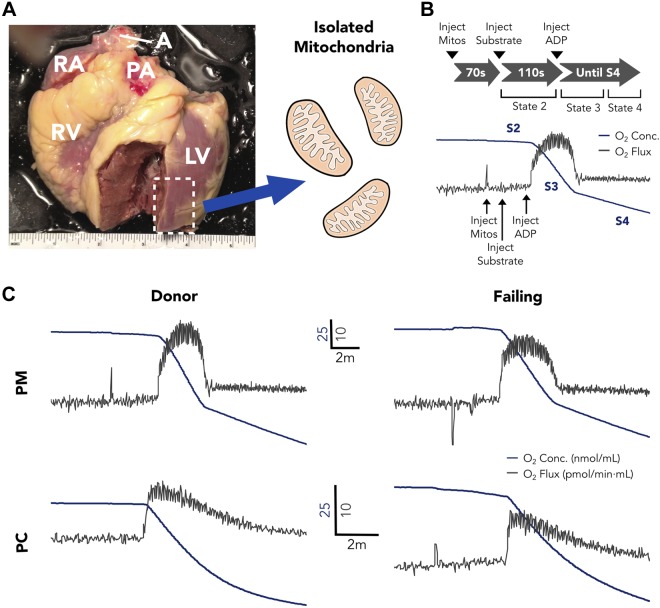
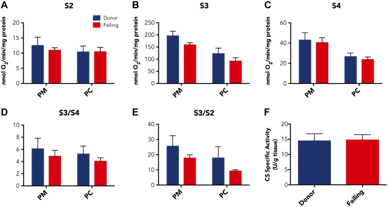
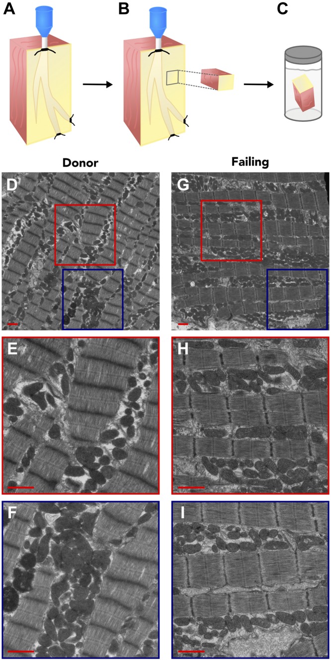
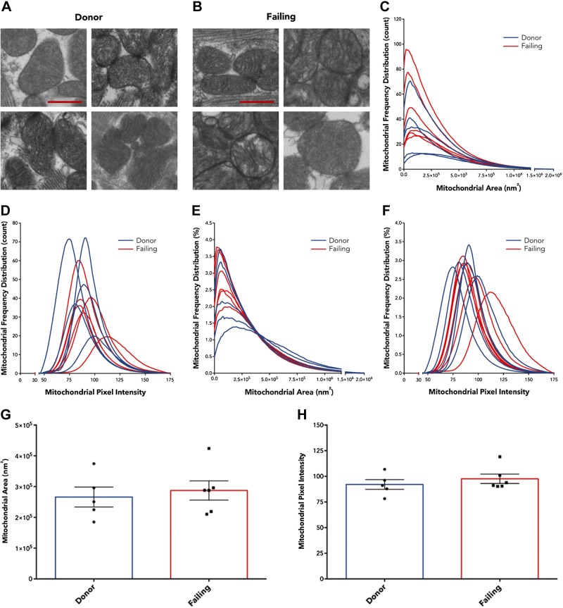
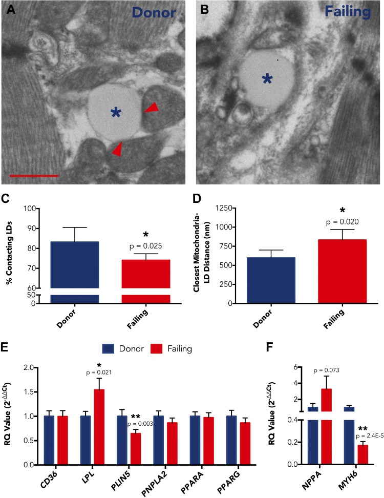
During human heart failure, the balance of cardiac energy use switches from predominantly fatty acids (FAs) to glucose. We hypothesized that this substrate shift was the result of mitochondrial degeneration; therefore, we examined mitochondrial oxidation and ultrastructure in the failing human heart by using respirometry, transmission electron microscopy, and gene expression studies of demographically matched donor and failing human heart left ventricular (LV) tissues. Surprisingly, respiratory capacities for failing LV isolated mitochondria (n = 9) were not significantly diminished compared with donor LV isolated mitochondria (n = 7) for glycolysis (pyruvate + malate)- or FA (palmitoylcarnitine)-derived substrates, and mitochondrial densities, assessed via citrate synthase activity, were consistent between groups. Transmission electron microscopy images also showed no ultrastructural remodeling for failing vs. donor mitochondria; however, the fraction of lipid droplets (LDs) in direct contact with a mitochondrion was reduced, and the average distance between an LD and its nearest neighboring mitochondrion was increased. Analysis of FA processing gene expression between donor and failing LVs revealed 0.64-fold reduced transcript levels for the mitochondrial-LD tether, perilipin 5, in the failing myocardium (P = 0.003). Thus, reduced FA use in heart failure may result from improper delivery, potentially via decreased perilipin 5 expression and mitochondrial-LD tethering, and not from intrinsic mitochondrial dysfunction.-Holzem, K. M., Vinnakota, K. C., Ravikumar, V. K., Madden, E. J., Ewald, G. A., Dikranian, K., Beard, D. A., Efimov, I. R. Mitochondrial structure and function are not different between nonfailing donor and end-stage failing human hearts.
Keywords: electron microscopy; energy substrate; lipid droplet; oxidative respiration; perilipin 5.
© FASEB.
Figures
Similar articles
-
Freshly isolated mitochondria from failing human hearts exhibit preserved respiratory function.J Mol Cell Cardiol. 2014 Mar;68:98-105. doi: 10.1016/j.yjmcc.2013.12.029. Epub 2014 Jan 9. J Mol Cell Cardiol. 2014. PMID: 24412531 Free PMC article.
-
Posttranslational modifications and dysfunction of mitochondrial enzymes in human heart failure.Am J Physiol Endocrinol Metab. 2016 Aug 1;311(2):E449-60. doi: 10.1152/ajpendo.00127.2016. Epub 2016 Jul 12. Am J Physiol Endocrinol Metab. 2016. PMID: 27406740
-
Endogenous activation of mitochondrial KATP channels protects human failing myocardium from hydroxyl radical-induced stunning.Circ Res. 2009 Oct 9;105(8):811-7. doi: 10.1161/CIRCRESAHA.109.206359. Epub 2009 Sep 3. Circ Res. 2009. PMID: 19729596
-
Mitochondrial Bioenergetics and Dysfunction in Failing Heart.Adv Exp Med Biol. 2017;982:65-80. doi: 10.1007/978-3-319-55330-6_4. Adv Exp Med Biol. 2017. PMID: 28551782 Review.
-
Energetic myocardial metabolism and oxidative stress: let's make them our friends in the fight against heart failure.Biomed Pharmacother. 2010 Mar;64(3):203-7. doi: 10.1016/j.biopha.2009.10.002. Epub 2009 Nov 13. Biomed Pharmacother. 2010. PMID: 19954925 Review.
Cited by
-
Comparison of the stage-dependent mitochondrial changes in response to pressure overload between the diseased right and left ventricle in the rat.Basic Res Cardiol. 2024 Aug;119(4):587-611. doi: 10.1007/s00395-024-01051-3. Epub 2024 May 17. Basic Res Cardiol. 2024. PMID: 38758338
-
Reduced Myocardial Mitochondrial ROS Production in Mechanically Unloaded Hearts.J Cardiovasc Transl Res. 2019 Apr;12(2):107-115. doi: 10.1007/s12265-018-9803-3. Epub 2018 Mar 27. J Cardiovasc Transl Res. 2019. PMID: 29589269
-
Lipotoxicity as a therapeutic target in obesity and diabetic cardiomyopathy.J Pharm Pharm Sci. 2024 Apr 19;27:12568. doi: 10.3389/jpps.2024.12568. eCollection 2024. J Pharm Pharm Sci. 2024. PMID: 38706718 Free PMC article. Review.
-
Lipid Droplet-a New Target in Ischemic Heart Disease.J Cardiovasc Transl Res. 2022 Aug;15(4):730-739. doi: 10.1007/s12265-021-10204-x. Epub 2022 Jan 4. J Cardiovasc Transl Res. 2022. PMID: 34984637 Review.
-
Engineering cardiac microphysiological systems to model pathological extracellular matrix remodeling.Am J Physiol Heart Circ Physiol. 2018 Oct 1;315(4):H771-H789. doi: 10.1152/ajpheart.00110.2018. Epub 2018 Jun 15. Am J Physiol Heart Circ Physiol. 2018. PMID: 29906229 Free PMC article. Review.
References
Publication types
MeSH terms
Grants and funding
LinkOut - more resources
Full Text Sources
Other Literature Sources
Medical
Research Materials
Miscellaneous
