. 2016 Apr 14:6:24297.
doi: 10.1038/srep24297.

Flotation Immunoassay: Masking the Signal from Free Reporters in Sandwich Immunoassays

Affiliations

Flotation Immunoassay: Masking the Signal from Free Reporters in Sandwich Immunoassays

Hui Chen et al. Sci Rep. .

Abstract

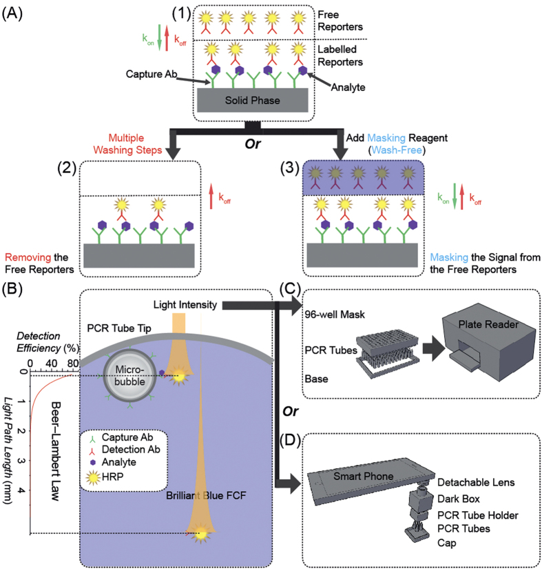
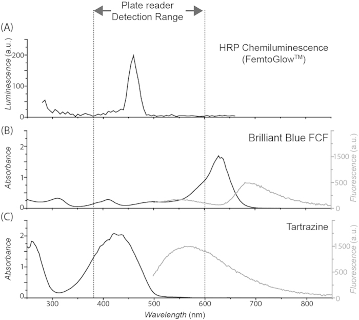
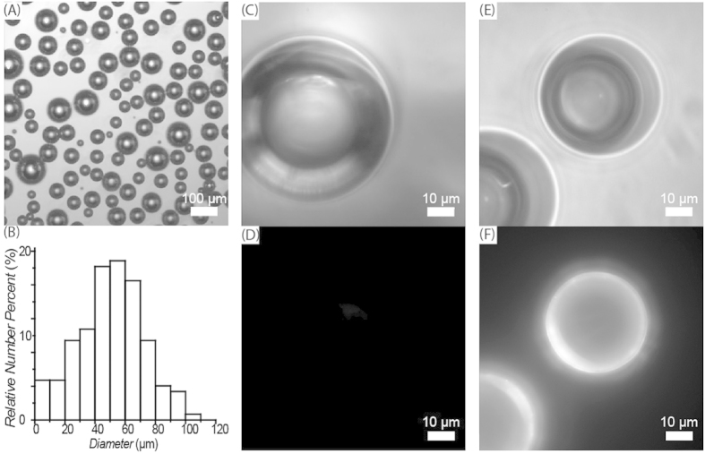
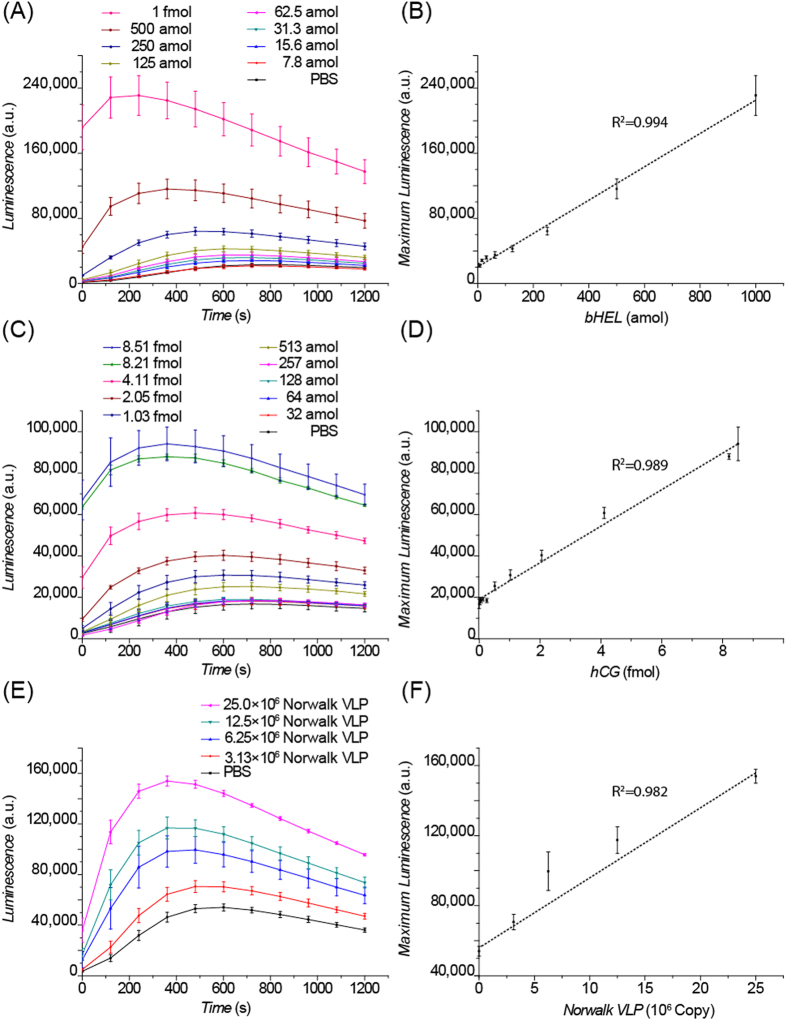
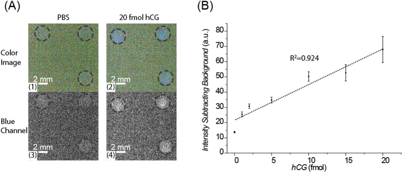
In this work, we demonstrate that signal-masking reagents together with appropriate capture antibody carriers can eliminate the washing steps in sandwich immunoassays. A flotation immunoassay (FI) platform was developed with horseradish peroxidase chemiluminescence as the reporter system, the dye Brilliant Blue FCF as the signal-masking reagent, and buoyant silica micro-bubbles as the capture antibody carriers. Only reporters captured on micro-bubbles float above the dye and become visible in an analyte-dependent manner. These FIs are capable of detecting proteins down to attomole levels and as few as 10(6) virus particles. This signal-masking strategy represents a novel approach to simple, sensitive and quantitative immunoassays in both laboratory and point-of-care settings.


Conflict of interest statement

RCW is an inventor on a pending patent application which potentially could cover aspects of this technology.

Figures

Figure 1
Figure 1
(A) Working principle of the signal-masking strategy for wash-free, flotation immunoassay (FI). 1) In a sandwich immunoassay, excess reporters are used to drive efficient labelling of the analytes. 2) In traditional sandwich immunoassays, free reporters are removed by multiple washings, also resulting in a decrease of the fraction of labelled analytes. 3) In the signal-masking based sandwich immunoassay, free reporters are not removed, instead, their signal is masked, without influencing the degree of labelling of the analytes. (B) Working principle of the flotation immunoassay (FI). Light-absorbing dyes are used to block the light from the free reporters dispersed in the bulk of the solution. Here, horseradish peroxidase (HRP) is used as the light-emitting reporter; Brilliant Blue FCF dye (BB-FCF) is used as the light-masking reagent; and silica micro-bubbles are used as the immuno-carriers to collect the labelled HRP molecules in the top detectable layer of the solution. (C) Design of the FI plate for plate reader read-out. (D) Design of the FI accessory for smart phone read-out.
Figure 2
Figure 2. Selection of Brilliant Blue FCF dye (BB-FCF) as the signal-masking reagent.
(A) Chemiluminescence emission spectrum of horseradish peroxidase (HRP) with FemtoGlow™ substrate; (B,C) Absorbance spectrum (black lines) and fluorescence emission spectrum (gray lines, excited at 460 nm) of (B) BB-FCF and (C) tartrazine. Vertical dashed lines indicate the luminescence detection range of the plate reader used in this work. The absorbance of BB-FCF is not as strong as those of some other dyes, but its fluorescence emission lies outside the range of sensitivity of the plate reader.
Figure 3
Figure 3
(A) Bright field image of micro-bubbles. (B) Size distribution of micro-bubbles, measured by microscopic imaging. (C) Bright field and (D) fluorescence images of BSA modified micro-bubbles labelled with biotinylated fluorescent reporters (biotin-M13-Alexa555). (E) Bright field and (F) fluorescence images of NeutrAvidin modified micro-bubbles labelled with biotinylated fluorescent reporters (biotin-M13-Alexa555).
Figure 4
Figure 4
(A) Signal profiles of the Flotation Immunoassay (FI) for the detection of biotinylated lysozyme (bHEL). (B) Linear response of the maximum chemiluminescence (CL) signal to the amount of bHEL. (C) Signal profiles of the FI for human chorionic gonadotropin (hCG) detection. (D) Linear response of the maximum CL signal to the amount of hCG. (E) Signal profiles of the FI for Norwalk VLP detection. (F) Linear response of the maximum CL signal to the amount of Norwalk VLP. Mean ± standard deviation; n = 3.
Figure 5
Figure 5
(A) Raw chemiluminescence images of the human chorionic gonadotropin flotation immunoassay (hCG FI), captured by a smart phone (iPhone 6 Plus) camera, using the SlowShutter app showing the color images of 1) PBS and 2) 20 fmol hCG and the isolated blue channel images of 3) PBS and 4) 20 fmol hCG. The PCR tubes holding the FI assay reagents are circled with dashed lines. (B) Quantitative analysis of the isolated blue channel images of the flotation immunoassay for hCG detection. Data represent the mean values ± the standard deviation obtained using three different PCR tubes.

References

    1. Pei X. et al.. Sandwich-type immunosensors and immunoassays exploiting nanostructure labels: A review. Analytica Chimica Acta 758, 1–18 (2013). - PubMed
    1. Klenin K. V., Kusnezow W. & Langowski J. Kinetics of protein binding in solid-phase immunoassays: Theory. The Journal of Chemical Physics 122, 214715 (2005). - PubMed
    1. Bielefeld-Sevigny M. Alphalisa immunoassay platform the no-wash high-throughput alternative to elisa. Assay and Drug Development Technologies 7, 90–92 (2009). - PubMed
    1. Park S.-H. & Raines R. T. Fluorescence polarization assay to quantify protein-protein interactions. In Protein-Protein Interactions 161–165 (Springer, 2004). - PubMed
    1. Degorce F. et al.. Htrf: a technology tailored for drug discovery-a review of theoretical aspects and recent applications. Current Chemical Genomics 3, 22 (2009). - PMC - PubMed

Publication types