. 2016 Apr 13;13(1):78.
doi: 10.1186/s12974-016-0543-5.

6-Mercaptopurine attenuates tumor necrosis factor-α production in microglia through Nur77-mediated transrepression and PI3K/Akt/mTOR signaling-mediated translational regulation

Affiliations

6-Mercaptopurine attenuates tumor necrosis factor-α production in microglia through Nur77-mediated transrepression and PI3K/Akt/mTOR signaling-mediated translational regulation

Hsin-Yi Huang et al. J Neuroinflammation. .

Abstract

Background: The pathogenesis of several neurodegenerative diseases often involves the microglial activation and associated inflammatory processes. Activated microglia release pro-inflammatory factors that may be neurotoxic. 6-Mercaptopurine (6-MP) is a well-established immunosuppressive drug. Common understanding of their immunosuppressive properties is largely limited to peripheral immune cells. However, the effect of 6-MP in the central nervous system, especially in microglia in the context of neuroinflammation is, as yet, unclear. Tumor necrosis factor-α (TNF-α) is a key cytokine of the immune system that initiates and promotes neuroinflammation. The present study aimed to investigate the effect of 6-MP on TNF-α production by microglia to discern the molecular mechanisms of this modulation.

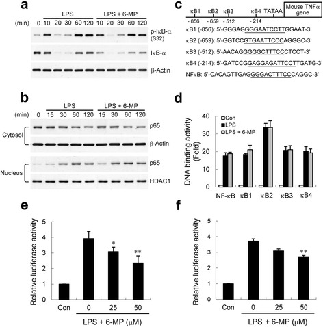
Methods: Lipopolysaccharide (LPS) was used to induce an inflammatory response in cultured primary microglia or murine BV-2 microglial cells. Released TNF-α was measured by enzyme-linked immunosorbent assay (ELISA). Gene expression was determined by real-time reverse transcription polymerase chain reaction (RT-PCR). Signaling molecules were analyzed by western blotting, and activation of NF-κB was measured by ELISA-based DNA binding analysis and luciferase reporter assay. Chromatin immunoprecipitation (ChIP) analysis was performed to examine NF-κB p65 and coactivator p300 enrichments and histone modifications at the endogenous TNF-α promoter.

Results: Treatment of LPS-activated microglia with 6-MP significantly attenuated TNF-α production. In 6-MP pretreated microglia, LPS-induced MAPK signaling, IκB-α degradation, NF-κB p65 nuclear translocation, and in vitro p65 DNA binding activity were not impaired. However, 6-MP suppressed transactivation activity of NF-κB and TNF-α promoter by inhibiting phosphorylation and acetylation of p65 on Ser276 and Lys310, respectively. ChIP analyses revealed that 6-MP dampened LPS-induced histone H3 acetylation of chromatin surrounding the TNF-α promoter, ultimately leading to a decrease in p65/coactivator-mediated transcription of TNF-α gene. Furthermore, 6-MP enhanced orphan nuclear receptor Nur77 expression. Using RNA interference approach, we further demonstrated that Nur77 upregulation contribute to 6-MP-mediated inhibitory effect on TNF-α production. Additionally, 6-MP also impeded TNF-α mRNA translation through prevention of LPS-activated PI3K/Akt/mTOR signaling cascades.

Conclusions: These results suggest that 6-MP might have a therapeutic potential in neuroinflammation-related neurodegenerative disorders through downregulation of microglia-mediated inflammatory processes.

Keywords: 6-Mercaptopurine; Histone H3 acetylation; Microglia; Nuclear factor-κB; Nur77; PI3K/Akt; TNF-α; mTOR.


Figures

Fig. 1
Fig. 1
6-MP attenuates TNF-α production in microglia. a Cells were pretreated (P) with vehicle or 50 μM 6-MP for the indicated times, cells were then stimulated with 100 ng/ml LPS for 6 h. TNF-α secretion into the culture medium was analyzed by ELISA. The relative differences between control and 6-MP pretreated groups were calculated and expressed as percent (%) of control. Data are presented as mean ± SEM for three independent experiments. *p < 0.05; **p < 0.01 compared with control. b, c Cells were pretreated with vehicle or various concentrations of 6-MP for 16 h followed by treatment with 100 ng/ml LPS for 6 h (b) or 2 h (c). Released TNF-α (b) was measured by ELISA. Expression of TNF-α mRNA (c) was quantified by real-time RT-PCR as described in the “Methods” section. Data are presented as mean ± SEM for three independent experiments. *p < 0.05; **p < 0.01 compared with control. The TNF-α content in untreated cultures was not detectable. The levels of TNF-α in LPS-treated alone BV-2 cells and primary microglia were 16.0 ± 0.4 and 2.8 ± 0.3 ng/ml, respectively. d The suppression of TNF-α mRNA by 6-MP requires de novo protein synthesis. BV-2 cells were pretreated with cyclohexamide (CHX, 0.5 μg/ml) or its vehicle 1 h before 16-h incubation without or with 6-MP (50 μM) and subsequent LPS stimulation (100 ng/ml) for 2 h. TNF-α mRNA expression was analyzed by real-time RT-PCR. Data are presented as mean ± SEM for three independent experiments. **p < 0.01 compared with control. e BV-2 cells were pretreated (P) with various concentrations of 6-MP for 4 h or 16 h followed by exposure to 100 ng/ml LPS for 2 h. Total RNA was then extracted for real-time RT-PCR analysis. Data are presented as mean ± SEM for three independent experiments. **p < 0.01 compared with respective control
Fig. 2
Fig. 2
6-MP does not inhibit LPS-induced activation of MAPKs signals. BV-2 cells (a) and primary microglia (b) were pretreated with vehicle or 6-MP (50 μM) for 16 h and stimulated with LPS (100 ng/ml) for the indicated times. Whole cell extracts were prepared. Western analysis was used to determine LPS-induced p38, ERK, and JNK activation in the absence or presence of 6-MP. The immunoblots are representative of three independent experiments
Fig. 3
Fig. 3
6-MP decreases LPS-induced transactivation of NF-κB and TNF-α promoter. BV-2 cells were preincubated with vehicle or 6-MP (50 μM) for 16 h before stimulation with 100 ng/ml LPS for the various times indicated. Whole cell lysates and nuclear extracts were prepared. a IκB-α degradation is not suppressed by 6-MP. Western analysis was used to determine total and phosphorylated IκB-α proteins in whole cell extracts. b Translocation of p65 from cytosol to the nucleus was determined by immunoblotting. β-Actin and histone deacetylase 1 (HDAC1) immunoblotting were performed to monitor loading for cytosol and nuclear proteins, respectively. c Putative 4-κB sites within the mouse TNF-α promoter (κB1 to 4) and a consensus κB sequence are schematically depicted. The underlines indicate the binding sequences for NF-κB. d Cells were pretreated with vehicle or 6-MP (50 μM) for 16 h before stimulation with 100 ng/ml LPS for 60 min. Nuclear proteins were isolated. ELISA-based measurement of p65 DNA binding was analyzed as described in the “Methods” section. e, f Cells were transfected with a 4XNF-κB (e) or TNF-α promoter (f) luciferase construct; 24 h post-transfection, cells were preincubated with vehicle or various concentrations of 6-MP for 16 h before stimulation with 100 ng/ml LPS for 6 h. Luciferase activity is presented as a fold of control. Data are presented as mean ± SEM for four independent experiments. *p < 0.05; **p < 0.01 compared with LPS alone
Fig. 4
Fig. 4
6-MP inhibits NF-κB p65 phosphorylation at Ser276 and acetylation at Lys310. BV-2 cells were pretreated with vehicle or 6-MP (50 μM) for 16 h and then stimulated with LPS (100 ng/ml) for the indicated times. Whole cell lysates were prepared and subjected to western blotting using antibodies specific for phosphorylated (Ser276, 468, and 536) (a), acetylated (Lys310) (b), or total forms of p65. Data are presented as mean ± SEM for three independent experiments. *p < 0.05; **p < 0.01 compared with respective cultures treated with LPS alone
Fig. 5
Fig. 5
6-MP reduces LPS-induced p65 and coactivator p300 recruitment and histone H3 acetylation at the TNF-α promoter. a–c BV-2 cells were stimulated with 100 ng/ml LPS for the indicated times and chromatin immunoprecipitation (ChIP) assays were performed as described in the “Methods” section. The TNF-α promoter regions encompassing κB1-κB4 were targeted for ChIP analyses assessing associations of p65 (a), p300 (b), and acetylated histone H3 (Ac-H3) (c). The abundance of the immunoprecipitated DNA in a sample was normalized to the amount of signal in the input DNA. The values of the untreated samples were set to 1.0. Data are presented as mean ± SEM for three independent experiments. d–f BV-2 cells were pretreated with vehicle or 6-MP (50 μM) for 16 h followed by exposure to 100 ng/ml LPS for 60 min. ChIP analyses were carried out with anti-p65 (d), anti-p300 (e), and anti-Ac-H3 (f) antibodies. The values of the LPS alone-treated samples were set to 100 %. Data are presented as mean ± SEM for five independent experiments. *p < 0.05; **p < 0.01 compared with respective LPS alone
Fig. 6
Fig. 6
Upregulated Nur77 is involved in 6-MP-mediated inhibitory effect on TNF-α production. BV-2 cells (a, b) and primary microglia (c, d) were treated with vehicle or various concentrations of 6-MP for 1 h (a, c) or 16 h (b, d). The expression of Nur77 mRNA (a, c) was quantified by real-time RT-PCR. Nuclear extracts were prepared and subjected to western blotting (b, d) using antibody specific for Nur77. HDAC1 immunoblotting was performed to monitor loading. Data are presented as mean ± SEM for three independent experiments. *p < 0.05; **p < 0.01 compared with control. e BV-2 cells were transfected with control or Nur77 siRNA for 48 h followed by treatment with 6-MP (50 μM) for 1 h (left) or 16 h (right). Total RNA and nuclear proteins were extracted. Expression levels of Nur77 mRNA and protein were analyzed by real-time RT-PCR (left) and western blotting (right), respectively. **p < 0.01 compared with 6-MP-treated control siRNA transfected cells. f, g After siRNA transfection, BV-2 cells were pretreated with 25 and 50 μM 6-MP for 16 h followed by treatment with LPS for another 2 h (f) or 6 h (g). Analyses of TNF-α mRNA expression and released TNF-α were performed as described in Fig. 1. Data are presented as mean ± SEM for three independent experiments. LPS-induced TNF-α production in control siRNA and Nur77 siRNA transfected cells were 25.1 ± 2.4 and 27.0 ± 3.5 ng/ml, respectively. *p < 0.05; **p < 0.01 compared with 6-MP-pretreated control siRNA transfected cells. h, i Nur77 contributes to 6-MP-mediated inhibition of NF-κB and TNF-α promoter transcriptional activities. After siRNA transfection, NF-κB (h) or TNF-α promoter (i) luciferase construct was transfected into BV-2 cells for 24 h. The transfected cells were pretreated with 50 μM 6-MP for 16 h followed by treatment with LPS for another 6 h. Luciferase activity from the non-stimulated cells transfected with control siRNA was arbitrarily set at 1.0 for the calculation of fold. Data are presented as mean ± SEM for four independent experiments. **p < 0.01 compared with 6-MP-pretreated control siRNA transfected cells
Fig. 7
Fig. 7
Nur77 is required for 6-MP-mediated inhibition of p65 post-translational modifications, p65 recruitment, and histone H3 acetylation. BV-2 cells were transfected with control or Nur77 siRNA for 48 h. Cells were pretreated with 50 μM 6-MP for 16 h followed by stimulation with 100 ng/ml LPS for 20 min (a) or 60 min (b–e). The levels of phosphorylated p65 (Ser276) (a) and acetylated p65 (Lys310) (b) were determined by western blotting. ChIP assays were performed with anti-p65 (c), anti-p300 (d), and anti-Ac-H3 (e) antibodies. The detection of the immunoprecipitated TNF-α promoter was analyzed by PCR with promoter-specific primers. *p < 0.05; **p < 0.01 compared with 6-MP-pretreated control siRNA transfected cells
Fig. 8
Fig. 8
6-MP blocks PI3K/Akt/mTOR signaling. BV-2 cells were pretreated with vehicle or 6-MP (50 μM) for 4 h and then stimulated with LPS (100 ng/ml) for the indicated times. Western analysis was used to determine total and phosphorylated Akt, S6K and 4E-BP1 proteins in whole cell extracts. Data are presented as mean ± SEM for three independent experiments. *p < 0.05; **p < 0.01 compared with respective LPS alone
Fig. 9
Fig. 9
Dose-dependently inhibitory effect of 6-MP on activation of PI3K/Akt/mTOR signaling cascades. BV-2 cells (a) or primary microglia (b) were pretreated with vehicle or various concentrations (a) or 50 μM (b) of 6-MP for 4 h followed by exposure to LPS (100 ng/ml) for 60 min. Western analysis was performed as described in Fig. 8. Data are presented as mean ± SEM of three independent experiments. *p < 0.05; **p < 0.01 compared with LPS alone
Fig. 10
Fig. 10
The mTOR inhibitor rapamycin reduces LPS-induced-TNF-α production through preventing mTOR signaling-mediated mRNA translation. BV-2 cells were pretreated with vehicle or various concentrations of rapamycin for 30 min followed by treatment with 100 ng/ml LPS for 60 min (western blotting), 2 h (TNF-α mRNA), or 6 h (TNF-α protein). Western analysis (a) was performed as described in Fig. 8. Released TNF-α (b) was measured by ELISA. The expression of TNF-α mRNA (c) was quantified by real-time RT-PCR. Data are presented as mean ± SEM of three independent experiments. **p < 0.01 compared with LPS alone. The level of TNF-α in cells treated with LPS alone was 18.1 ± 2.3 ng/ml. d. LPS-induced NF-κB or TNF-α promoter activity is not altered by rapamycin. Following transfection of BV-2 cells with NF-κB or TNF-α promoter luciferase construct, cells were pretreated with various concentrations of rapamycin for 30 min prior to treatment with LPS (100 ng/ml) for 6 h. Luciferase activity is presented as a fold of control. Data are presented as mean ± SEM of three independent experiments
Fig. 11
Fig. 11
Proposed model for 6-MP inhibition of LPS-induced TNF-α expression. Release of NF-κB dimmers following LPS stimulation is linked to phosphorylation of p65 at Ser276, either in the cytoplasm by PKAc or in the nucleus by MSK1. In microglia, 6-MP upregulates Nur77 expression. Nur77 subsequently attenuates p65 phosphorylation (S276) by inhibiting a yet unidentified kinase and acetylation (K310) by dampening p300 activity to limit transcriptional activation of p65. Furthermore, Nur77 blocks the acetylation of histone H3 at TNF-α promoter, ultimately leading to compacting chromatin structures and impairing binding of p65. Both events reduce the LPS-induced TNF-α gene transcription. On the other hand, the inhibitory action of 6-MP also occurs via inactivation of PI3K/Akt/mTOR signaling to downregulate translation

References

    1. Hanisch UK, Kettenmann H. Microglia: active sensor and versatile effector cells in the normal and pathologic brain. Nat Neurosci. 2007;10:1387–94. doi: 10.1038/nn1997. - DOI - PubMed
    1. Block ML, Zecca L, Hong JS. Microglia-mediated neurotoxicity: uncovering the molecular mechanisms. Nat Rev Neurosci. 2007;8:57–69. doi: 10.1038/nrn2038. - DOI - PubMed
    1. Glass CK, Saijo K, Winner B, Marchetto MC, Gage FH. Mechanisms underlying inflammation in neurodegeneration. Cell. 2010;140:918–34. doi: 10.1016/j.cell.2010.02.016. - DOI - PMC - PubMed
    1. McCoy MK, Tansey MG. TNF signaling inhibition in the CNS: implications for normal brain function and neurodegenerative diseases. J Neuroinflammation. 2008;5:45. doi: 10.1186/1742-2094-5-45. - DOI - PMC - PubMed
    1. Probert L. TNF and its receptors in the CNS: the essential, the desirable and the deleterious effects. Neuroscience. 2015;302:2–22. doi: 10.1016/j.neuroscience.2015.06.038. - DOI - PubMed

Publication types

MeSH terms

LinkOut - more resources