Review
. 2016;7(5):319-34.
doi: 10.4155/tde-2016-0006.

mRNA vaccine delivery using lipid nanoparticles

Affiliations
Review

mRNA vaccine delivery using lipid nanoparticles

Andreas M Reichmuth et al. Ther Deliv. 2016.

Erratum in

  • Corrigendum.
    [No authors listed] [No authors listed] Ther Deliv. 2016 Jun;7(6):411. doi: 10.4155/tde-2016-0006c1. Epub 2016 Jun 10. Ther Deliv. 2016. PMID: 27282962 Free PMC article. No abstract available.

Abstract

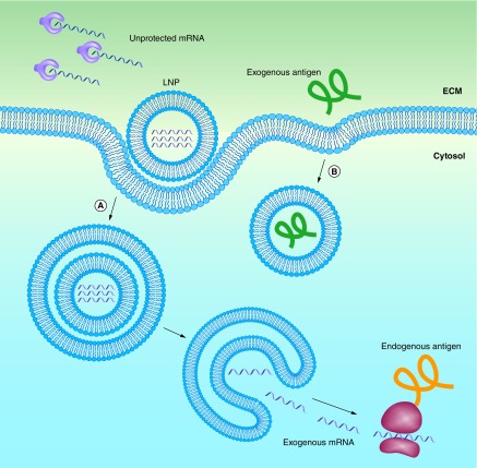
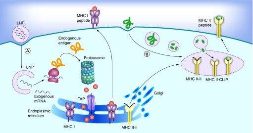
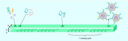
mRNA vaccines elicit a potent immune response including antibodies and cytotoxic T cells. mRNA vaccines are currently evaluated in clinical trials for cancer immunotherapy applications, but also have great potential as prophylactic vaccines. Efficient delivery of mRNA vaccines will be key for their success and translation to the clinic. Among potential nonviral vectors, lipid nanoparticles are particularly promising. Indeed, lipid nanoparticles can be synthesized with relative ease in a scalable manner, protect the mRNA against degradation, facilitate endosomal escape, can be targeted to the desired cell type by surface decoration with ligands, and as needed, can be codelivered with adjuvants.

Keywords: adjuvant; cancer immunotherapy; cationic lipid; drug delivery; lipid nanoparticle; mRNA; oligonucleotide; therapeutic vaccine; vaccine.


Conflict of interest statement

Financial & competing interests disclosure

This work was funded by the National Institutes of Health (Grant# EB 000244). Robert Langer is a co-founder and member of the board of directors of Moderna therapeutics. The authors have no other relevant affiliations or financial involvement with any organization or entity with a financial interest in or financial conflict with the subject matter or materials discussed in the manuscript apart from those disclosed.

No writing assistance was utilized in the production of this manuscript.

Figures

<b>Figure 1.</b>
Figure 1.. Lipid nanoparticles protect mRNA from degradation, and facilitate endocytosis and endosomal escape.
(A) mRNA can be encapsulated in lipid nanoparticles (LNPs) for protection from enzymatic degradation. A positively charged LNP favors localization of mRNA at the negatively charged cell membrane, including subsequent endocytosis into the cytosol. In order to be transcribed, the mRNA must escape both the LNP and the endosome. (B) Extracellular proteins based vaccines are endocytosed in a similar manner, but do not need to escape from the endosome to be presented on MHCII.
<b>Figure 2.</b>
Figure 2.. Antigen presentation on MHC I and II pathways in dendritic cells.
(A) Endogenous proteins with pathogen or self origin are primarily displayed on the MHC I pathway. These proteins are degraded into smaller peptides by the proteasome. The peptides are transported into the endoplasmic reticulum for loading onto the MHC class I molecules. This MHC I–peptide complex is then displayed at the cell surface to CD8 T-cells. (B) On the other hand, proteins that enter the cell on the endocytic route are displayed on the MHC II pathway. For this purpose, the MHC class II molecules are protected with the invariant chain (Ii) from binding to endogenous peptides in the endoplasmic reticulum. The MHC II-Ii complex is then exported through the Golgi to the MIIC/CIIV compartment, where the invariant chain is replaced with antigens. The MHC II–peptide complex is then displayed at the cell surface to CD4 T-cells.
<b>Figure 3.</b>
Figure 3.. Staggered herringbone mixer for lipid nanoparticle synthesis.
Lipids dissolved in ethanol and an aqueous buffer of mRNA are pumped into the two primary inlets of the microfluidic mixer using syringe pumps. The herringbone structures induce chaotic advection in the laminar flow that enables rapid mixing of ethanol and the aqueous phase. Although the mixing time depends on the flow rate, approximately 15 cycles are needed for complete mixing. The optional secondary inlet can be used to prevent lipid nanoparticle fusion by further dilution with buffer, or to add water-soluble lipid derivatives to the lipid nanoparticles. Approximate dimension are w = 200 μm, h = 77 μm, a = 18 μm.
<b>Figure 4.</b>
Figure 4.. Injection of a cationic lipid nanoparticle formulation into the SC induces inflammation.
The control figure is a mouse skin section 24 h after a saline injection. The injected mouse figure corresponds to a mouse skin section 24 h after lipid nanoparticle injection, coding for green fluorescent protein. An infiltration of monocytes, characterized by a higher density of blue dots, is visible below the cutaneous muscle layer. The lipid nanoparticle consisted of C12-200, DOPE, cholesterol, and a PEGylated lipid.

Similar articles

Cited by

References

    1. Crawford NW, Bines JE, Royle J, Buttery JP. Optimizing immunization in pediatric special risk groups. Expert Rev. Vaccines. 2011;10(2):175–186. - PubMed
    1. Liu MA. Immunologic basis of vaccine vectors. Immunity. 2010;33(4):504–515. - PubMed
    1. Hilleman MR. Recombinant vector vaccines in vaccinology. Dev. Biol. Stand. 1994;82:3–20. - PubMed
    1. Deering RP, Kommareddy S, Ulmer JB, Brito LA, Geall AJ. Nucleic acid vaccines: prospects for non-viral delivery of mRNA vaccines. Expert Opin. Drug Deliv. 2014;11(6):885–899. - PubMed
    1. Pascolo S. Vaccination with messenger RNA (mRNA) In: Bauer PDS, Hartmann PDG, editors. Toll-Like Receptors (TLRs) and Innate Immunity. Springer Berlin Heidelberg; Germany: 2008. pp. 221–235.

Publication types