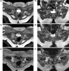
1.0 s Ultrafast MRI in non-sedated infants after reduction with spica casting for developmental dysplasia of the hip: a feasibility study
- PMID: 27075984
- PMCID: PMC4909649
- DOI: 10.1007/s11832-016-0734-8
1.0 s Ultrafast MRI in non-sedated infants after reduction with spica casting for developmental dysplasia of the hip: a feasibility study
Abstract
Purpose: The aim of this study was to first develop and use 1.0 s ultrafast magnetic resonance imaging (MRI) to confirm the location of the femoral head in non-sedated infants with developmental dysplasia of the hip (DDH) after reduction with spica cast application in clinical settings.
Methods: The ultrafast acquisition was achieved by employing a balanced steady-state free precession sequence and immobilizing the patient with dedicated sandbags. On completion of the ultrafast MRI study, all infants were sedated for conventional MRI scanning. Two orthopaedic surgeons retrospectively evaluated the image quality, result of the reduction and total MRI study time (including patient immobilization, coil setup, and scanning) in 14 DDHs of 13 infants (one with bilateral DDHs).
Results: Both reviewers stated that there were no motion artefacts for non-sedated infants during the ultrafast MRI and that the quality of both the ultrafast and conventional MRI images were acceptable to assess the femoral head location. Assessment of the reduction procedure resulted in two hips being categorized as 'incomplete reduction' requiring a re-reduction procedure. The total study time of ultrafast and conventional MRI was 6 ± 1 min and 14 ± 3 min, respectively (P < 0.001). No complications due to sedation, such as hypoxia, were reported. The average sedation waiting time was 1 h 25 min ± 34 min.
Conclusion: The ultrafast MRI procedure reported here can be readily employed to confirm the location of the femoral head in infants with DDHs, without the use of any sedation.
Keywords: Developmental dysplasia of the hip; Femoral head location; Reduction; Spica casting; Ultrafast magnetic resonance imaging.
Figures


Similar articles
-
Reliability of indices measured on infant hip MRI at time of spica cast application for dysplasia.Hip Int. 2014 Jul-Aug;24(4):405-16. doi: 10.5301/hipint.5000143. Epub 2014 May 30. Hip Int. 2014. PMID: 24970320
-
Axial STIR MRI: a faster method for confirming femoral head reduction in DDH.J Child Orthop. 2009 Jun;3(3):223-7. doi: 10.1007/s11832-009-0170-0. Epub 2009 May 5. J Child Orthop. 2009. PMID: 19415363 Free PMC article.
-
Does Perfusion MRI After Closed Reduction of Developmental Dysplasia of the Hip Reduce the Incidence of Avascular Necrosis?Clin Orthop Relat Res. 2016 May;474(5):1153-65. doi: 10.1007/s11999-015-4387-6. Clin Orthop Relat Res. 2016. PMID: 26092677 Free PMC article.
-
Outcome Prognostic Factors in MRI during Spica Cast Therapy Treating Developmental Hip Dysplasia with Midterm Follow-Up.Children (Basel). 2022 Jul 7;9(7):1010. doi: 10.3390/children9071010. Children (Basel). 2022. PMID: 35883994 Free PMC article. Review.
-
Cochrane Review: Screening programmes for developmental dysplasia of the hip in newborn infants.Evid Based Child Health. 2013 Jan;8(1):11-54. doi: 10.1002/ebch.1891. Evid Based Child Health. 2013. PMID: 23878122 Review.
Cited by
-
Docking phenomenon and subsequent acetabular development after gradual reduction using overhead traction for developmental dysplasia of the hip over six months of age.J Child Orthop. 2021 Dec 1;15(6):554-563. doi: 10.1302/1863-2548.15.210143. J Child Orthop. 2021. PMID: 34987665 Free PMC article.
-
Magnetic resonance imaging evaluation of the labrum to predict acetabular development in developmental dysplasia of the hip: A STROBE compliant study.Medicine (Baltimore). 2017 May;96(21):e7013. doi: 10.1097/MD.0000000000007013. Medicine (Baltimore). 2017. PMID: 28538419 Free PMC article.
-
Magnetic resonance imaging follow-up can screen for soft tissue changes and evaluate the short-term prognosis of patients with developmental dysplasia of the hip after closed reduction.BMC Pediatr. 2021 Mar 8;21(1):115. doi: 10.1186/s12887-021-02587-2. BMC Pediatr. 2021. PMID: 33685416 Free PMC article.
-
Intra- and interobserver variability of novel magnetic resonance imaging parameters for hip screening and treatment outcomes at age 5 years.Pediatr Radiol. 2023 Mar;53(3):415-425. doi: 10.1007/s00247-022-05565-7. Epub 2023 Jan 9. Pediatr Radiol. 2023. PMID: 36622404
References
-
- Fukiage K, Futami T, Ogi Y, Harada Y, Shimozono F, Kashiwagi N, Takase T, Suzuki S (2015) Ultrasound-guided gradual reduction using flexion and abduction continuous traction for developmental dysplasia of the hip: a new method of treatment. Bone Joint J 97-B:405–411. doi:10.1302/0301-620X.97B3.34287 - PubMed
-
- Gardner RO, Bradley CS, Howard A, Narayanan UG, Wedge JH, Kelley SP (2014) The incidence of avascular necrosis and the radiographic outcome following medial open reduction in children with developmental dysplasia of the hip: a systematic review. Bone Joint J 96-B:279–286. doi:10.1302/0301-620X.96B2.32361 - PubMed
LinkOut - more resources
Full Text Sources
Other Literature Sources
Research Materials
Miscellaneous

