. 2016 Apr 12;23(4):622-34.
doi: 10.1016/j.cmet.2016.03.005.

ERRγ Is Required for the Metabolic Maturation of Therapeutically Functional Glucose-Responsive β Cells

Affiliations

ERRγ Is Required for the Metabolic Maturation of Therapeutically Functional Glucose-Responsive β Cells

Eiji Yoshihara et al. Cell Metab. .

Abstract

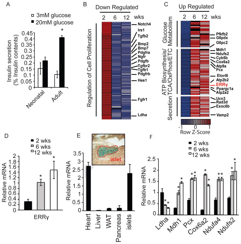
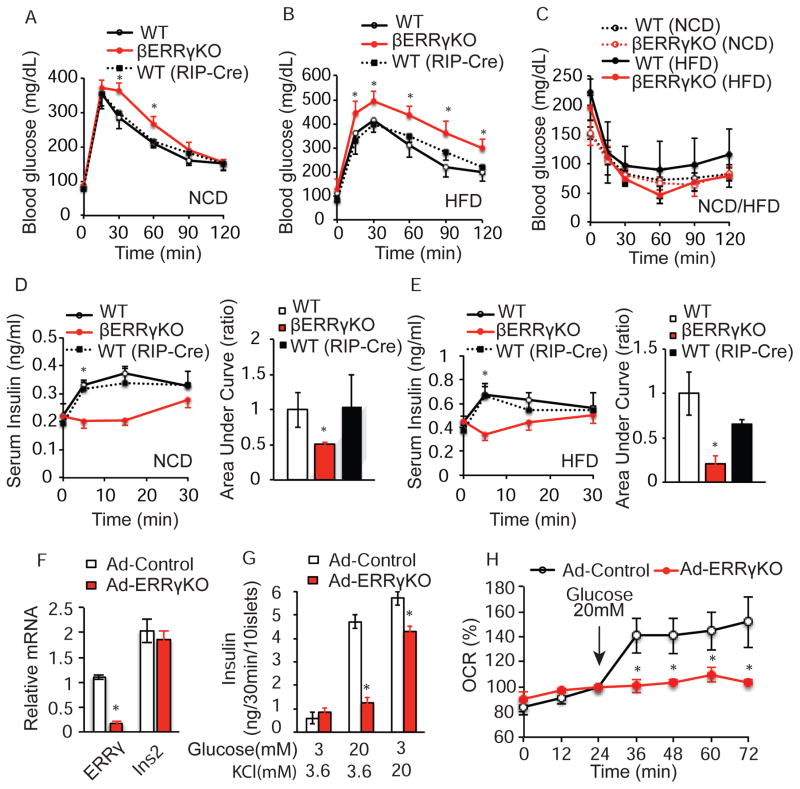
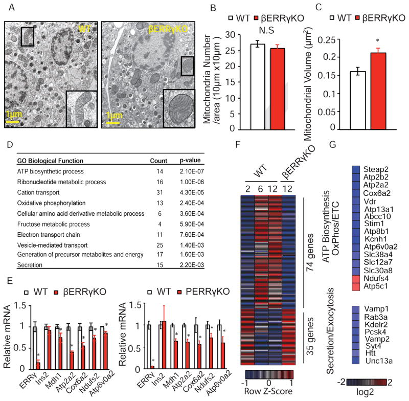
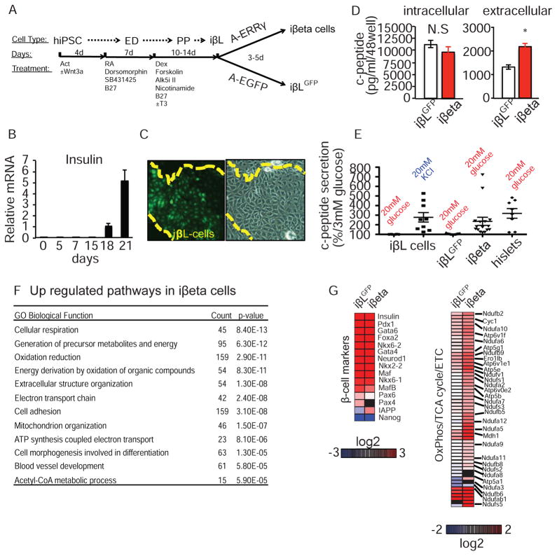
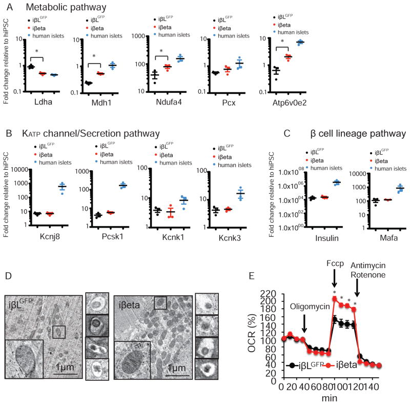
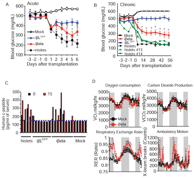
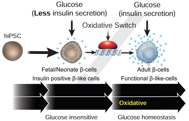
Pancreatic β cells undergo postnatal maturation to achieve maximal glucose-responsive insulin secretion, an energy intensive process. We identify estrogen-related receptor γ (ERRγ) expression as a hallmark of adult, but not neonatal β cells. Postnatal induction of ERRγ drives a transcriptional network activating mitochondrial oxidative phosphorylation, the electron transport chain, and ATP production needed to drive glucose-responsive insulin secretion. Mice deficient in β cell-specific ERRγ expression are glucose intolerant and fail to secrete insulin in response to a glucose challenge. Notably, forced expression of ERRγ in iPSC-derived β-like cells enables glucose-responsive secretion of human insulin in vitro, obviating in vivo maturation to achieve functionality. Moreover, these cells rapidly rescue diabetes when transplanted into β cell-deficient mice. These results identify a key role for ERRγ in β cell metabolic maturation, and offer a reproducible, quantifiable, and scalable approach for in vitro generation of functional human β cell therapeutics.


Figures

Figure 1
Figure 1. Islets acquire oxidative features postnatally
(A) Glucose-stimulated insulin secretion from freshly isolated mouse neonatal (< 14 days) and adult (> 12 weeks) islets after sequential perfusion with 3mM and 20mM glucose for 30 min (reported as % insulin content, 10 islets per assay, n=6). (B–C) Heatmaps of transcriptional changes in islets during postnatal maturation. Row Z-score of down-regulated (B), and up-regulated (C) genes. (D) Relative Errγ expression in isolated islets measured by qPCR. (E) Relative Errγ expression in murine 12 wk old heart, liver, white adipose (WAT), pancreas and islets (n=4). X-gal staining indicating ERRγ expression in islets from 12 wk old ERRγ knock-in mice (top panel). (F) Relative expression of Ldha and selected mitochondrial metabolic genes during postnatal islet maturation, as measured by qPCR. (n=3). Data represent the mean ±s.e.m. *p<0.01 Student’s unpaired t-test. See also Figure S1.
Figure 2
Figure 2. ERRγ expression in β cells is required for glucose homeostasis
(A–B) Intra-peritoneal glucose tolerance test (IP-GTT, 2g/kg, 8 wk old mice) of ERRγfl/fl (WT n=8), βERRγKO (n=6), and RIP-Cre (WT (RIP-Cre), n= 3) mice on a normal chow diet (NCD) (A), and a high fat diet (HFD, 4 weeks of HFD after weaning) (B). (C) Intra-peritoneal insulin tolerance test (IP-ITT) of WT and βERRγKO mice on NCD (9 wk old, WT n=4, βERRγKO n=5) and HFD (5 wk HFD after weaning; WT n=8, βERRγKO n=8). (D–E) Serum insulin level during IP-GTT in 8 wk old mice under NCD (WT n=4, βERRγKO n=12, WT(RIP-Cre) n=5) (D), and after 4 wks HFD (WT n=6, βERRγKO n=6, and WT(RIP-Cre), n=6) (E). Bar graphs indicate relative area under the curve (AUC). (F) Relative Errγ and Insulin 2 (Ins2) expression, (G) ex vivo glucose-stimulated (20mM) or KCl-stimulated (20mM) insulin secretion, and (H) oxygen consumption rate (OCR) in adenoviral-EGFP (Ad-Control) and adenoviral-Cre (Ad-ERRγKO) infected ERRγfl/fl islets from 12 wk old mice. Data represent the mean ±s.e.m. *p<0.05 Student’s unpaired t-test. See also Figures S2 and S3.
Figure 3
Figure 3. ERRγ is required for functional maturation of β cells
(A) Electron microscopy images showing mitochondrial morphology, (B) mitochondria number, and (C) mitochondrial volume in β cells from ERRγfl/fl (WT) and βERRγKO islets (n=8, 12 wk old mice). (D) Functional annotation of dysregulated gene categories in βERRγKO islets identified by Gene Ontology (GO). (E) Relative expression of metabolic genes in βERRγKO (left panel) and pancreatic ERRγKO (ERRγfl/fl x PDX1-Cre, PERRγKO, right panel) islets, as measured by qPCR. (F) Heatmap of gene expression levels (Row Z-score) during functional maturation of islets compared to βERRγKO islets. (G) Heatmap of expression changes in selected metabolic and secretion/exocytosis pathway genes in βERRγKO islets. Data represent the mean ±s.e.m. *p<0.05 Student’s unpaired t-test. See also Figure S4.
Figure 4
Figure 4. ERRγ promotes maturation of human iPSC-derived β-like cells
(A) Schematic protocol for iPSC-derived β-like cell generation (ED, endoderm; PP, pancreatic progenitors; iβL, iPSC-derived β-like cells; iβeta, ERRγ-expressing iPSC-derived β-like cells). Growth factors and small molecules were added at each stage. Vitamin C, GABA and 13.6 mM glucose were in the base media for all stages. (B) Relative expression of human insulin during iPSC differentiation (C) Human insulin reporter-driven GFP expression (left panel) and phase contrast image (right) of day 22 iβL cells. (D) Intracellular (left) and extracellular (right) c-peptide concentrations of iβL cells after adenoviral infection: Ad-EGFP infected, iβLGFP cells, open bars; Ad-ERRγ infected, iβeta cells, red bars. (E) Induced c-peptide secretion in iβL cells, iβLGFP cells, iβeta cells, and human islets. (F) Functional annotation of upregulated gene categories in iβeta cells identified by Gene Ontology (GO). (G) Heatmap of expression changes in known β cell maker genes (left) and metabolic genes (right) in iβLGFP and iβeta cells (log2 ratio relative to undifferentiated iPSC). Data represent the mean ±s.e.m. *p<0.05 Student’s unpaired t-test. See also Figure S5.
Figure 5
Figure 5. ERRγ regulates oxidative capacity through metabolic gene expression inβ-like cells
(A–C) Relative expression of metabolic (A), KATP channel related (B) and β cell lineage genes (C) in iβLGFP cells, iβeta cells and human islets, as determined by qPCR (n=3). (D) Electron microscopy images showing mitochondrial morphology (left panel) and crystallized insulin granules (right panels) in iβLGFP and iβeta cells, respectively. (E) Oxidative capacity of iβLGFP and iβeta cells, as measured by oxygen consumption rate (OCR). *p<0.05 Student’s unpaired t-test.
Figure 6
Figure 6. iβeta cells restore glucose homeostasis in diabetic mice
(A) Acute effects on ad lib fed blood glucose levels in STZ-induced hyperglycemic NOD-SCID mice after mock transplantation (n=3), transplantation of iβLGFP cells (n=8), iβeta cells (n=7) and mouse islets (n=5). (B) Chronic effects on ad lib fed blood glucose levels of mock transplantation (n=3), transplantation of iβLGFP cells (n=14/12; 2 mice died 2 wks after transplantation), iβeta cells (n=13), mouse islets (n=5) and human islets (n=2). (C) Human c-peptide levels before and 15 minutes after a glucose challenge (2g/kg) in mice 2 months after the indicated transplantation (c-peptide ELISA limit of detection indicated by dotted line). (D) Oxygen consumption (VO2), carbon dioxide production (VCO2), Respiratory Exchange Ratio (RER), and ambulatory motion of STZ treated NOD-SCID mice 56 days after mock (n=4) or iβeta cell transplantation (mice with blood glucose levels <250mg/dL, n=4). See also Figures S6 and S7.
Figure 7
Figure 7. ERRγ is required for the functional maturation of β cells and iβeta cells
Schematic describing the role of ERRγ in regulating an oxidative switch necessary for glucose stimulated insulin secretion in β and iβeta cells.

Comment in

References

    1. Alaynick WA, Kondo RP, Xie W, He W, Dufour CR, Downes M, Jonker JW, Giles W, Naviaux RK, Giguere V, et al. ERRgamma directs and maintains the transition to oxidative metabolism in the postnatal heart. Cell metabolism. 2007;6:13–24. - PubMed
    1. Anello M, Lupi R, Spampinato D, Piro S, Masini M, Boggi U, Del Prato S, Rabuazzo AM, Purrello F, Marchetti P. Functional and morphological alterations of mitochondria in pancreatic beta cells from type 2 diabetic patients. Diabetologia. 2005;48:282–289. - PubMed
    1. Ashcroft FM, Rorsman P. Diabetes mellitus and the beta cell: the last ten years. Cell. 2012;148:1160–1171. - PMC - PubMed
    1. Bliss CR, Sharp GW. Glucose-induced insulin release in islets of young rats: time-dependent potentiation and effects of 2-bromostearate. The American journal of physiology. 1992;263:E890–896. - PubMed
    1. Blum B, Hrvatin SS, Schuetz C, Bonal C, Rezania A, Melton DA. Functional beta-cell maturation is marked by an increased glucose threshold and by expression of urocortin 3. Nature biotechnology. 2012;30:261–264. - PMC - PubMed

Publication types

MeSH terms