. 2016 Apr 14:6:24090.
doi: 10.1038/srep24090.

Tuning of AKT-pathway by Nef and its blockade by protease inhibitors results in limited recovery in latently HIV infected T-cell line

Affiliations

Tuning of AKT-pathway by Nef and its blockade by protease inhibitors results in limited recovery in latently HIV infected T-cell line

Amit Kumar et al. Sci Rep. .

Abstract

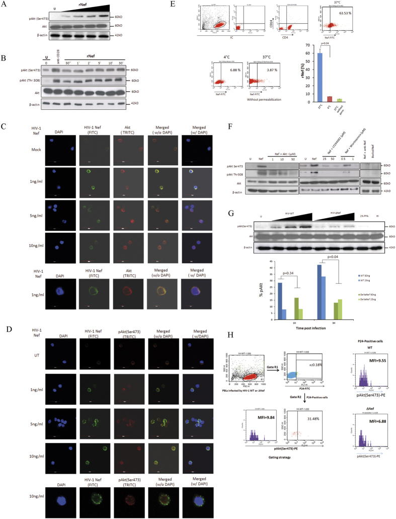
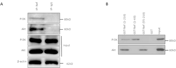
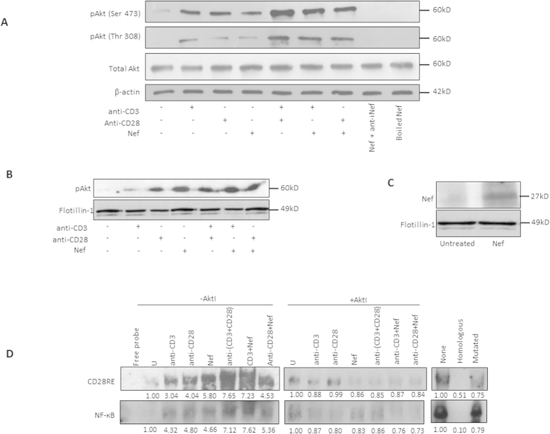
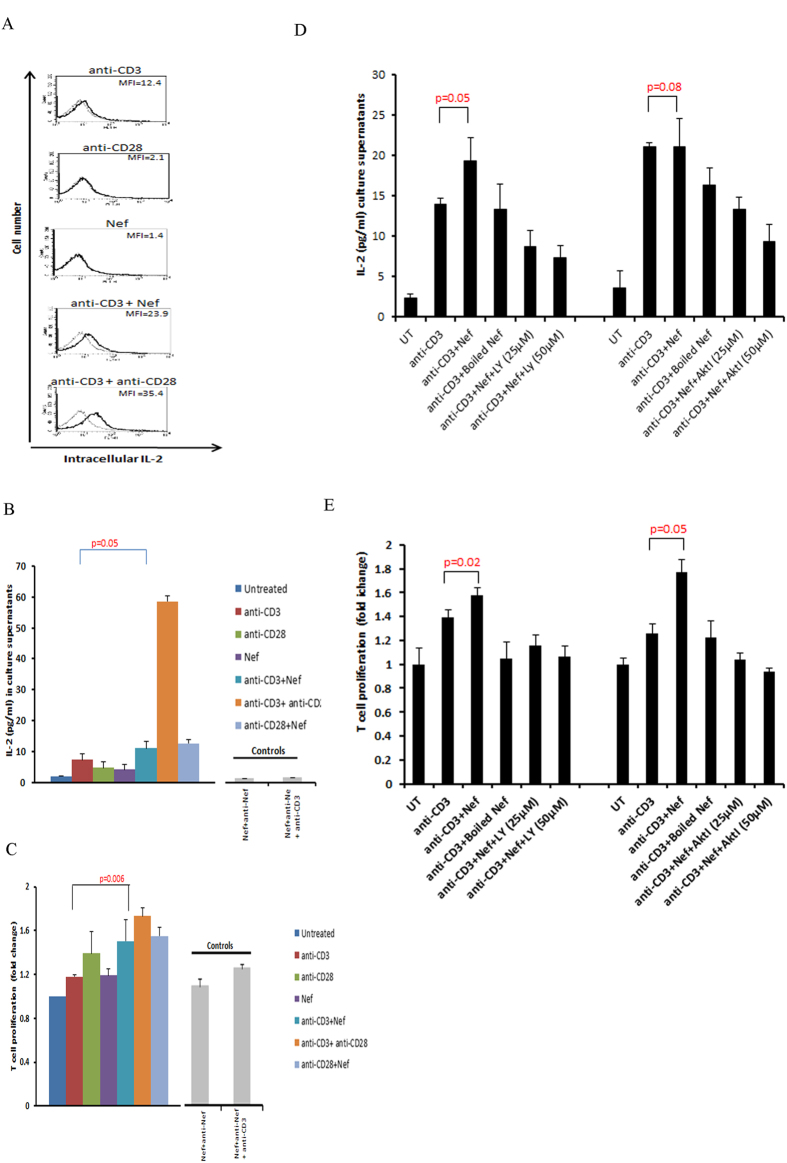
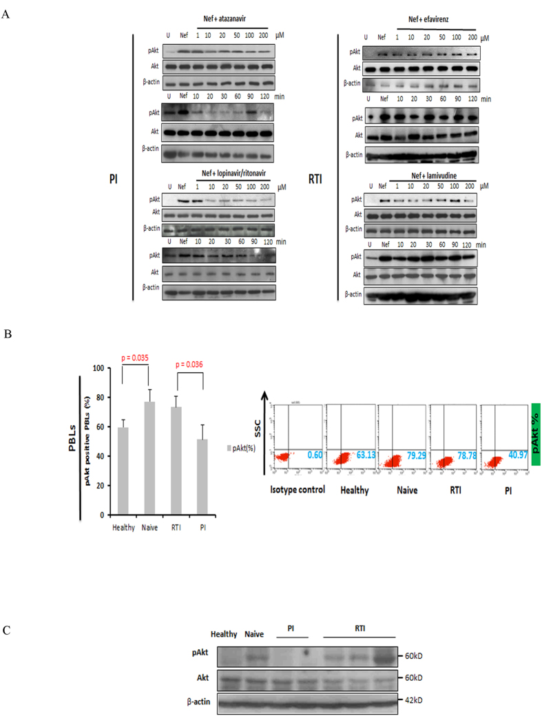
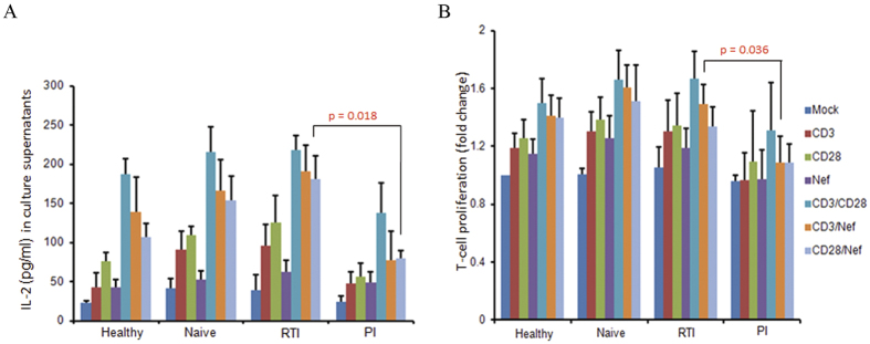
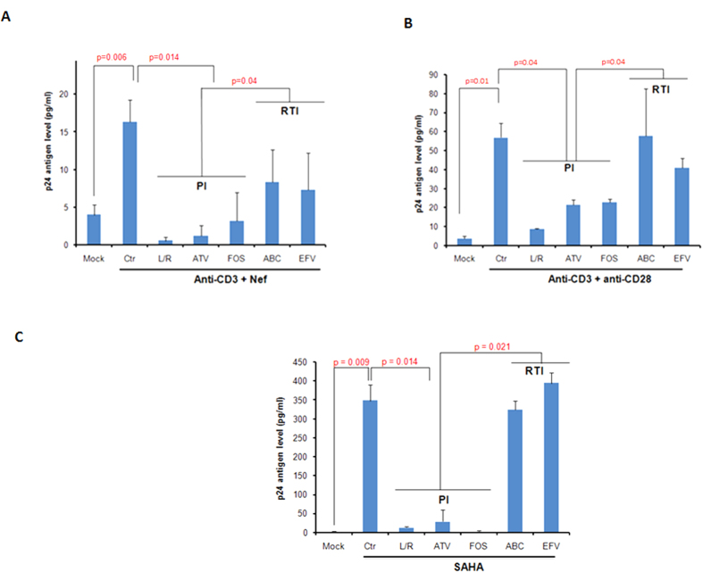
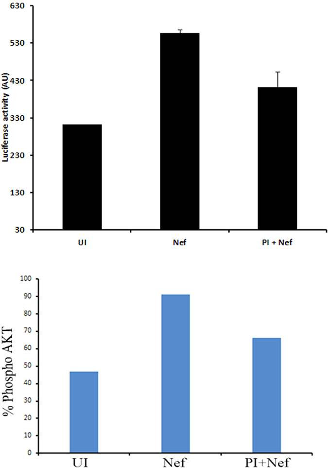
Akt signaling plays a central role in many biological processes, which are key players in human immunodeficiency virus 1 (HIV-1) pathogenesis. We found that Akt interacts with HIV-1 Nef protein. In primary T cells treated with exogenous Nef or acutely infected with Nef-expressing HIV-1 in vitro, Akt became phosphorylated on serine(473) and threonine(308). In vitro, Akt activation mediated by Nef in T-cells was blocked by HIV protease inhibitors (PI), but not by reverse transcriptase inhibitors (RTI). Ex vivo, we found that the Akt pathway is hyperactivated in peripheral blood lymphocytes (PBLs) from cART naïve HIV-1-infected patients. PBLs isolated from PI-treated patients, but not from RTI-treated patients, exhibited decreased Akt activation, T-cell proliferation and IL-2 production. We found that PI but not RTI can block HIV-1 reactivation in latently infected J-Lat lymphoid cells stimulated with various stimuli. Using luciferase measurement, we further confirmed that Nef-mediated reactivation of HIV-1 from latency in 1G5 cells was blocked by PI parallel to decreased Akt activation. Our results indicate that PI-mediated blockade of Akt activation could impact the HIV-1 reservoir and support the need to further assess the therapeutic use of HIV-1 PI in order to curtail latently infected cells in HIV-1-infected patients.


Conflict of interest statement

G.H. and O.R. are editorial board members. The authors declare no other conflict of interest.

Figures

Figure 1
Figure 1. HIV-1 Nef is internalized by CD4 + T cells and activates Akt in PBLs which is mediated via PI3K in a dose and time dependent manners.
(A), Dose-dependent (n = 3) and (B), Time-dependent activation of Akt (pAkt(Ser473)) in PBLs treated with rNef (n = 3). (B) Five million PBLs were either left untreated or treated with rNef (100 ng/ml) for various period of time (30 seconds to 30 minutes). As a positive control, PBLs were treated with anti-CD28 antibody. Expression of pAkt (Ser473, Thr308), total Akt and β-actin were detected by standard western blotting method as described in materials and methods (n = 3). (C,D) A series of confocal images showing internalization and colocalization of HIV-1 Nef and Akt (C) at serum concentration of Nef (1 to 10 ng/ml) and a dose response of rNef treatment on Akt activation (D) in PBLs isolated from healthy donors. (E), Internalization of rNef by CD4+ T cells determined by flow cytometry. Five million CD4+ T cells were treated with rNef for 30 min at 37 °C and 4 °C with and without permeabilization. Expression of rNef was determined by confocal microscopy (n = 3).(F), Activation of Akt in PBLs treated with rNef is mediated via PI3K. Western blot detection of activated pAkt (Ser473, Thr308) in the lysates derived from 5 × 106 PBLs treated with 100 ng/ml of Nef with or without Akt (Akt inhibitor VIII) and PI3K inhibitors (LY294002 and Wortmannin) (n = 3). (G), Akt activation in PBLs by wild-type HIV-1, but not by isogenic HIV-1∆Nef. Five million PBLs were infected with various doses (of p24) of either wild type HIV-1 or ∆Nef virus infectious clones. Post infection lysates were prepared and expression of pAkt(Ser473), was determined by western blotting (upper panel) and flow cytometry (n = 2) (lower panel). (H), Left panel: Gating strategy for flow cytometry analysis. In this sample gating cells were first gated for PBLs (Gate R1). R1 gate is further analysed for p24 positive PBLs (Gate R2). Gate R2 is analyzed for the expression of pAkt. Right panel is the representative data of pAKt positive cells in p24 positive population obtained by infection of PBLs with WT and Delta Nef virus.
Figure 2
Figure 2. HIV-1 Nef interacts with Akt and PI3K.
(A), Nef interacts with Akt and PI3K in PBLs treated with rNef as detected by co-immunoprecipitation. Five million PBLs isolated from healthy donor were left untreated or treated with HIV-1 rNef (100 ng/ml) and subjected to immunoprecipitation with anti-HIV Nef-Ab or isotype control antibody followed by western blot using indicated antibodies (n = 3). (B), Nef interacts with Akt and PI3K via its C-terminus and N-terminus respectively, as measured by pull-down assays. Representative western blot analysis of pull down assay using 1500 μg of PBLs lysates with 20 μg of different HIV-Nef constructs [N-terminus, 1-60aa, C-terminus 55-210aa and full length Nef, 1-210 aa] expressed in BL21DE as indicated. Input corresponds to 10% of the material used for pull down. Presence of PI3K and Akt was detected by respective antibodies mentioned in Material and Method section (n = 3).
Figure 3
Figure 3. Nef triggers the activation of NF-ĸB and CD28RE via lipid raft mediated Akt signaling.
(A), Exogenous Nef activates Akt in TCR stimulated PBLs as measured by the detection of pAkt(Ser473). Five million PBLs were either left untreated or stimulated in various combinations: rNef only, anti-CD3-mAb only, anti-CD3-mAb + rNef (100 ng/ml), anti-CD28-mAb only, anti-CD28-mAb + rNef (100 ng/ml) and anti-CD3-mAb + anti-CD28-mAb) for 30 min. Expression of pAkt(Ser473 Thr308), total Akt and β-actin was determined by western blotting (n = 2). As control boiled rNef and rNef incubated with anti-Nef antibody were also included. (B,C), Detection of pAkt(Ser473) (B) and rNef (C) in lipid raft fractions isolated from PBLs treated with rNef. Five million PBLs were either left untreated or stimulated in various combinations: rNef only, anti-CD3-mAb only, anti-CD3-mAb + rNef (100 ng/ml), anti-CD28-mAb only, anti-CD28-mAb + rNef (100 ng/ml) and anti-CD3-mAb + anti-CD28-mAb) for 30 min. Cell lysates were prepared in 1% triton X-100 and subjected to ultracentrifugation over a sucrose gradient. Lipid rafts fractions were isolated and expression of pAkt (Ser 473), Nef and flotillin-1 were determined by western blotting (n = 5). Flotillin-1 was used as a lipid raft associated marker. (D), Akt-dependent NF-κB and CD28RE activation in PBLs treated with rNef. Five million PBLs were either left untreated or treated with rNef (100 ng/ml) for 30 min in the presence and absence of TCR stimulation (anti-CD3-mAb, anti-CD28-mAb), and pre-treated or not with the Akt inhibitor VIII for 2 hrs. Electrophoretic mobility shift assays was performed to measured CD28 responsive element and NF-kB activation in response to stimuli on a 6% native polyacrylamide gel as described in the materials and methods (n = 3).
Figure 4
Figure 4. In vitro rNef-mediated Akt-dependent hyperactivation of T cells favors increased IL-2 production and T-cell proliferation.
(A,B) Exogenous HIV-1 Nef triggers IL-2 production in TCR-stimulated T-cells. (A), Five million PBLs were stimulated with anti-CD3-mAb (5 μg/ml), anti-CD3-mAb + rNef, anti-CD28-mAb (5 μg/ml), anti-CD28-mAb + anti-CD3-mAb or rNef (100 ng/ml) alone for 24 h. Post treatment cells were fixed, permeabilized and expression of intracellular IL-2 was determined by flow cytometry. Flow cytometric data indicating the mean fluorescence intensity (MFI) for intracellular IL-2 production. Data are representative of 5 independent experiments. (B), Five million PBLs were either left untreated or treated with rNef (100 ng/ml), anti-CD3-mAb (5 μg/ml), anti-CD28-mAb (5 μg/ml), anti-CD3-mAb + rNef, anti-CD28-mAb + rNef, and antiCD3-mAb + antiCD28-mAb for 24 h. Post 24 h supernatants were collected and IL-2 production was measured in supernatants by ELISA. Means ± s.d. (n = 3). (C), Exogenous HIV-1 Nef triggers cell proliferation in TCR-stimulated T cells. Five million PBLs were either left untreated or treated with rNef (100 ng/ml), anti-CD3-mAb (5 μg/ml), anti-CD28-mAb (5μg/ml), anti-CD3-mAb + rNef, anti-CD28-mAb + rNef and antiCD3-mAb+ antiCD28-mAb in the absence of Akt I and PI3K inhibitor for 24 h. Cell proliferation was measured using MTT cell proliferation assay. Means ± s.d. (n = 3). (D,E), Exogenous HIV-1 Nef triggers IL-2 production and T cell proliferation in CD3 stimulated cells in PI3K/Akt dependent manner. Five million PBLs were either left untreated or treated with Akt I (25 μM, 50 μM) and PI3K inhibitor LY294002 (25 μM, 50 μM) for 2 h followed by treatment with rNef (100 ng/ml), heat denatured rNef (100 ng/ml), anti-CD3-mAb (5 μg/ml) and anti-CD3-mAb + rNef, for 24 h. After 24 h supernatants were collected and IL-2 production and T cell proliferation was determined by ELISA and MTT assay respectively. Means ± s.d. (n = 3). LY: LY294002; AktI: Akt inhibitor VIII.
Figure 5
Figure 5. HIV-1 protease inhibitors block Akt activation in rNef-treated PBLs in vitro and ex vivo in PBLs isolated from cART-treated patients.
(A), HIV protease inhibitors, but not RTIs, inhibit pAkt(Ser473) activation in a dose dependent manner in PBLs treated with rNef as determined by western blot. Five million PBLs were treated with rNef (100 ng/ml) for 30 min. Expression of pAkt(Ser473), total Akt and β-actin was determined by western blotting (n = 8). (B), Decreased Akt activation in PBLs from PI-treated patients compared to RTI-treated and cART naive patients as measured by flow cytometric analysis. Mean values ± s.d. (n = 5). (C), Decreased Akt activation in PBLs from PI-treated patients compared to RTI-treated and cART naive patients as measured by western-blotting.
Figure 6
Figure 6. Reduced IL-2 production and T-cell proliferation in PBLs isolated from PI-treated patients.
(A,B), Lower IL-2 production (A) and decreased proliferation of PBLs (B) in PI-treated patients compared to RTI-treated and cART naive patients, respectively. PBLs were isolated from healthy, naïve and HIV-1 infected individuals under PI and RTI treatment. Five million PBLs were either left untreated or treated with rNef (100 ng/ml), anti-CD3-mAb (5 μg/ml), anti-CD28-mAb (5 μg/ml), anti-CD3-mAb + rNef, anti-CD8-mAb + rNef, and anti-CD3-mAb + anti-CD28-mAb for 24 h. IL-2 production (A) and cell proliferation (B) was measured using ELISA and MTT cell proliferation assay respectively. Means ± s.d. (n = 5).
Figure 7
Figure 7. HIV-1 protease inhibitors block HIV-1 recovery from latently infected J-Lat 8.4 cells.
(AC), J-Lat cells were either left untreated or treated with anti-CD3-mAb (2 μg/ml) +rNef (100 ng/ml) (A), anti-CD28-mAb (2.67 μg/ml) +anti-CD3-mAb (2 μg/ml) (B) and SAHA (2.5 μM) (C) in the absence or presence of different PI and RTI compounds as indicated. The viral production was determined by measuring the level of p24 antigen using ELISA. Means ± s.d. (n = 6). Ctr: Control; L/R, lopinavir/ritonavir; ATV: atazanavir; FOS: fosamprenavir, ABC: abacavir; EFV: efavirenz.
Figure 8
Figure 8. rNef induces HIV-1 expression in a latent model cell line 1G5 and protease inhibitor can subsequently block it.
Five million 1G5 cells were either left untreated or treated with rNef in the presence or absence of protease inhibitor. Post 24 h of treatment cells, the expression of luciferase was determined (upper panel) and expression of pAkt was determined by flow cytometry (lower panel). The data shown are representative of two independent experiments.

Similar articles

Cited by

References

    1. Fayard E., Xue G., Parcellier A., Bozulic L. & Hemmings B. A. Protein kinase B (PKB/Akt), a key mediator of the PI3K signaling pathway. Curr. Top. Microbiol. Immunol. 346, 31–56 (2010). - PubMed
    1. Malim M. H. & Emerman M. HIV-1 accessory proteins–ensuring viral survival in a hostile environment. Cell Host. Microbe 3, 388–398 (2008). - PubMed
    1. Maartens G., Celum C. & Lewin S. R. HIV infection: epidemiology, pathogenesis, treatment, and prevention. Lancet 384, 258–271 (2014). - PubMed
    1. Deng K. & Siliciano R. F. HIV: Early treatment may not be early enough. Nature 512, 35–36 (2014). - PubMed
    1. Liu X. & Kumar A. Differential signaling mechanism for HIV-1 Nef-mediated production of IL-6 and IL-8 in human astrocytes. Sci Rep. 5, 9867 (2015). - PMC - PubMed

Publication types

MeSH terms

Substances