. 2016 Apr 13;36(15):4182-95.
doi: 10.1523/JNEUROSCI.4317-15.2016.

Monocyte-Derived Macrophages Contribute to Spontaneous Long-Term Functional Recovery after Stroke in Mice

Affiliations

Monocyte-Derived Macrophages Contribute to Spontaneous Long-Term Functional Recovery after Stroke in Mice

Somsak Wattananit et al. J Neurosci. .

Abstract

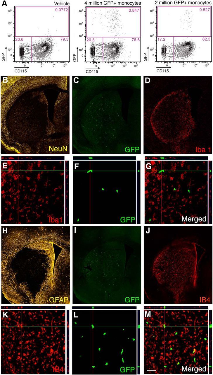
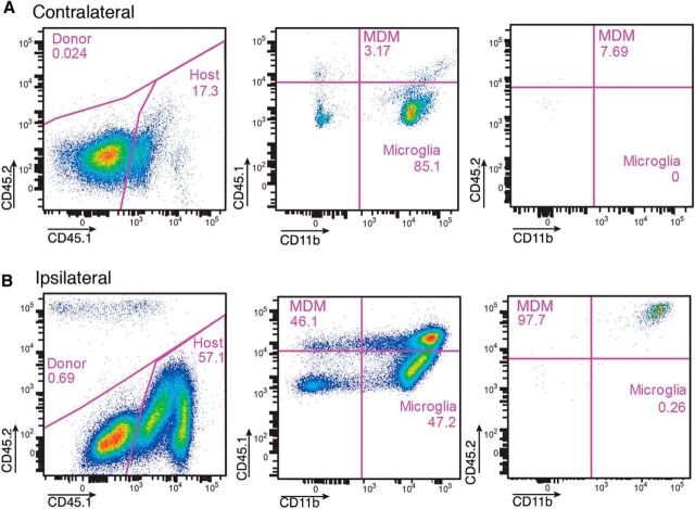
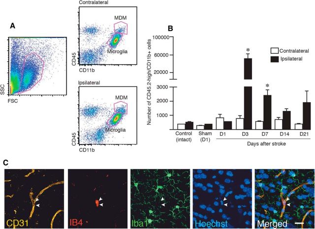
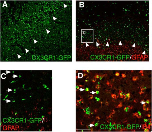
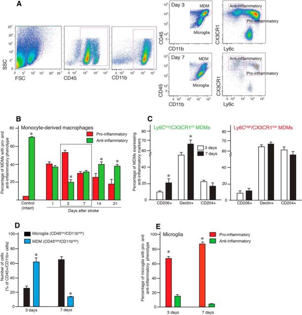
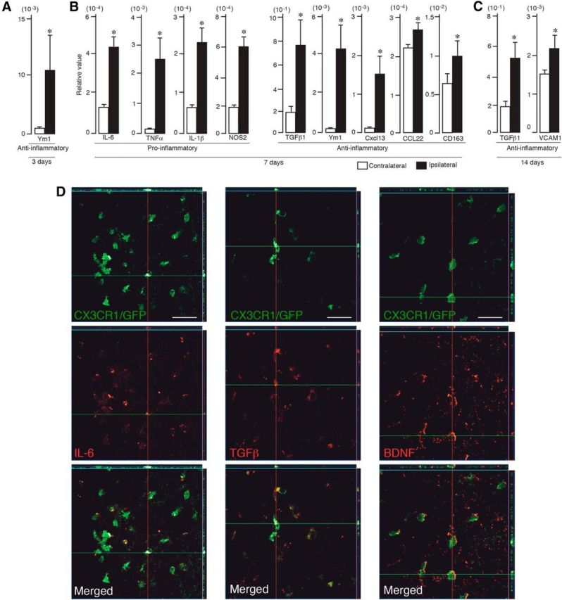
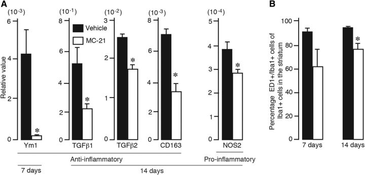
Stroke is a leading cause of disability and currently lacks effective therapy enabling long-term functional recovery. Ischemic brain injury causes local inflammation, which involves both activated resident microglia and infiltrating immune cells, including monocytes. Monocyte-derived macrophages (MDMs) exhibit a high degree of functional plasticity. Here, we determined the role of MDMs in long-term spontaneous functional recovery after middle cerebral artery occlusion in mice. Analyses by flow cytometry and immunocytochemistry revealed that monocytes home to the stroke-injured hemisphere., and that infiltration peaks 3 d after stroke. At day 7, half of the infiltrating MDMs exhibited a bias toward a proinflammatory phenotype and the other half toward an anti-inflammatory phenotype, but during the subsequent 2 weeks, MDMs with an anti-inflammatory phenotype dominated. Blocking monocyte recruitment using the anti-CCR2 antibody MC-21 during the first week after stroke abolished long-term behavioral recovery, as determined in corridor and staircase tests, and drastically decreased tissue expression of anti-inflammatory genes, including TGFβ, CD163, and Ym1. Our results show that spontaneously recruited monocytes to the injured brain early after the insult contribute to long-term functional recovery after stroke.

Significance statement: For decades, any involvement of circulating immune cells in CNS repair was completely denied. Only over the past few years has involvement of monocyte-derived macrophages (MDMs) in CNS repair received appreciation. We show here, for the first time, that MDMs recruited to the injured brain early after ischemic stroke contribute to long-term spontaneous functional recovery through inflammation-resolving activity. Our data raise the possibility that inadequate recruitment of MDMs to the brain after stroke underlies the incomplete functional recovery seen in patients and that boosting homing of MDMs with an anti-inflammatory bias to the injured brain tissue may be a new therapeutic approach to promote long-term improvement after stroke.

Keywords: macrophage; microglia; monocyte; neuroinflammation; stroke.


Figures

Figure 1.
Figure 1.
Transplanted and endogenous monocytes are recruited to injured brain tissue after stroke. A, Flow cytometry analysis of blood samples from animals injected intravenously 1 d after MCAO with either vehicle (n = 2) or with 2 (n = 4) or 4 (n = 4) million GFP+ monocytes and killed 2 d later. B–G, Fluorescence microscopic images of mouse brain coronal sections showing the ischemic lesion in the striatum visualized by NeuN staining (B), distribution of grafted GFP+ monocytes within the lesion (C, F), and expression of Iba1 (D, E) by cells within the injured striatum. E–G, Confocal images showing GFP+ grafted monocytes in the lesioned striatum (F) not expressing Iba1 (E) with merged image in G. H–K, Fluorescence microscopic images of mouse brain coronal sections showing extensive GFAP staining mostly outside the lesion (H), distribution of grafted GFP+ monocytes within the lesion (I), and expression of IB4 (J, K) by cells within the injured striatum. L, M, Confocal images showing GFP+ grafted monocytes in the lesioned striatum (L) expressing activation marker IB4 (I) with merged image in M. Scale bar (in M): B–D, H–J, 420 μm; E–G, K–M, 50 μm.
Figure 2.
Figure 2.
Flow cytometry analysis of brain hemispheres [contralateral (A) and ipsilateral (B) to the lesion] from CD45.1 mice subjected to MCAO and injected intravenously with 4 million monocytes from CD45.2 mice on the day after the insult and killed 2 d later. Note the presence of high numbers of grafted CD45.2high/CD11bhigh and endogenous CD45.1high/CD11bhigh monocytes ipsilateral to the ischemic lesion. The CD45.1low/CD11bhigh cells are microglia.
Figure 3.
Figure 3.
Spontaneous infiltration of circulating monocytes to the sites of lesion peaks at 3 d after stroke. A, Examples of flow cytometry analysis of brain hemispheres (contralateral and ipsilateral to lesion) of mice subjected to MCAO, identifying MDMs and microglia as CD45high/CD11bhigh and CD45low/CD11bhigh, respectively. B, Time course of numbers of MDMs based on flow cytometry analysis in hemispheres contralateral and ipsilateral to MCAO or sham treatment and in control hemisphere. Numbers of animals: Control, n = 4; Sham, n = 3; D1, n = 3; D3, n = 12; D7, n = 7; D14, n = 5; and D21, n = 6. Data are means ± SEMs; *p < 0,05, paired t test between contralateral and ipsilateral sides for each group. C, Fluorescence microscopic images of mouse brain showing CD31+ vessel, IB4+ activated monocytes, Iba1+ microglia, Hoechst+ nuclei in the striatum of sham-treated animal, and merged image. D, Day; SSC, side scatter; FSC, forward scatter. Scale bar, 20 μm.
Figure 4.
Figure 4.
MC-21 antibody efficiently depletes circulating monocytes and MDMs in the brain. A, Numbers of circulating CCR2+ monocytes in sham-operated and stroke-subjected mice, injected with either vehicle or MC-21 antibody. Number of animals: Sham-vehicle, n = 9; MCAO-vehicle, n = 9; Day 4, n = 13; Day 7, n = 5; Day 10, n = 7; and Day 14, n = 8. Data are means ± SEMs; *p < 0.05, one-way ANOVA. B, Correlation graph showing CCR2+ circulating monocytes expressed as percentage of CD45high/CD11bhigh blood monocytes and MDMs expressed as percentage of all CD45+/CD11b+ macrophages in brains of stroke-subjected mice, injected with either vehicle or MC-21 antibody (n = 7). Correlation analysis, R2 = 0.90, p < 0.05.
Figure 5.
Figure 5.
Depletion of circulating CCR2+ monocytes impairs long-term spontaneous behavioral recovery after stroke. A–C, Comparison between sham-treated and vehicle-injected (sham, n = 10), stroke-subjected and vehicle-injected (vehicle, n = 9), and stroke-subjected and MC-21-injected (MC-21, n = 10) in performance in corridor (A) and staircase (B, C) tests. Performance in the corridor test was calculated by dividing the number of contralateral retrievals by the total number of retrievals from both sides. Performance in the staircase test was calculated as the number of retrieved or eaten pellets on the impaired side divided by the total number of pellets on both sides and expressed as percentage of performance at baseline. Data are means ± SEMs; *p < 0.05, repeated-measures ANOVA. D, Location and pattern of ischemic injury, mainly confined to the lateral and dorsolateral parts of striatum, shown by NeuN staining in brain sections from stroke-subjected mice, treated with vehicle or MC-21, at 18 weeks after insult. Insets are enlargements from respective coronal sections. Scale bar, 1 mm. E, Mean volume of ischemic lesion treated with vehicle (n = 9) or MC-21 (n = 10), at 18 weeks after insult. Data are means ± SEMs; *p < 0,05, unpaired t test.
Figure 6.
Figure 6.
CX3CR1–GFP+ MDMs infiltrate the lesion site of chimeric mice subjected to ischemic stroke. A, B, Fluorescence microscopic images of mouse brain coronal sections showing distribution of GFP+ MDMs and GFAP+ astrocytes within and outside the ischemically injured tissue, respectively. Arrowheads depict the lesion border. C, Enlargement of inset depicted in B. Arrows point to individual GFP+ MDMs. D, Fluorescence microscopic image showing double-immunostaining of GFP+ MDMs (green) with the activation marker IB4 (red). The majority of MDMs are immunopositive for IB4 (arrows). Scale bar (in D): A, B, 250 μm; C, 100 μm; D, 50 μm.
Figure 7.
Figure 7.
MDMs switch from a proinflammatory to anti-inflammatory phenotype during the first weeks after stroke. A, Flow cytometry analysis of the brain hemisphere ipsilateral to the lesion in mice subjected to stroke and killed 3 and 7 d thereafter. CD45/CD11b immunoreactivity is used to distinguish MDMs and microglia and CX3CR1/Ly6C to define proinflammatory and anti-inflammatory phenotype of MDMs. B, Changes as a function of time in percentage of MDMs with a proinflammatory and anti-inflammatory phenotype defined by flow cytometry analysis in the ischemically injured brains; intact hemispheres were used as controls. C, Estimation of the percentage of CD204+, CD206+, and Declin+ cells within the MDM population in the injured hemisphere of mice subjected to stroke and killed 3 and 7 d thereafter. D, Estimation of the percentage of microglia and MDMs in injured hemisphere of mice subjected to stroke and killed 3 and 7 d thereafter. E, Estimation of the percentage of microglia with a proinflammatory and anti-inflammatory phenotype in the injured hemisphere of mice subjected to stroke and killed 3 and 7 d thereafter. SSC, Side scatter; FSC, forward scatter. Number of animals: Control, n = 4; Day 1, n = 3; Day 3, n = 12; Day 7, n = 7; Day 14, n = 5; and Day 21, n = 6. Data are means ± SEMs; *p < 0,05, unpaired t test between the proinflammatory and anti-inflammatory phenotype (B, E), 3 and 7 d (C), and microglia and MDMs (D).
Figure 8.
Figure 8.
Proinflammatory and anti-inflammatory factors are expressed in the stroke-injured hemisphere. A–C, Quantitative PCR shows increased expression in the injured (ipsilateral) hemisphere of anti-inflammatory factor (Ym1) at 3 d (A), proinflammatory (IL-6, TNFα, IL-1β, NOS2) and anti-inflammatory (TGFβ1, Ym1, CXCL113, CCL22, CD163) factors at 7 d (B), and anti-inflammatory factors (TGFβ1, VCAM1) at 14 d (C) after stroke (n = 7). Data are means ± SEMs; *p < 0.05, unpaired t test. D, Fluorescence microscopic images of CXCR3–GFP+ chimeric mouse brain coronal sections showing double-immunostaining of MDMs (green) and IL-6, TGFβ, and BDNF (all red) at 3 and 7 d after stroke. Note the decreased immunoreactivity for IL-6 and increased staining for TGFβ and BDNF at 7 d compared with 3 d. Scale bar, 150 μm.
Figure 9.
Figure 9.
Depletion of circulating monocytes preferentially reduces anti-inflammatory factor expression in stroke-injured brain. A, Quantitative PCR showing decreased expression of anti-inflammatory (Ym1, TGFβ1, TGFβ2, CD163) and proinflammatory (NOS2) factors in the injured hemisphere of animals treated with vehicle or MC-21 and killed at 3 (vehicle, n = 5; MC-21, n = 5), 7 (vehicle, n = 6; MC-21, n = 5), or 14 (vehicle, n = 5; MC-21, n = 5) days after stroke. Data are means ± SEMs; *p < 0.05, unpaired t test. B, Number of ED1+ cells expressed as percentage of total number of Iba1+ cells in the injured hemisphere of animals treated with vehicle or MC-21 and killed at 7 or 14 d after stroke. Data are means ± SEMs; *p < 0.05, unpaired t test.

Comment in

References

    1. Andsberg G, Kokaia Z, Lindvall O. Upregulation of p75 neurotrophin receptor after stroke in mice does not contribute to differential vulnerability of striatal neurons. Exp Neurol. 2001;169:351–363. doi: 10.1006/exnr.2001.7646. - DOI - PubMed
    1. Baird AL, Meldrum A, Dunnett SB. The staircase test of skilled reaching in mice. Brain Res Bull. 2001;54:243–250. doi: 10.1016/S0361-9230(00)00457-3. - DOI - PubMed
    1. Benakis C, Garcia-Bonilla L, Iadecola C, Anrather J. The role of microglia and myeloid immune cells in acute cerebral ischemia. Front Cell Neurosci. 2015;8:461. doi: 10.3389/fncel.2014.00461. - DOI - PMC - PubMed
    1. Bose S, Cho J. Role of chemokine CCL2 and its receptor CCR2 in neurodegenerative diseases. Arch Pharm Res. 2013;36:1039–1050. doi: 10.1007/s12272-013-0161-z. - DOI - PubMed
    1. Campanella M, Sciorati C, Tarozzo G, Beltramo M. Flow cytometric analysis of inflammatory cells in ischemic rat brain. Stroke. 2002;33:586–592. doi: 10.1161/hs0202.103399. - DOI - PubMed

Publication types

MeSH terms