. 2016 May 13;118(10):1512-24.
doi: 10.1161/CIRCRESAHA.116.308332. Epub 2016 Apr 13.

Endogenous Transmembrane TNF-Alpha Protects Against Premature Senescence in Endothelial Colony Forming Cells

Affiliations

Endogenous Transmembrane TNF-Alpha Protects Against Premature Senescence in Endothelial Colony Forming Cells

Linden A Green et al. Circ Res. .

Abstract

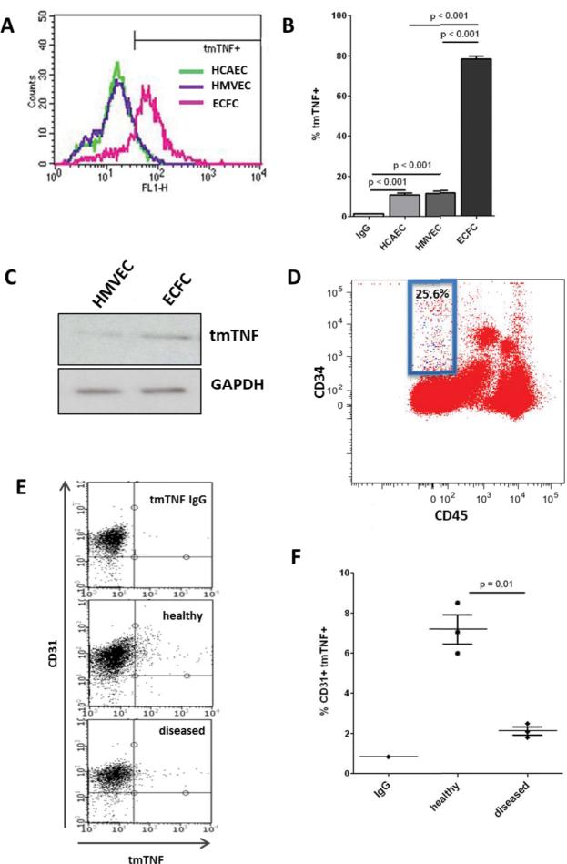
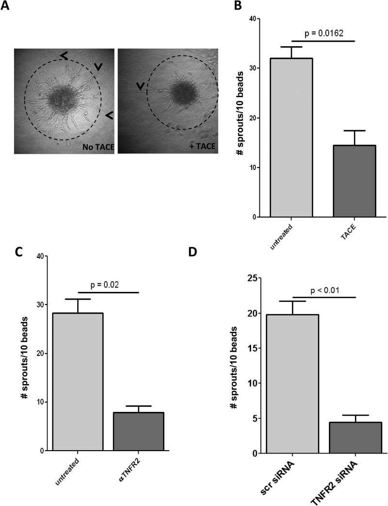
Rationale: Transmembrane tumor necrosis factor-α (tmTNF-α) is the prime ligand for TNF receptor 2, which has been shown to mediate angiogenic and blood vessel repair activities in mice. We have previously reported that the angiogenic potential of highly proliferative endothelial colony-forming cells (ECFCs) can be explained by the absence of senescent cells, which in mature endothelial cells occupy >30% of the population, and that exposure to a chronic inflammatory environment induced premature, telomere-independent senescence in ECFCs.

Objective: The goal of this study was to determine the role of tmTNF-α in the proliferation of ECFCs.

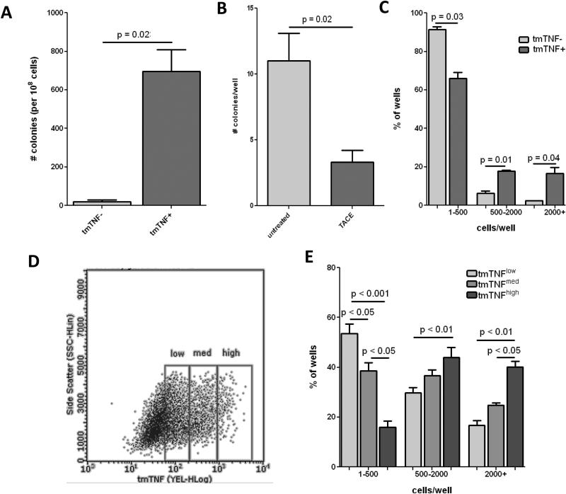
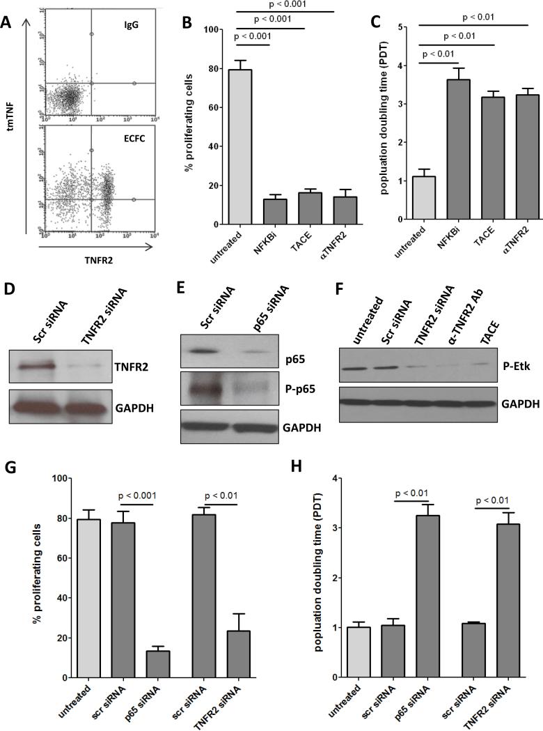
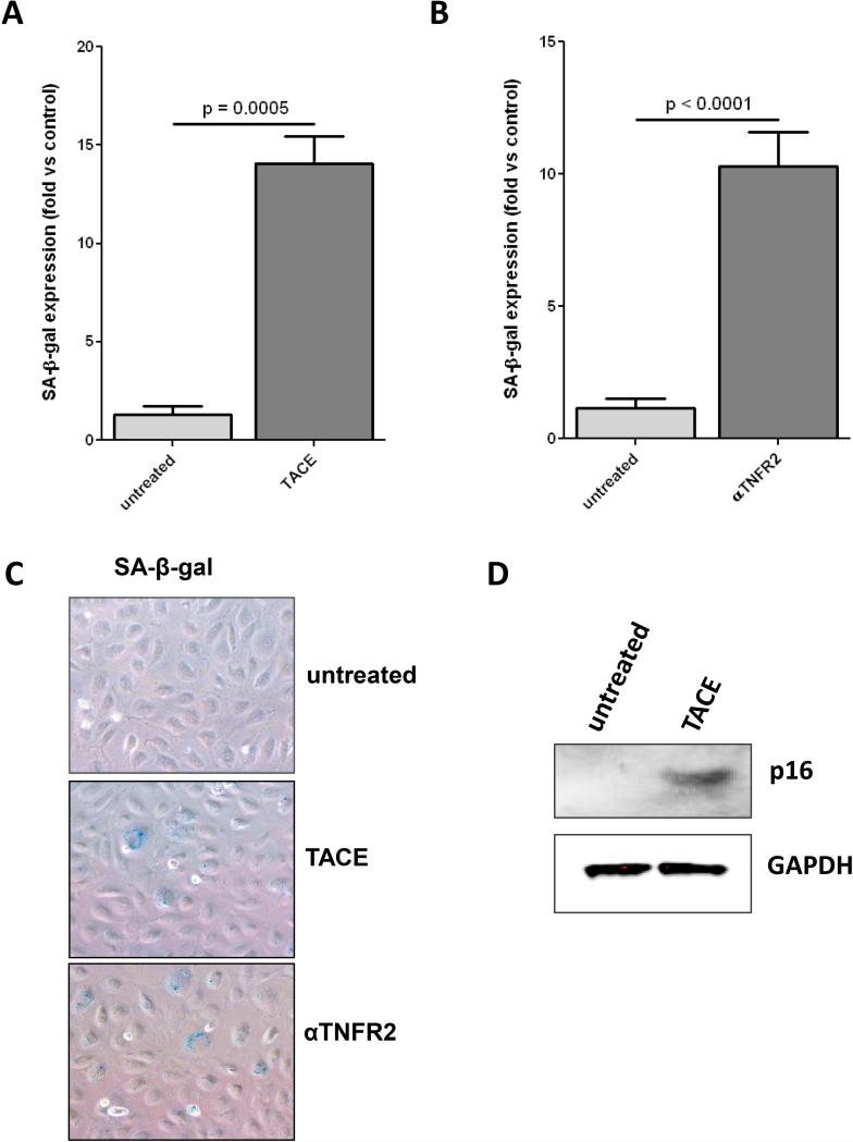
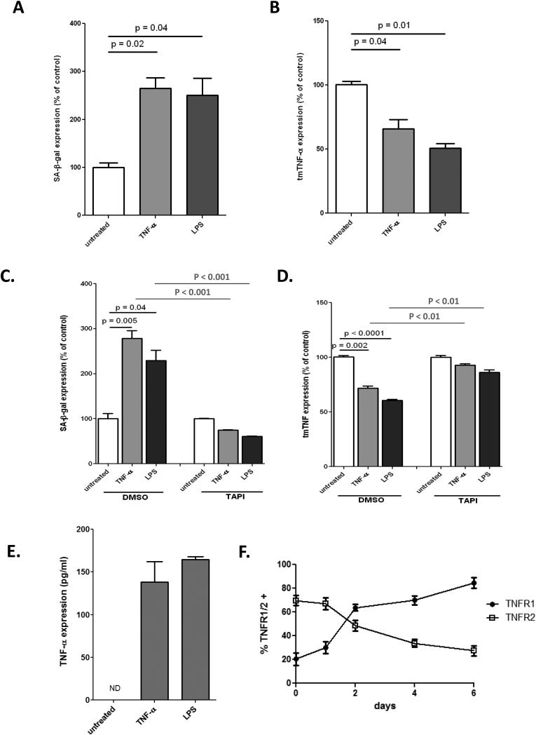
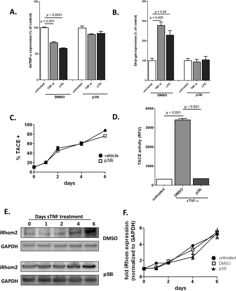
Methods and results: Here, we show that tmTNF-α expression on ECFCs selects for higher proliferative potential and when removed from the cell surface promotes ECFC senescence. Moreover, the induction of premature senescence by chronic inflammatory conditions is blocked by inhibition of tmTNF-α cleavage. Indeed, the mechanism of chronic inflammation-induced premature senescence involves an abrogation of tmTNF/TNF receptor 2 signaling. This process is mediated by activation of the tmTNF cleavage metalloprotease TNF-α-converting enzyme via p38 MAP kinase activation and its concurrent export to the cell surface by means of increased iRhom2 expression.

Conclusions: Thus, we conclude that tmTNF-α on the surface of highly proliferative ECFCs plays an important role in the regulation of their proliferative capacity.

Keywords: apoptosis; endothelium; inflammation; metalloprotease; vasodilation.


Figures

Figure 1
Figure 1. tmTNF is expressed on a subset of EC
A-B. ECFC, HCAEC, or HMVEC (passage 3) were stained for tmTNF and analyzed by FACS, with percentage tmTNF+ determined based on IgG controls. C. tmTNF was detected in HMVEC and ECFC cell lysates by Western blot. D. Freshly isolated cord blood MNCs were stained for CD45, CD34, and tmTNF, and the percentage of CD45−/CD34+ cells that were positive for tmTNF was determined (blue dots in blue box ). E-F. Age-matched internal mammary arteries (healthy) were obtained from patients undergoing cardiac bypass surgery and tibial arteries (diseased) from patients with critical limb ischemia. Endothelial lining was gently removed from the vessel wall and co-stained for CD31 and tmTNF-α then analyzed by FACS. Data are representative of 3-4 independent experiments.
Figure 2
Figure 2. tmTNF is associated with highly proliferative ECFC
A. Freshly isolated cord blood MNCs were sorted into tmTNF− or tmTNF+ fractions using MACS beads and plated with equal density on collagen-coated plates. ECFC colony formation was determined after 3 weeks. B. Freshly isolated cord blood MNCs were plated on collagen-coated plates and incubated with or without the addition of TACE. ECFC colony formation was determined after 3 weeks. C. Colonies of equal size were picked from tmTNF− or tmTNF+ plates and reseeded onto 96-well plates with 1 cell/well. After 14 days the number of cells/well were counted to determine low (1-500), medium (500-2000), and high (2000+) proliferative potential. D. Colonies from the tmTNF+ fraction were FACS sorted into tmTNFlow, tmTNFmed, and tmTNFhigh fractions and replated onto 96-well plates with 1 cell/well. E. After 14 days the number of cells/well were counted to determine low, medium, and high proliferative potential. Data are representative of 3-4 independent experiments.
Figure 3
Figure 3. Loss of tmTNF/TNFR2 axis results in less ECFC proliferation
A. ECFCs were stained with antibodies for tmTNF and TNFR2 or IgG controls and expression analyzed by FACS. B. ECFCs were labeled with 5 μM CFSE for 10 minutes at RT and incubated for 4 days with NFκB inhibitor (100nM), TACE, or αTNFR2 (500ng/ml). The percentage of proliferating cells was determined by dilution of the CFSE signal. C. ECFCs were seeded into 12-well plates and cultured for 4 days with NFκB inhibitor (100nM), TACE, or αTNFR2 (500ng/ml), after which cell number was determined and the population doubling time (pdt) was calculated. ECFCs were transfected with siRNA targeted to TNFR2 (D) or the p65 subunit of NFκB (E) and knockdown confirmed by Western blot. F. ECFCs were treated with TNFR2 siRNA, αTNFR2, or TACE and phosphorylation of TNFR2-specific Etk determined by Western blot. Percent proliferation (G) and pdt (H) were determined two days after transfection with p65 NFκB siRNA or TNFR2 siRNA. Data are representative of 3-4 independent experiments.
Figure 4
Figure 4. Loss of tmTNF/TNFR2 axis diminished the angiogenic capacity of ECFCs
A-D. ECFCs were grown to confluency on collagen-coated CytoDex beads, embedded in fibrin gel and incubated in ECFC media +/− TACE (0.2 μg/ml) (B) or αTNFR2 (500ng/ml) for 5 days. Sprouts longer than the diameter of the beads (dashed line, A) were counted. Representative pictures of untreated (left panel) and TACE (right panel) sprouts are shown in A. C. Sprouting was determined using ECFCs transfected with TNFR2 siRNA. Data are representative of 3-4 independent experiments.
Figure 5
Figure 5. Loss of tmTNF/TNFR2 axis induces premature senescence in ECFCs
A-D. ECFCs were treated with TACE (10 ng/ml) (A.) or αTNFR2 (500ng/ml) (B.) for 6 days, then stained for SA-β-gal and senescent cells were quantified. C. Representative SA-β-gal staining. D. To confirm senescence with another marker of senescence, p16ink (p16) ECFCs were treated with TACE for 6 days and p16 expression was determined by Western blot. Data are representative of 3-4 independent experiments.
Figure 6
Figure 6. Inflammation-induced premature senescence correlates with loss of tmTNF
ECFCs were treated with soluble TNF (10 ng/ml) or LPS (100 ng/ml) for 6 days, then stained for SA-β-gal. (A.) or tmTNF expression was determined by FACS (B). Soluble TNF or LPS treatment was carried out in the presence of TAPI (10μM), then stained for SA-β-gal (A) or tmTNF (B.) and normalized to untreated controls. E. Soluble TNF or LPS treatment was carried out and soluble TNF levels in the supernatant determined by ELISA, with final amounts adjusted for the initial TNF treatment. F. ECFCs were treated with soluble TNF and surface expression of TNFR1 and TNFR2 determined by FACS over the course of 6 days. Data are representative of 3-4 independent experiments.
Figure 7
Figure 7. TACE expression is upregulated during chronic inflammatory conditions and required p38 activity and iRhom expression
ECFC were treated with soluble TNF (10 ng/ml) or LPS (100 ng/ml) for 6 days +/− p38 inhibitor (10nM), then stained for tmTNF (A.) or SA-β-gal (B). C. ECFC were treated with soluble TNF (10 ng/ml) for 6 days +/− p38 inhibitor. Every 2 days cells were harvested and stained for surface TACE and analyzed by FACS. D. ECFC were treated with soluble TNF (10 ng/ml) for 6 days +/− p38 inhibitor and TACE activity was determined using a fluorescence-based kit (Anaspec). E.ECFC were treated with soluble TNF (10 ng/ml) for 6 days +/− p38 inhibitor. Cell lysates were harvested every 2 days and probed for iRhom2 and GAPDH expression. F. Densitometry analysis of iRhom2, normalized to GAPDH. Data are representative of 3-4 independent experiments.
Figure 8
Figure 8. Schematic of tmTNF/TNFR2 regulation in ECFC
Under normal conditions tmTNF signaling through TNFR2 results in NFκB-dependent proliferation in the presence of growth factor receptor (GFRs) mediated signaling (green arrows). Upon cultivation in chronic inflammatory conditions, signaling through TNFR1 results in an upregulation of iRhom2 and activation of p38 MAPK, which translocate TACE to the cell surface and activate it, respectively. TACE then cleaves tmTNF, resulting in a loss of tmTNF/TNFR2 signaling and subsequent development of senescence (red arrows).

Similar articles

Cited by

References

    1. Wang YX, Fitch RM. Vascular stiffness: measurements, mechanisms and implications. Curr Vasc Pharmacol. 2004;2:379–84. - PubMed
    1. Op den Buijs J, Musters M, Verrips T, Post JA, Braam B, van Riel N. Mathematical modeling of vascular endothelial layer maintenance: the role of endothelial cell division, progenitor cell homing, and telomere shortening. Am J Physiol Heart Circ Physiol. 2004;287:H2651–8. - PubMed
    1. Ingram DA, Mead LE, Tanaka H, Meade V, Fenoglio A, Mortell K, Pollok K, Ferkowicz MJ, Gilley D, Yoder MC. Identification of a novel hierarchy of endothelial progenitor cells using human peripheral and umbilical cord blood. Blood. 2004;104:2752–60. - PubMed
    1. Yoder MC, Mead LE, Prater D, Krier TR, Mroueh KN, Li F, Krasich R, Temm CJ, Prchal JT, Ingram DA. Redefining endothelial progenitor cells via clonal analysis and hematopoietic stem/progenitor cell principals. Blood. 2007;109:1801–9. - PMC - PubMed
    1. Asahara T, Murohara T, Sullivan A, Silver M, van der Zee R, Li T, Witzenbichler B, Schatteman G, Issner JM. Isolation of putative progenitor endothelial cells for angiogenesis. Science. 1997;275:964–967. - PubMed

Publication types

Substances