. 2016 Jun 24;90(14):6174-6186.
doi: 10.1128/JVI.02877-15. Print 2016 Jul 15.

A Single Amino Acid Substitution in Poliovirus Nonstructural Protein 2CATPase Causes Conditional Defects in Encapsidation and Uncoating

Affiliations

A Single Amino Acid Substitution in Poliovirus Nonstructural Protein 2CATPase Causes Conditional Defects in Encapsidation and Uncoating

Emmanuel Asare et al. J Virol. .

Abstract

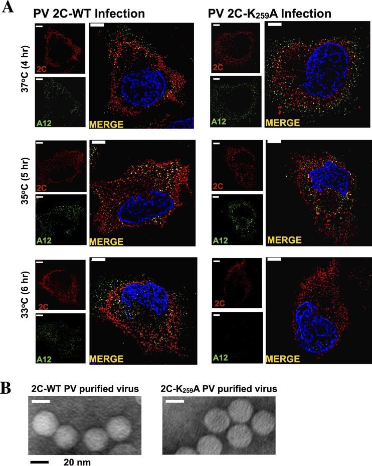
The specificity of encapsidation of C-cluster enteroviruses depends on an interaction between capsid proteins and nonstructural protein 2C(ATPase) In particular, residue N252 of poliovirus 2C(ATPase) interacts with VP3 of coxsackievirus A20, in the context of a chimeric virus. Poliovirus 2C(ATPase) has important roles both in RNA replication and encapsidation. In this study, we searched for additional sites in 2C(ATPase), near N252, that are required for encapsidation. Accordingly, segments adjacent to N252 were analyzed by combining triple and single alanine mutations to identify residues required for function. Two triple alanine mutants exhibited defects in RNA replication. The remaining two mutations, located in secondary structures in a predicted three-dimensional model of 2C(ATPase), caused lethal growth phenotypes. Most single alanine mutants, derived from the lethal variants, were either quasi-infectious and yielded variants with wild-type (wt) or temperature-sensitive (ts) growth phenotypes or had a lethal growth phenotype due to defective RNA replication. The K259A mutation, mapping to an α helix in the predicted structure of 2C(ATPase), resulted in a cold-sensitive virus. In vivo protein synthesis and virus production were strikingly delayed at 33°C relative to the wt, suggesting a defect in uncoating. Studies with a reporter virus indicated that this mutant is also defective in encapsidation at 33°C. Cell imaging confirmed a much-reduced production of K259A mature virus at 33°C relative to the wt. In conclusion, we have for the first time linked a cold-sensitive encapsidation defect in 2C(ATPase) (K259A) to a subsequent delay in uncoating of the virus particle at 33°C during the next cycle of infection.

Importance: Enterovirus morphogenesis, which involves the encapsidation of newly made virion RNA, is a process still poorly understood. Elucidation of this process is important for future drug development for a large variety of diseases caused by these agents. We have previously shown that the specificity of encapsidation of poliovirus and of C-cluster coxsackieviruses, which are prototypes of enteroviruses, is dependent on an interaction of capsid proteins with the multifunctional nonstructural protein 2C(ATPase) In this study, we have searched for residues in poliovirus 2C(ATPase), near a presumed capsid-interacting site, important for encapsidation. An unusual cold-sensitive mutant of 2C(ATPase) possessed a defect in encapsidation at 37°C and subsequently in uncoating during the next cycle of infection at 33°C. These studies not only reveal a new site in 2C(ATPase) that is involved in encapsidation but also identify a link between encapsidation and uncoating.


Figures

FIG 1
FIG 1
Poliovirus genome organization and functional motifs in the PV 2CATPase protein. (A) Poliovirus RNA contains a long 5′ nontranslated region (5′NTR), a single open reading reading frame, a short 3′NTR, and a poly(A) tail. (B) The locations of the known functional domains of the 2CATPase protein are illustrated. (C) Previously identified mutations in 2CATPase involved in encapsidation or uncoating are shown in detail. These include the hydantoin-resistant mutations (9), N252, the capsid-interacting site in the PV/CAV chimera (10), the residues involved in encapsidation derived from alanine scanning mutagenesis (12, 13), and the mutations leading to an uncoating defect in a mutant containing a nearby linker insertion (1).
FIG 2
FIG 2
Phyre2-predicted model of the PV 2CATPase polypeptide structure and the locations of the FMI/AAA and GKL/AAA mutations on the structure. (A) The amino acid sequence of PV1(M) 2CATPase was submitted to the Phyre2 online server to generate a 3D structural model (Materials and Methods). The structure of the N-terminal domain, highlighted in black, is highly unreliable. Sequences highlighted in gold are predicted with good confidence. The 2CATPase structure shown in light green is predicted with 100% confidence. (B) The predicted three domains in the 2CATPase structure. The N-terminal domain (red) consists of helical structures, the central helicase domain (blue) contains the NTP binding/helicase boxes A, B, and C, and the C-terminal domain (green) consist mostly of helical structures. The flexible region between the central and C-terminal domains contains residue N252. (C and D) Locations of the triple alanine mutations on the predicted 2CATPase structure are illustrated. The QVM residues are in a flexible domain, while the EYS residues are partly in an α-helical structure. The FMI residues are predicted to be located in a β sheet, and the GKL residues are in an α-helix structure. (E) Alignment of picornavirus 2CATPase proteins surrounding residues N252. (Top) The locations of the triple alanine mutants in the 2CATPase polypeptide are shown. The growth phenotypes of the mutants are also indicated. (Below) The amino acid sequence of picornavirus 2CATPase proteins, surrounding N252, is shown. The highly conserved residues are indicated by stars over a black background, and the less conserved residues are shown with a gray background. Dashes indicate the absence of certain residues in the sequences alignment. The size and location of the flexible region are also indicated. The enterovirus 2CATPase proteins are boxed to separate them from the more distantly related picornaviruses (FMDV, Aphtovirus genus; EMCV [encephalomyocarditis virus], Cardiovirus genus).
FIG 3
FIG 3
Characterization of PV 2CATPase triple alanine mutants. (A) Growth properties of mutants at different temperatures. The locations of N252 and of the triple alanine mutants on the 2CATPase sequence are illustrated on top. RNA transcripts of wt and mutant constructs were transfected into HeLa R19 monolayers and incubated at 33, 37, and 39.5°C for 48 h or until CPE (Materials and Methods). Freeze-thawed supernatants were used for passage on fresh HeLa R19 monolayers. The genotypes of recovered progeny viruses from 33°C passages are indicated. (B) Virus titers and plaque phenotypes of viable mutants QVM/AVA and EYS/AAA. Viruses derived from 33°C transfections were plaqued at 33, 37, and 39.5°C, and the titers were determined (Materials and Methods). The plaque phenotypes of the viruses at different temperatures are shown at the indicated dilutions. The lysates derived from the 33°C transfections were passaged 10 times, and the sequences of the full-length genomes of the progeny were determined. No additional genetic changes were observed. (C) In vitro translation of wt and mutant transcript RNAs. Transcript RNAs of the wt and of the lethal mutants were translated in HeLa cell extracts (Materials and Methods). (D) RNA levels in HeLa cells infected with mutant EYS/AAA and variant QVM/AVA. HeLa cells were infected at an MOI of 5 with wt and mutant viruses obtained from 33°C transfections, and the titer was determined at 37°C. The infected cells were harvested at various time points after infection, and total RNA was isolated from the lysates. RNA levels were determined by qPCR, as described in Materials and Methods.
FIG 4
FIG 4
Growth phenotypes of single alanine mutants. (A) Virus viability and the genotype of progeny at 37°C. Single alanine mutants derived from the two nonviable triple amino acid mutants FMI/AAA and GKL/AAA were generated. They were tested for viability at 37°C, and the genotypes of the progeny were determined. (B) Virus titers and plaque phenotypes of single alanine mutants. Titers of viruses grown at 37°C were determined at 33, 37, and 39.5°C by plaque assay (Materials and Methods). The plaque phenotypes are shown on the right panel. Subscripts indicate the virus dilution at which the image was taken. It should be noted that the mixed plaque sizes seen with the lysate derived from M246A (39.5°C) presumably contain a mixture of the original alanine mutant (small) and of the V variant (large). Lysates derived from 37°C transfections were passaged 4 times, and the sequences of the full-length genomes were determined. No additional genetic changes were observed.
FIG 5
FIG 5
The 2CATPase K259A mutant is delayed in virus production and protein synthesis at the restrictive temperatures (35 and 33°C). (A) Growth curves of wt and K259A mutant polioviruses at the permissive (37°C) and restrictive (35 and 33°C) temperatures. HeLa cells were infected at an MOI of 5 with viruses derived from 37°C transfections. The titer of the viral progeny was determined by plaque assay at different times postinfection (Materials and Methods). Black lines indicate growth curves at 37°C, and gray lines indicate growth curves at 33°C. (B) Protein synthesis by the wt and K259A mutant measured by Western analysis. HeLa cells were infected at either 33, 35, or 37°C at an MOI of 5 with viruses derived from 37°C transfections. The infected cells were isolated at various times postinfection and lysed. The level of 2CATPase-related proteins and of capsid protein VP3 were measured by Western analysis using a monoclonal antibody to 2CATPase and a polyclonal antibody to VP3, respectively, as described in Materials and Methods. Tubulin was used as a loading control. The experiment was carried out three times.
FIG 6
FIG 6
The K259A 2CATPase mutant possesses an encapsidation defect at 33°C. (A) Genome structure of a Renilla luciferase (R-Luc) reporter virus (R-Luc-PPP). The R-Luc gene was fused between the 5′NTR and P1 structural proteins, flanked by a 3CDpro cleavage site. Wild-type and mutant K259A 2CATPase reporter virus RNA transcripts were transfected into HeLa cells at 33 or 37°C, in both the absence and presence of GnHCl (Materials and Methods). R-Luc assays were performed at 8 h posttransfection. Aliquots of the lysates from the transfections were used to infect fresh HeLa cells, in both the absence and presence of GnHCl. R-Luc assays were performed at 8 h postinfection. R-Luc ratios were calculated by dividing the raw R-Luc values in the absence by the R-Luc values in the presence of GnHCl. (B) The genome structure of firefly luciferase PV1(M) replicons used in the experiment is shown above. A time course of RNA replication was measured with the wt and the mutant 2CATPase K259A mutant using F-Luc replicons. Monolayer HeLa R19 cells were transfected with 3 to 5 μg of firefly Renilla luciferase replicon transcript RNAs. Transfected cells were incubated for 2, 4, 6, and 8 h at 33°C in the presence or absence of 2 mM GnHCl. Luciferase activity was determined on the cell supernatants after three freeze-thawing steps. F-Luc activity with the wt virus is taken as 100%. The experiment was carried out three times.
FIG 7
FIG 7
Immunofluorescence imaging of PV 2C wt and PV 2C K259A mutant virus-infected HeLa cells. HeLa cells were infected with wt or PV 2C K259A virus at an MOI of 5. Cells were incubated for 4 (37°C), 5 (35°C), or 6 (33°C) h and were fixed with paraformaldehyde. Infected cells were probed with primary antibody against 2CATPase and mature virus (monoclonal antibody A12), followed by Alexa Fluor 555 (red)- and 488 (green)-conjugated antibodies, respectively. The cell nucleus was stained with DAPI (4′,6-diamidino-2-phenylindole), shown in blue. (B) Electron microscopy of purified wt and K259A 2CATPase mutant viruses. Wild-type and mutant viruses were grown at 37°C and purified on cesium chloride density gradients (see Materials and Methods).

References

    1. Li J-P, Baltimore D. 1990. An intragenic revertant of a poliovirus 2C mutant has an uncoating defect. J Virol 64:1102–1107. - PMC - PubMed
    1. Cho MW, Teterina N, Egger D, Bienz K, Ehrenfeld E. 1994. Membrane rearrangement and vesicle induction by recombinant poliovirus 2C and 2BC in human cells. Virology 202:129–145. doi:10.1006/viro.1994.1329. - DOI - PubMed
    1. Aldabe R, Carrasco L. 1995. Induction of membrane proliferation by poliovirus proteins 2C and 2BC. Biochem Biophys Res Commun 206:64–76. doi:10.1006/bbrc.1995.1010. - DOI - PubMed
    1. Teterina NL, Kean KM, Gorbalenya AE, Agol VI, Girard M. 1992. Analysis of the functional significance of amino acid residues in the putative NTP-binding pattern of the poliovirus 2C protein. J Gen Virol 73:1977–1986. - PubMed
    1. Barton DJ, Flanegan JB. 1997. Synchronous replication of poliovirus RNA: initiation of negative-strand RNA synthesis requires the guanidine-inhibited activity of protein 2C. J Virol 71:8482–8489. - PMC - PubMed

Publication types

MeSH terms