. 2016 May 27;90(12):5830-5844.
doi: 10.1128/JVI.00394-16. Print 2016 Jun 15.

Cell-Free Hepatitis B Virus Capsid Assembly Dependent on the Core Protein C-Terminal Domain and Regulated by Phosphorylation

Affiliations

Cell-Free Hepatitis B Virus Capsid Assembly Dependent on the Core Protein C-Terminal Domain and Regulated by Phosphorylation

Laurie Ludgate et al. J Virol. .

Abstract

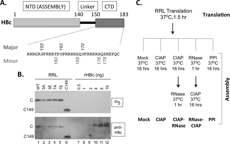
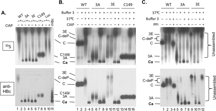
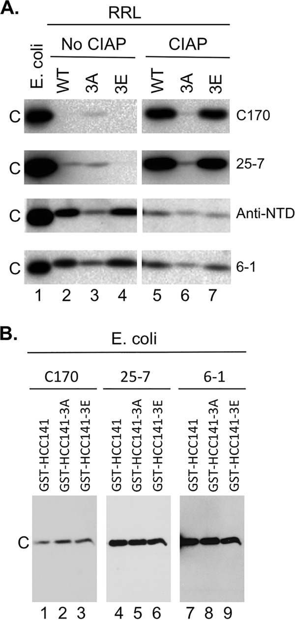
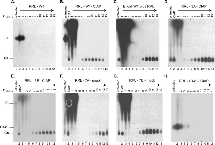
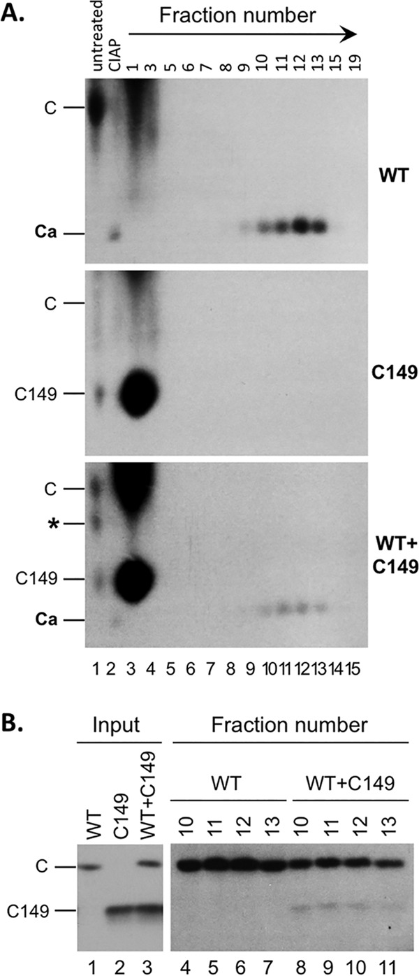
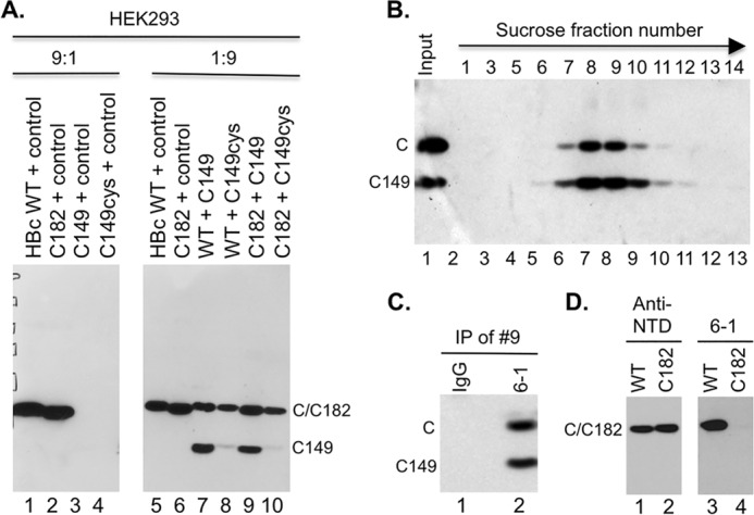
Multiple subunits of the hepatitis B virus (HBV) core protein (HBc) assemble into an icosahedral capsid that packages the viral pregenomic RNA (pgRNA). The N-terminal domain (NTD) of HBc is sufficient for capsid assembly, in the absence of pgRNA or any other viral or host factors, under conditions of high HBc and/or salt concentrations. The C-terminal domain (CTD) is deemed dispensable for capsid assembly although it is essential for pgRNA packaging. We report here that HBc expressed in a mammalian cell lysate, rabbit reticulocyte lysate (RRL), was able to assemble into capsids when (low-nanomolar) HBc concentrations mimicked those achieved under conditions of viral replication in vivo and were far below those used previously for capsid assembly in vitro Furthermore, at physiologically low HBc concentrations in RRL, the NTD was insufficient for capsid assembly and the CTD was also required. The CTD likely facilitated assembly under these conditions via RNA binding and protein-protein interactions. Moreover, the CTD underwent phosphorylation and dephosphorylation events in RRL similar to those seen in vivo which regulated capsid assembly. Importantly, the NTD alone also failed to accumulate in mammalian cells, likely resulting from its failure to assemble efficiently. Coexpression of the full-length HBc rescued NTD assembly in RRL as well as NTD expression and assembly in mammalian cells, resulting in the formation of mosaic capsids containing both full-length HBc and the NTD. These results have important implications for HBV assembly during replication and provide a facile cell-free system to study capsid assembly under physiologically relevant conditions, including its modulation by host factors.

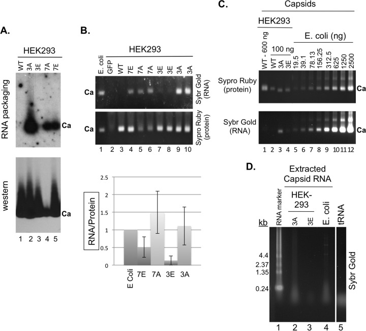
Importance: Hepatitis B virus (HBV) is an important global human pathogen and the main cause of liver cancer worldwide. An essential component of HBV is the spherical capsid composed of multiple copies of a single protein, the core protein (HBc). We have developed a mammalian cell-free system in which HBc is expressed at physiological (low) concentrations and assembles into capsids under near-physiological conditions. In this cell-free system, as in mammalian cells, capsid assembly depends on the C-terminal domain (CTD) of HBc, in contrast to other assembly systems in which HBc assembles into capsids independently of the CTD under conditions of nonphysiological protein and salt concentrations. Furthermore, the phosphorylation state of the CTD regulates capsid assembly and RNA encapsidation in the cell-free system in a manner similar to that seen in mammalian cells. This system will facilitate detailed studies on capsid assembly and RNA encapsidation under physiological conditions and identification of antiviral agents that target HBc.


Figures

FIG 1
FIG 1
Expression of WT and mutant HBc proteins in RRL. (A) Schematic diagram of HBc domain structure and the CTD sequence. The three major phosphorylation sites in CTD (S155, S162, and S170) are marked above the sequence and the four minor sites (T160, S168, S176, and S178) below. pCI-HBc-3A (3A) and 3E have the three major sites changed to A and E, respectively, whereas pCI-HBc-7A (7A) and 7E have all seven sites substituted. (B) Estimation of HBc concentration as expressed in RRL. Each lane contained 3 μl of the translation reaction mixture. The 35S-labeled HBc proteins were detected by autoradiography (top) and by Western blot analysis using the anti-HBc NTD MAb (bottom). (C) Translation in RRL and capsid assembly schemes. The five different assembly conditions are listed at the bottom. See the text for details. C, WT HBc protein; C149, C-terminally truncated HBc protein (terminated at position 149); CIAP, calf intestine alkaline phosphatase; PPI, phosphatase inhibitors.
FIG 2
FIG 2
HBV capsid assembly in RRL and effects of exogenous phosphatase and phosphatase inhibitors on assembly. The WT and mutant HBc proteins or the control luciferase (Luc) was translated in RRL. All samples were resolved by agarose gel electrophoresis. (A) The indicated protein translated in RRL was incubated overnight at 37°C in 1× NEB restriction digestion buffer 3 alone (lanes 1, 3, 5, 7, and 9) or with CIAP (lanes 2, 4, 6, 8, and 10) before resolution on the gel. The recombinant HBV capsid (rHBc) purified from E. coli was loaded in lane 11. (B) The indicated translation reaction mixture was loaded directly following translation upon dilution in double-distilled water (dH2O) and without the overnight incubation (i.e., no assembly reaction) (lanes 1, 5, 9, and 13), after dilution in NEB buffer 3 (buffer 3) but without the overnight incubation (lanes 2, 6, 10, and 14), after dilution in buffer 3 and with incubation overnight at 37°C (lanes 3, 7, 11, and 15), or after overnight incubation in buffer 3 and with CIAP (lanes 4, 8, 12, and 16). (C) The indicated translation reaction mixture was loaded directly following translation upon dilution in dH2O and without the overnight incubation (i.e., no assembly reaction) (lanes 1, 5, and 9), after dilution in dH2O and with incubation overnight at 37°C (lanes 2, 6, and 10), after dilution in buffer 3 and with incubation overnight at 37°C (lanes 3, 7, and 11), or after overnight incubation at 37°C in buffer 3 and with a mixture of phosphatase inhibitors (PPI) (lanes 4, 8, and 12). Each lane contained 3 μl translation product except that 3.125 ng rHBc was loaded in lane 11 of panel A. 35S signals were detected by autoradiography (top). The HBc proteins were also detected by the MAb antibody against the NTD (bottom). C/3A/3E, WT, 3A, or 3E HBc subunits (i.e., not present in the capsid); C149, C-terminally truncated HBc protein (terminated at position 149); C-deP, dephosphorylated WT HBc subunits; Ca, capsids.
FIG 3
FIG 3
Confirmation of HBc phosphorylation in RRL and dephosphorylation by CIAP using CTD antibodies. (A) WT HBc (lanes 2 and 5), 3A (lanes 3 and 6), or 3E (lanes 4 and 7) was expressed in RRL and resolved by SDS-PAGE, with prior CIAP treatment (lanes 5 to 7) or without prior CIAP treatment (lanes 2 to 4), and was detected by the anti-CTD antibodies (C170, top panels; 25-7, upper middle panels; 6-1, bottom panels) or the anti-NTD MAb (lower middle panels). WT HBc expressed in E. coli (lane 1) was analyzed in parallel. (B) Equal amounts (15 ng) of the indicated GST-CTD fusion proteins purified from E. coli (thus all nonphosphorylated) were resolved by SDS-PAGE and detected by the C170 (lanes 1 to 3), 25-7 (lanes 4 to 6), or 6-1 (lanes 7 to 9) antibody. GST-HCC141 contains HBc CTD sequences from 141-183, and GST-HCC141–3A or –3E contains the same sequences except that the three major S phosphorylation sites (Fig. 1A) are substituted with A or E, respectively. C, WT or mutant HBc.
FIG 4
FIG 4
Effects of exogenous phosphatase and RNase treatment on capsid assembly in RRL. The indicated HBc proteins were translated in RRL, and the translation reaction mixtures were resolved by agarose gel electrophoresis (top panels) or SDS-PAGE (bottom panels) without any further treatment (lanes 1, 7, 13, 19, 25, and 31) or were treated with NEB buffer 3 alone overnight at 37°C (buffer) (lanes 2, 8, 14, 20, 26, and 32), with buffer 3 plus CIAP overnight at 37°C (CIAP) (lanes 3, 9, 15, 21, 27, and 33), with buffer 3 plus CIAP overnight at 37°C followed by RNase treatment for one additional hour (CIAP-RNase) (lanes 4, 10, 16, 22, 28, and 34), with RNase for 1 h followed by buffer 3 plus CIAP overnight at 37°C (lanes 5, 11, 17, 23, 29, and 35), or with the mixture of phosphatase inhibitors overnight at 37°C (lanes 6, 12, 18, 24, 30, and 36). All lanes contained 2 μl translation products. 35S-labeled HBc proteins were detected by autoradiography. C, 3A, and 3E/7A, WT or mutant HBc subunits; C149, C-terminally truncated HBc protein (terminated at position 149); C-deP, dephosphorylated HBc subunits; Ca, capsids.
FIG 5
FIG 5
Analysis of capsid assembly in RRL by sucrose gradient centrifugation. The indicated RRL translation reaction mixtures (100 μl), either left untreated or treated as indicated, were separated over a linear 15% to 30% sucrose gradient spun in an SW55 rotor at 27,000 rpm for 4 h at 4°C. The indicated sucrose fractions (10 μl each) or input RRL translation mixtures (either left untreated or treated as indicated) (0.5 μl) were resolved by agarose gel electrophoresis. The direction of centrifugation is indicated by the arrows. 35S-labeled HBc proteins were detected by autoradiography. Capsids purified from E. coli (unlabeled) in panel C were detected by Western blot analysis using the anti-HBc NTD MAb. Fract #, sucrose fraction number; mock, overnight incubation in the assembly buffer alone; C and 3E, full-length HBc proteins; C149, C-terminally truncated HBc protein (terminated at position 149); Ca, capsids.
FIG 6
FIG 6
Rescue of capsid assembly of CTD-deleted HBc by WT HBc in RRL. WT or C149 HBc was translated alone (A, top or middle, respectively; B, lane 1 or lane 2, respectively), or WT HBc and C149 HBc were translated together (A, bottom; B, lanes 3) in RRL. The capsids were induced to assemble using CIAP treatment, and the assembled capsids were resolved by sucrose gradient centrifugation as described for Fig. 4. The translation reaction mixtures (A, lane 1, 0.5 μl), the assembly reaction mixtures (i.e., the input to the gradient; CIAP) (A, lane 2, 0.5 μl; B, lanes 1 to 3, 5 μl), and the indicated sucrose fractions (A, lanes 3 to 15, 10 μl; B, lanes 4 to 11, 50 μl) were resolved by agarose gel electrophoresis (A) or SDS-PAGE (B) and detected by autoradiography. C, WT HBc subunits; C149, C-terminally truncated HBc protein (terminated at position 149); Ca, capsids; *, novel band appearing only in the mixed translation and migrating between WT and C149 HBc subunits. The direction of centrifugation is indicated by the arrow in panel A.
FIG 7
FIG 7
Rescue of expression and assembly of CTD-deleted HBc by WT HBc in HEK293 cells. (A) Cytoplasmic lysate (20 μl) from HEK293 cells transiently transfected with the indicated plasmids was resolved by SDS-PAGE and detected by Western blot analysis using the anti-HBc NTD MAb. The control plasmid expresses the neomycin resistance gene and was used as a filler. The ratios (9:1 and 1:9) refer to the amount of the first plasmid (always expressing an HBc protein) relative to the amount of the second plasmid (expressing either no HBc or the indicated HBc protein). (B) Capsids in cytoplasmic lysate of HEK293 cells cotransfected with both the WT HBc- and C149-expressing plasmids were fractionated on a linear 15% to 30% sucrose gradient in an SW32 rotor at 27,000 rpm for 4 h at 4°C (the direction of centrifugation is indicated by the arrow). The indicated fractions (lanes 2 to 13), along with the input lysate (lane 1), were resolved by SDS-PAGE, and WT HBc and C149 were detected by Western blot analysis using the anti-HBc NTD MAb. (C) Coimmunoprecipitation of C149 and WT HBc in mosaic capsids. The capsid peak fraction (fraction 9) from the sucrose gradient was subjected to immunoprecipitation using the anti-CTD MAb (6-1) (lane 2) or a control IgG (lane 1). The immunoprecipitates were detected by the anti-NTD MAb following SDS-PAGE and Western blot analysis. (D) Cytoplasmic lysate from HEK293 cells transiently transfected with WT (lanes 1 and 3) or C182 (lanes 2 and 4) was resolved by SDS-PAGE and detected by Western blot analysis using the anti-NTD MAb (lanes 1 and 2) or anti-CTD MAb 6-1 (lanes 3 and 4). C, WT HBc monomer; C149 monomer, C-terminally truncated HBc protein (terminated at position 149); IP, immunoprecipitation. C182 comigrated with WT HBc.
FIG 8
FIG 8
Analysis of nonspecific RNA packaging by capsids assembled in mammalian and bacterial cells. The indicated WT and mutant HBc expression constructs were transfected into HEK293 cells. (A) Cytoplasmic lysates (5 μl) from transfected cells were resolved on an agarose gel and transferred to a nitrocellulose membrane. Packaged HBc mRNA was detected by 32P-labeled antisense HBV riboprobe (top) and the capsid (Ca) by the anti-HBc NTD MAb (bottom). (B) Total RNA packaged in capsids in the HEK293 lysates (15 μl), along with the capsid standard purified from E. coli, was also detected by Sypro gold staining and the capsid protein signal detected by Sypro ruby staining following treatment of the lysate with micrococcal nuclease and proteinase K (see Materials and Methods for details) and agarose gel electrophoresis (gel images). Green fluorescent protein (GFP) (lane 2) represented a negative control for the staining that contained the lysate from cells transfected with a GFP-expressing plasmid. Note that duplicate 3E (lanes 7 and 8), 3A (lanes 9 and 10), and 7A (lanes 5 and 6) samples from two separate transfections were loaded. The RNA staining signals normalized to the protein signals are presented in the graph. (C) Capsids purified by sucrose gradient centrifugation from transfected HEK293 cells (lanes 1 to 5) or E. coli (lanes 6 to 12) were resolved on an agarose gel and detected by Sypro ruby staining (top) and their associated nucleic acids by Sybr gold staining (bottom). (D) Nucleic acid isolated from the purified capsids from HEK293 cells (3A, lane 2; 3E, lane 3) or E. coli (lane 4) was isolated and resolved on an agarose gel and detected by Sybr gold staining. The RNA marker and tRNA were also loaded as size standards (lanes 1 and 5, respectively). Ca, capsids; kb, sizes of RNA markers.
FIG 9
FIG 9
Regulation of specific versus nonspecific RNA packaging of HBV capsids by the degree of CTD phosphorylation and dephosphorylation. The HBc CTD state of phosphorylation is proposed to be regulated dynamically by the host protein kinase(s) and phosphatase(s). The letter P on the capsids denotes phosphorylation. The specific viral pgRNA is depicted as a thick and long wavy line inside the capsid (diamond), and the nonspecific RNAs are depicted as thin and short wavy lines. The dashed arrow indicates that under natural replication conditions in human cells, HBc is unlikely to be in a nonphosphorylated state upon expression and thus that few capsids with nonspecific RNA packaging would form, in contrast to capsid assembly in E. coli. See the text for details. For clarity, RT packaging is not shown.

Similar articles

Cited by

References

    1. Trépo C, Chan HL, Lok A. 2014. Hepatitis B virus infection. Lancet 384:2053–2063. doi:10.1016/S0140-6736(14)60220-8. - DOI - PubMed
    1. Seeger C, Zoulim F, Mason WS. 2013. Hepadnaviruses, p 2185–2221. In Knipe DM, Howley PM, Cohen JI, Griffin DE, Lamb RA, Martin MA, Racaniello VR, Roizman B (ed), Fields virology, 6th ed Lippincott, Williams & Wilkins, Philadelphia, PA.
    1. Hu J. 2016. Hepatitis B virus virology and replication, p 1–34. In Liaw Y-F, Zoulim F (ed), Hepatitis B virus in human diseases. Humana Press, New York, NY.
    1. Summers J, Mason WS. 1982. Replication of the genome of a hepatitis B–like virus by reverse transcription of an RNA intermediate. Cell 29:403–415. doi:10.1016/0092-8674(82)90157-X. - DOI - PubMed
    1. Hu J, Seeger C. 2015. Hepadnavirus genome replication and persistence. Cold Spring Harb Perspect Med 5:a021386. doi:10.1101/cshperspect.a021386. - DOI - PMC - PubMed

Publication types

MeSH terms