. 2016 Mar;8(3):553-74.
doi: 10.21037/jtd.2016.01.63.

Complete video-assisted thoracoscopic surgery (VATS) bronchial sleeve lobectomy

Affiliations

Complete video-assisted thoracoscopic surgery (VATS) bronchial sleeve lobectomy

Jun Huang et al. J Thorac Dis. 2016 Mar.

Abstract

Background: To explore the effectiveness of video-assisted thoracoscopic surgery (VATS) bronchial sleeve resection and reconstruction.

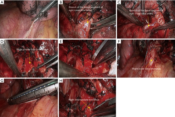
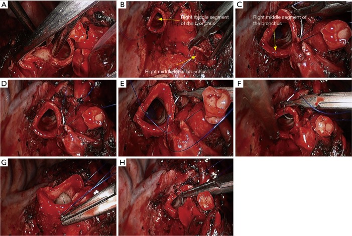
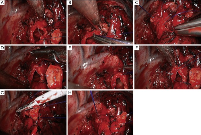
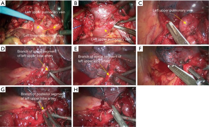
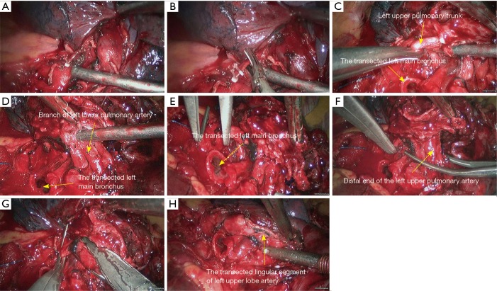
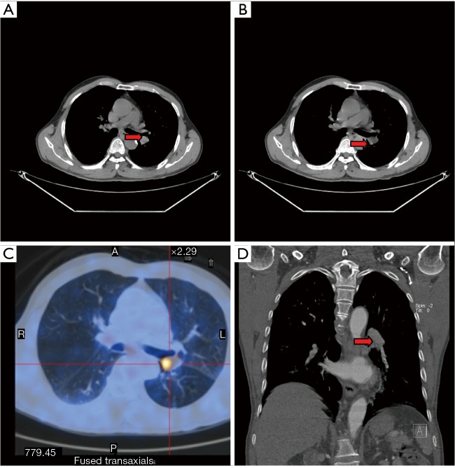
Methods: The clinical data of patients who had received VATS bronchial sleeve lobectomy in our center from January 2008 to February 2015 were retrospectively analyzed.

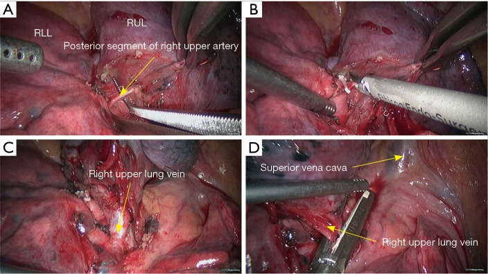
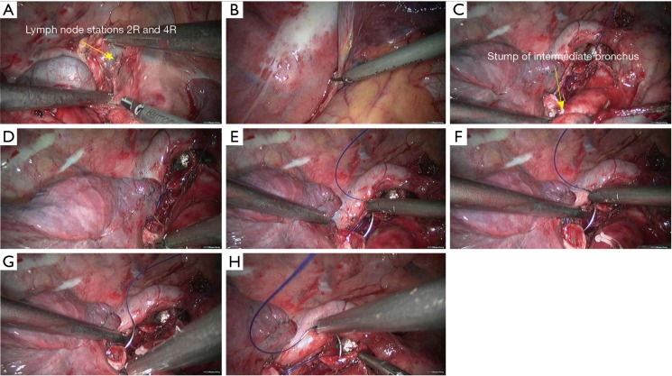
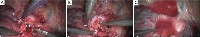
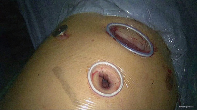
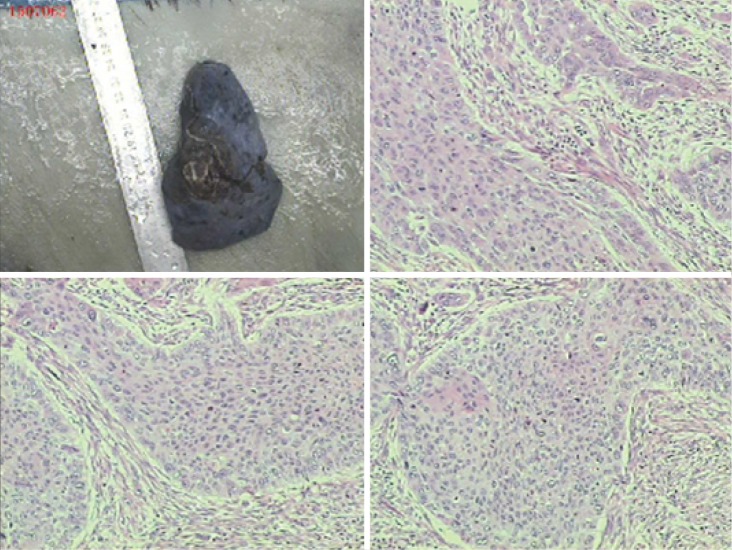
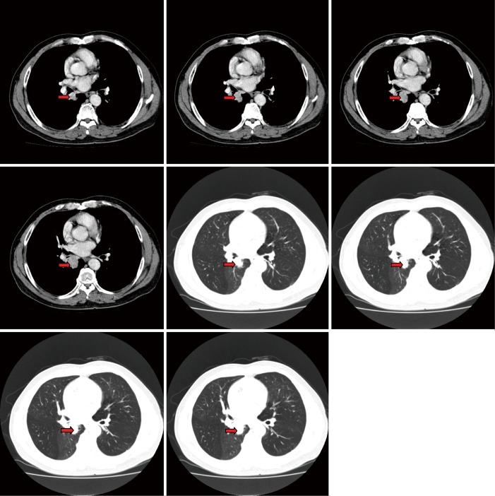
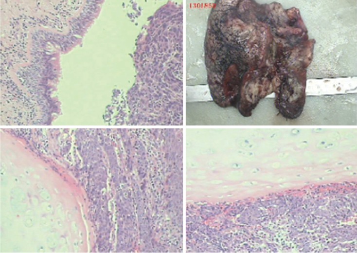
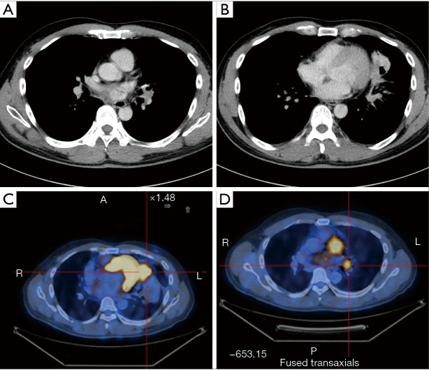
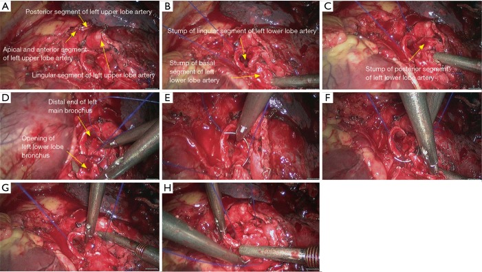
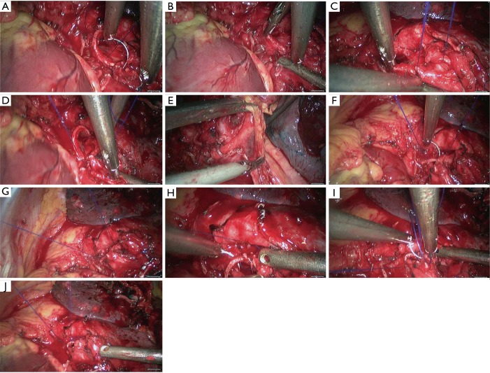
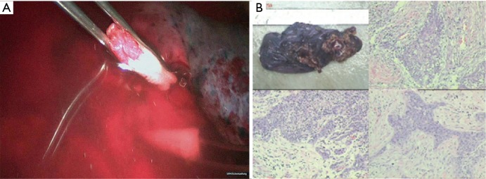
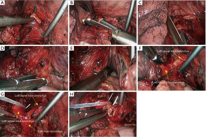
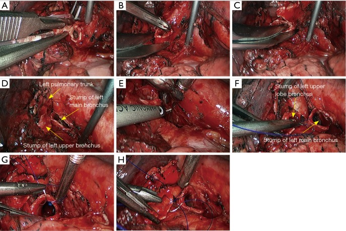
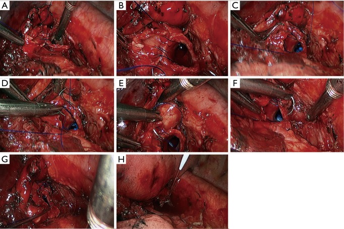
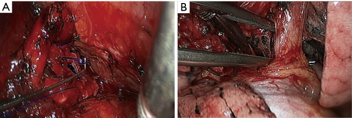
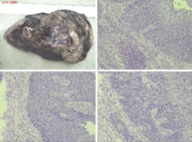
Results: Totally 118 patients (105 men and 13 women) received the VATS bronchial sleeve lobectomy. The procedures included sleeve resection of right upper lobe (n=59), right middle lobe (n=7), right lower lobe (n=8), left upper lobe (n=34), and left lower lobe (n=10). The lesions were confirmed to be squamous cell carcinoma (n=68), adenocarcinoma (n=16), mucoepidermoid carcinoma (n=8), adenosquamous carcinoma (n=7), large cell carcinoma (n=1), carcinoids (n=5), and others (n=13; including small cell carcinoma, pleomorphic carcinoma, and inflammatory myofibroblastic tumor). Operations lasted 118-223 min [mean ± standard deviations (SD): 124.00±31.75 min]. The length of removed bronchus was 1.50-2.00 cm (mean ± SD: 1.75±0.26 cm). The duration of bronchial anastomosis (from the first puncture to the completion of knotting) was 15-42 min (mean ± SD: 30.20±7.97 min). The number of dissected lymph node stations (at least three mediastinal lymph node stations, including station 7) was 5-9 stations (mean ± SD: 6.50±1.18 min). The number of dissected lymph nodes was 10-46 (mean ± SD: 26.00±10.48). The intraoperative blood loss was 20-400 mL (mean ± SD: 71.00±43.95 mL), and no blood transfusion was performed. All patients were observed in intensive care unit (ICU) for 1 day. Postoperative drainage was performed for 3-8 days (mean ± SD: 5.00±1.49 days). Postoperative hospital stay was 3-8 days (mean ± SD: 5.10±2.07 days).

Conclusions: VATS bronchial sleeve resection and reconstruction is a safe and feasible technique.

Keywords: Video-assisted thoracoscopic surgery (VATS); lobectomy; lung cancer; sleeve resection; surgery.


Conflict of interest statement

Conflicts of Interest: The authors have no conflicts of interest to declare.

Figures

Figure 1
Figure 1
Surgical position.
Figure 2
Figure 2
Distribution of incisions.
Figure 3
Figure 3
CT scan of right upper lobe mass: contrast-enhanced chest spiral CT shows the presence of a neoplasm at the opening of right upper lobe bronchus (arrow).
Figure 4
Figure 4
Right upper sleeve lobectomy. (A) Separate the lung fissure to expose the posterior segment of right upper lung artery (recurrent branch); (B) block the posterior segment of right upper lung artery (recurrent branch) using the Hemo Lock and then transect the distal end using HIFU; (C) dissect to separate the right upper lung vein; (D) transect the right upper lung vein using the endoscopic cutter/stapler. RLL, right lower lobe; RUL, right upper lobe; HIFU, high intensity focused ultrasound.
Figure 5
Figure 5
Right upper sleeve lobectomy. (A) Transect the right upper lung vein to expose the apical anterior segment of right upper lung artery; (B) dissect to expose the apical anterior segment of right upper lung artery; (C) transect the apical anterior segment of right upper lung artery using endoscopic cutter/stapler; (D) the right upper lobe bronchus is then exposed; (E) dissect to separate the intermediate bronchus in right lung; (F) divide its cartilaginous part using a pair of right-angled scissors; (G) divide its membrane part using HIFU; (H) after the sleeve resection of right upper lobe, a tracheal tube is visible via the opened airway. RLL, right lower lobe; RUL, right upper lobe; HIFU, high intensity focused ultrasound.
Figure 6
Figure 6
Right upper sleeve lobectomy. (A) Dissect the superior mediastinal lymph nodes; (B) release the inferior pulmonary ligament; (C,D) continuous suture of the bronchial membranes using 3-0 Prolene sutures; (E–H) continuous suture of the posterior wall of the bronchus using 3-0 Prolene sutures.
Figure 7
Figure 7
Right upper sleeve lobectomy. (A–D) Continuous suture of the posterior wall of the bronchus using 3-0 Prolene sutures; (E,F) continuous suture of the lateral wall of the bronchus using 3-0 Prolene sutures; (G,H) continuous suture of the bronchial cartilage at the right side using 3-0 Prolene sutures.
Figure 8
Figure 8
Right upper sleeve lobectomy. (A,B) Continuous suture of the anterior wall of the bronchus using 3-0 Prolene sutures; (C) anastomotic leak testing is negative.
Figure 9
Figure 9
Design of incision(s): VATS 3-port method was applied: operation port (3.5 cm in length) was located in the 3rd intercostal space in the right axillary line (with incision protector); the observation port (12 mm in length) was located in the 6th intercostal space in the midaxillary line; and the auxiliary port (5 mm in length) was located in the 6th intercostals space in the posterior axillary line (with trocar). VATS, video-assisted thoracoscopic surgery.
Figure 10
Figure 10
Moderately-differentiated squamous cell carcinoma. Magnification ×100.
Figure 11
Figure 11
CT scans of right lower lobe mass: contrast-enhanced chest spiral CT show the presence of a neoplasm at the opening of right lower lobe bronchus (arrows).
Figure 12
Figure 12
Right lower sleeve lobectomy. (A) Release the inferior pulmonary ligament; (B) dissect to expose the basal segment of right lower lobe artery, which was then transected after having been ligated using a silk suture; (C) dissect to expose the posterior segment of right lower lobe artery, which was then transected after having been ligated using a silk suture; (D) dissect to expose the right middle lobe bronchus; (E) transect the right middle lobe bronchus using a pair of scissors; (F) dissect to expose the right lower lobe vein; (G) transect the right lower lobe vein using endoscopic cutter/stapler; (H) transect the right intermediate bronchus using a scalpel.
Figure 13
Figure 13
Right lower sleeve lobectomy. (A) Transect the right middle segment of the bronchus using a pair of scissors; (B–H) continuous end-to-end anastomosis of the right middle segment of the bronchus with the residual cartilage of right middle lobar bronchus using 3-0 Prolene sutures.
Figure 14
Figure 14
Right lower sleeve lobectomy. (A–C) Continuous end-to-end anastomosis of the right middle segment of the bronchus with the residual cartilage of right middle lobar bronchus using 3-0 Prolene sutures; (D–G) a continuous end-to-end anastomosis of the right middle segment of the bronchus with the residual cartilage of right middle lobar bronchus using 3-0 Prolene sutures; (H) effectiveness after sleeve reconstruction.
Figure 15
Figure 15
Anastomotic leak testing is negative.
Figure 16
Figure 16
Poorly-differentiated squamous cell carcinoma. Magnification ×100.
Figure 17
Figure 17
Under general anesthesia with double lumen tube, video-assisted thoracoscopic right upper lobe sleeve lobectomy (12). Available online: http://www.asvide.com/articles/906
Figure 18
Figure 18
Under general anesthesia with double lumen tube, video-assisted thoracoscopic right lower lobe sleeve lobectomy (13). Available online: http://www.asvide.com/articles/907
Figure 19
Figure 19
CT scans of left upper lobe mass: contrast-enhanced whole-body PET-CT scans show the presence of a neoplasm at the opening of left upper lobe bronchus.
Figure 20
Figure 20
Left upper sleeve lobectomy. (A,B) Pericardiectomy was performed to separate the left upper pulmonary vein; (C) transect the left upper vein using the endoscopic linear cutter stapler; (D,E) dissect to expose the apical segment of left upper lobe artery; (F) transect the apical segment of left upper lobe artery using the endoscopic linear cutter stapler; (G) dissect to expose the posterior segment of left upper lobe artery; (H) block the root of the posterior segment of left upper lobe artery using Hemo Lock.
Figure 21
Figure 21
Left upper sleeve lobectomy. (A) Block the root of the posterior segment of left upper lobe artery using Hemo Lock; (B) transect the distal end of the posterior segment of left upper lobe artery using HIFU; (C) dissect to expose the posterior segment of left upper pulmonary trunk; (D) dissect to expose the left lower pulmonary artery; (E) block the proximal end of the left upper pulmonary artery using a vascular blocking forceps; (F) block the distal end of the left upper pulmonary artery using a vascular blocking forceps; (G,H) transect the lingular segment of left upper lobe artery using the endoscopic linear cutter stapler. HIFU, high intensity focused ultrasound.
Figure 22
Figure 22
Left upper sleeve lobectomy. (A) Transect the lingular segment of left upper lobe artery using the endoscopic linear cutter/stapler; (B–E) continuous suture of the bronchus using 3-0 Prolene sutures; (F–H) continuous suture of the posterior segment and basal segment of left lower lobe bronchus using 3-0 Prolene sutures.
Figure 23
Figure 23
Left upper sleeve lobectomy. (A–D) Continuous suture of the posterior segment and basal segment of left lower lobe bronchus using 3-0 Prolene sutures; (E) release the left inferior pulmonary ligament; (F–J) continuous suture using 3-0 Prolene sutures; sleeve reconstruction of left upper lobe bronchus.
Figure 24
Figure 24
Left upper sleeve lobectomy. (A) Air-leak test; (B) moderately-and poorly-differentiated squamous cell carcinoma. Magnification ×100.
Figure 25
Figure 25
Under general anesthesia with double lumen tube, video-assisted thoracoscopic left upper lobe sleeve lobectomy (14). Available online: http://www.asvide.com/articles/908
Figure 26
Figure 26
CT scan of left lower lobe mass: contrast-enhanced whole-body PET-CT scan shows the presence of a neoplasm at the opening of left lower lobe bronchus (arrow).
Figure 27
Figure 27
Left lower sleeve lobectomy. (A) Dissect to expose the basal segment of left lower pulmonary artery; (B,C) transect the basal segment of left lower lobe artery using the endoscopic linear cutter stapler; (D,E) transect the left lower pulmonary vein using the endoscopic linear cutter/stapler; (F) thoroughly expose the left lower lobe bronchus; (G) thoroughly expose the left lower lobe bronchus and left upper lobe bronchus; (H) transect the left upper lobe bronchus using a scalpel.
Figure 28
Figure 28
Left lower sleeve lobectomy. (A) Transect the left upper lobe bronchus using a scalpel; (B,C) transect the left main bronchus using a pair of tissue scissors; (D) the exposed stumps of left main bronchus and left upper lobe bronchus; (E) dissect the lymph node station 7; (F–H) continuous suture of the bronchus using 3-0 Prolene sutures.
Figure 29
Figure 29
Left lower sleeve lobectomy. (A–H) Continuous suture of the bronchus using 3-0 Prolene sutures.
Figure 30
Figure 30
Left lower sleeve lobectomy. (A) Continuous suture of the bronchus using 3-0 Prolene sutures; (B) thoroughly release the left upper pulmonary vein.
Figure 31
Figure 31
Moderately differentiated squamous cell carcinoma. Magnification ×100.
Figure 32
Figure 32
Under general anesthesia with double lumen tube, video-assisted thoracoscopic left lower lobe sleeve lobectomy (15). Available online: http://www.asvide.com/articles/909

References

    1. Siegel RL, Miller KD, Jemal A. Cancer statistics, 2016. CA Cancer J Clin 2016;66:7-30. 10.3322/caac.21332 - DOI - PubMed
    1. Torre LA, Siegel RL, Jemal A. Lung Cancer Statistics. Adv Exp Med Biol 2016;893:1-19. 10.1007/978-3-319-24223-1_1 - DOI - PubMed
    1. Swanson SJ, Herndon JE, 2nd, D'Amico TA, et al. Video-assisted thoracic surgery lobectomy: report of CALGB 39802--a prospective, multi-institution feasibility study. J Clin Oncol 2007;25:4993-7. 10.1200/JCO.2007.12.6649 - DOI - PubMed
    1. Hennon M, Sahai RK, Yendamuri S, et al. Safety of thoracoscopic lobectomy in locally advanced lung cancer. Ann Surg Oncol 2011;18:3732-6. 10.1245/s10434-011-1834-9 - DOI - PubMed
    1. He J, Shao W, Cao C, et al. Long-term outcome of hybrid surgical approach of video-assisted minithoracotomy sleeve lobectomy for non-small-cell lung cancer. Surg Endosc 2011;25:2509-15. 10.1007/s00464-011-1576-6 - DOI - PubMed

LinkOut - more resources